





































Specialty Wireless Ultrasound Probes Cardiac Ophthalmic
A specialty ultrasound probe does one job well. It reads that one job to a high standard. Three such probes cover the heart and the eye. The first reads the heart’s fast jets with continuous-wave Doppler. Full handheld echocardiography runs on the second. The eye sits shallow under the lid. It takes a high-frequency probe of its own. Each packs the reach of a cart machine into a wireless shell that fits a coat pocket.
What makes a probe a specialty probe
The build of a probe follows its one job. A specialty probe bends every part of its design toward a single organ and a single kind of question. Its footprint, its frequency, and its modes all serve that one task. The narrowing buys the depth a hard case needs. A clinic reaches for such a probe the day a particular job, a valve gradient or a blind eye, earns the extra tool. The cost of the probe comes back over the cases it opens up. A clinic that knows its caseload knows which probes earn a place on the shelf.
Frequency sets how deep a beam reaches and how fine a picture it draws. A higher frequency packs more wave cycles into each inch, so it draws finer lines on the screen. A probe holds a row of tiny crystals, each one sending and catching a pulse of sound. The machine times those pulses to steer and focus the beam, with no moving part inside the probe. Its strength stays in the shallow reach, a few centimetres into tissue. The eye lies a few centimetres under a closed lid, so a high frequency suits it well, often well above ten megahertz. At that frequency the thin layers of the retina come clear. A lower frequency carries its strength deeper, far enough to cross the chest wall and the edge of the lung. The heart sits a hand’s depth behind the ribs, so it takes a low-frequency probe, down at a few megahertz, to reach the chambers at all. The footprint answers the same need. A cardiac probe wears a small face that slips into the gap between two ribs. An ophthalmic probe rests a flat face on a clear path to the eye. The modes answer it too. The heart’s fast jets take a Doppler mode that reads high speeds with no ceiling. The eye takes a clean grey-scale picture at a shallow depth. Each organ sets the frequency, the footprint, and the mode of its probe. The three together fix a probe to its one job.
The working numbers divide the three areas as sharply as the organs do. The table below lines them up by the probe each uses, the frequency it runs at, the depth it reaches, and the mode it leans on. A clinician reads down it to see why one probe cannot stand in for another. The eye probe’s high frequency fades long before it reaches the heart’s depth in the chest. Each probe earns its place by fitting one organ.
| Area | Probe and frequency | Depth reached | What the mode centres on |
|---|---|---|---|
| Cardiac CW Doppler | Phased array, ~1–5 MHz | ~15–20 cm | Fast-jet velocities, no speed ceiling |
| Advanced cardiac echo | Phased array, ~1–5 MHz | ~15–20 cm | Chambers, walls, and valves in motion |
| Ophthalmic | Linear, ~10–15 MHz | ~3–5 cm | Fine grey-scale of a shallow eye |
The set sits ready for the case that walks in. A clinician names the scan the question needs. That scan points to the one probe that fits it. The choice rarely takes more than a moment. A clinic shapes its set around the work it sees and leaves out the probe it will never reach for. The shelf comes to mirror the caseload over the years. A wider range of work fills it out.
The decision to add a probe is a plain sum. A clinic counts how often a job comes up and weighs it against the price of the tool. A probe used every week pays for itself fast. The wireless probes make the sum easier, since each costs a fraction of the cart machine it stands in for. A clinic adds one area at a time, in step with the work and the budget, and holds off where the caseload stays thin. A probe earns its place by the patients it reads each week.
The wireless handheld shell
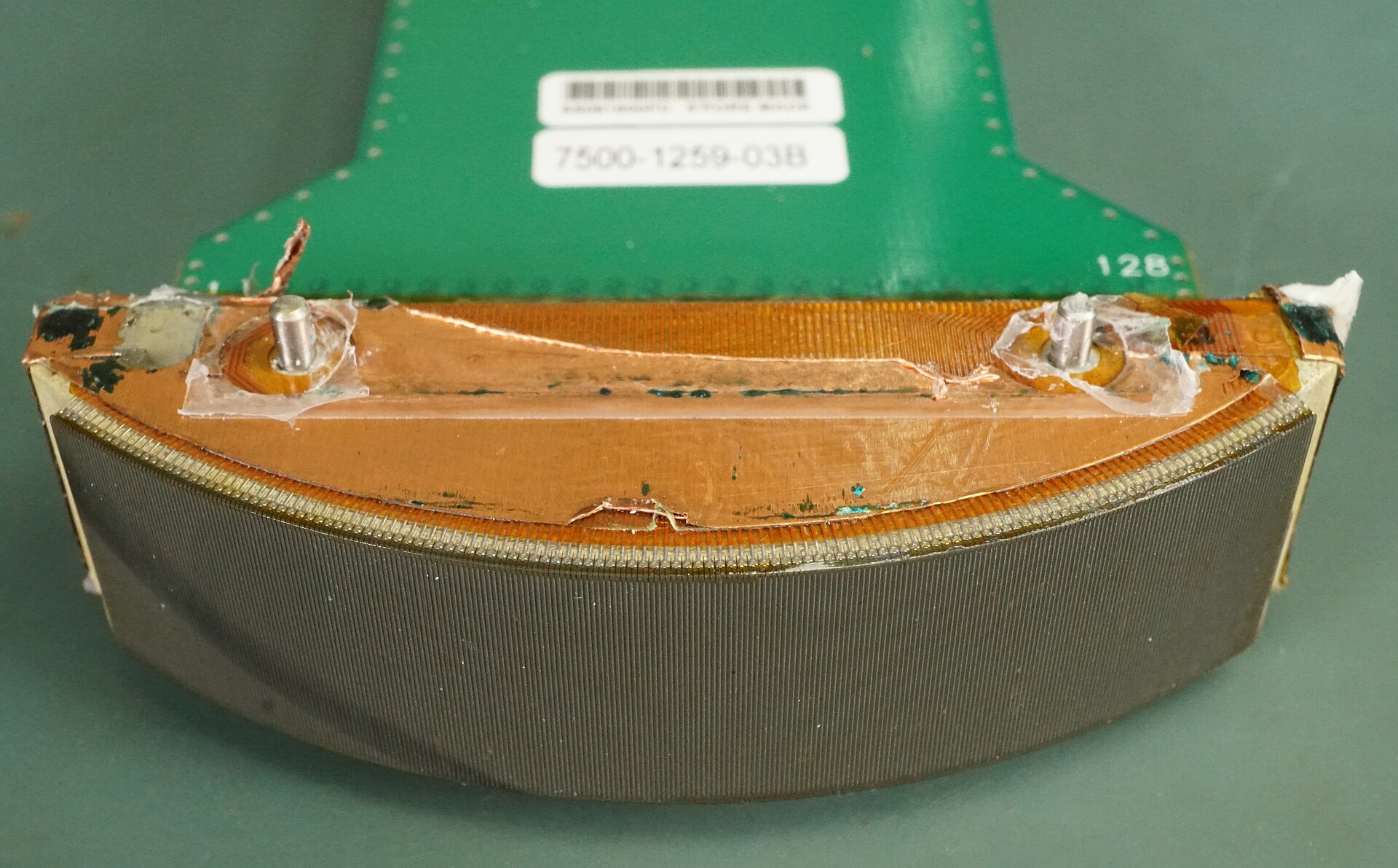
Each of the three is a wireless handheld. The probe holds the beam-former, the processor, and the battery inside one shell. It sends the picture it builds over the air to a phone or a tablet in the clinician’s hand. No cart rolls in behind it. No cable runs to a socket in the wall. The shrink came from chips that now do in a small shell the work a rack of boards once did across a lab. A clinician wakes the probe, lays it on the skin, and reads within seconds. The image lands on the screen in real time over a wireless link. A clip and its measurements save to the patient’s record from the same screen. A clinician pulls up the last study for a side-by-side look or sends a trace to a specialist across the network for a second read. The whole machine weighs little and rides in a coat pocket between patients, charged off a phone or a small cell. A single charge lasts a clinic session. A spare cell covers a long list.
The form decides where the reading happens. A specialty scan that once waited on a booked lab slot now travels to the bedside in a pocket. A clinician at a rural post reads a heart or an eye in a clinic no cart could reach. The depth travels to the patient now. It gives its answer in the same visit. A power bank charges the probe between clinics, so a field team carries it into a tent or the back of a truck. The reading happens at the chair, the ward, or the roadside, wherever the patient already is. A wipe cleans the probe between patients, with no gel cart to wheel and no console to scrub down. The kit packs into a bag a clinician carries on one shoulder.
The heart’s fast jets
The first area reads the fast blood the heart drives through a faulty valve. A narrowed or a leaking valve sends the blood through at speed. The jet it throws, caught by a continuous-wave Doppler probe, becomes the pressure the heart works against. That pressure grades the valve. A clinician reads the speed off the trace and turns it into a number a whole team can act on. This is the one cardiac job that needs a Doppler mode with no speed ceiling. The handheld shows the jet as a bright streak on the colour map first. A clinician lays the beam down that streak to read its speed.
The continuous-wave mode is the line between this probe and the rest of its size. It sends and listens at once. That asks for split crystals and a steady stream of processing. Packing all of it into a pocket shell is the hard part of the build. The probe that carries the mode reads the fast jet whole and grades the valve from its speed. The mode is the one the basic pocket scanners leave out. The rarity is the point of the difference, since the fast jet is exactly where the grade lives.
The mode opens the high-pressure work of the heart at the chair. A clinician grades a tight aortic valve, sizes a leaking mitral one, and reads the pressure in the lungs from a small backward jet at the tricuspid valve. Each of these rests on a fast jet that only the continuous-wave mode reads whole. The probe catches the jet and hands back a number in a minute. A clinic with the probe settles a murmur on the spot, turning a heard sound into a measured load with no referral to the echo lab. A clinician who once wrote a referral and waited a week now reads the valve, grades it, and books the follow-up before the patient stands up. The mode reads a replaced valve too, tracking its gradient over the years for a rising number that warns of trouble.
The number lands while the patient is still in the room. A clinician reads a valve on the first visit and plans the next step the same hour. The referral goes only when the grade calls for one.
A full look at the heart
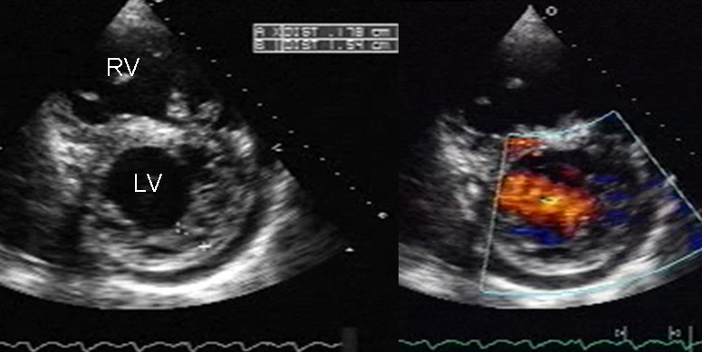
The second area takes in the whole heart at work. A phased-array cardiac probe slips its beam between the ribs and brings the chambers, the walls, and the valves onto the screen in motion. Handheld echocardiography works from a few set windows on the chest, each one opening onto a part of the heart. A clinician finds the standard views and reads them in turn. A clinician sweeps from the apex to the edge of the sternum, reading one window at a time. The set of windows covers the heart from a handful of spots on the chest. The same low-frequency probe that carries the fast-jet Doppler builds this picture, so one device covers both cardiac areas on the one probe.
The study answers a sick heart’s questions in the first minutes at the bedside. A clinician checks the size of each chamber and the strength of the squeeze. A wall that barely moves, a chamber swollen out of shape, a dark rim of fluid around the heart: each shows on the scan within those first minutes. The finding points the next step of care before the bloods come back. A clinician marks the chamber sizes and the wall motion on the spot, building a record the next reader can follow. A clinician reaches for this read at the side of a breathless patient or a crashing one, where a few minutes change the outcome. A clean heart on the screen clears the worst worry in seconds, so the same scan calms as readily as it warns.
The work follows the echo guidelines that set out what a full study covers and how each view is taken. A handheld reaches the parts its probe can manage at the chair. A clinician knows that reach before scanning and sends the complex case on for a complete study in the lab. The bedside read settles the common questions early and flags the few that need the cart. It aims at speed and reach. A hard case still ends with a full lab study, on the larger probe with its three-dimensional views. The handheld opens the assessment in the clinic.
The area carries its own standards and its own roster of findings. A pericardial effusion pressing on the heart, a wall left still by a blocked artery, a right heart strained by a clot in the lung: each has its own read and its own clock. A clinician learns them as a set and runs through them at the bedside. The depth here is the depth of a focused echo, carried to where the patient lies, with no wait for a lab slot a week away.
A clinic gains a cardiac study it once had to send away. The patient is scanned where they lie. The answer rarely waits on another appointment.
The eye
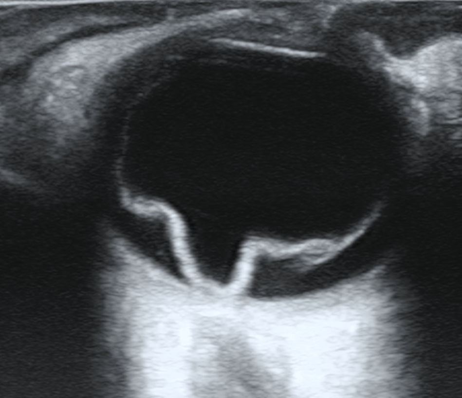
The third area reads the eye, a small ball of fluid that sits a few centimetres under a closed lid. The shallow depth lets the probe run at a high frequency. The finest detail comes clear at that frequency. An ophthalmic probe rests a flat face on the lid and reads the eye lying behind it. The scan maps the retina, the vitreous, the lens, and the optic nerve in a matter of seconds. A drop of gel on the closed lid is all the preparation it asks of the patient. A clinician asks the patient to look up, down, and to each side. After that, the whole back of the globe passes under the beam.
The scan reads an eye the doctor cannot see into. The globe shows as a dark circle on the screen, its back wall a bright curve behind the fluid. A cataract clouds the lens. Blood clouds the vitreous. Ultrasound reads straight through both clouds to the back wall of the eye. A retina peeled off its bed, a vitreous full of blood, a metal splinter lodged in the globe: each shows on the scan when the view through the front of the eye is lost. The picture stands in for a look the doctor can no longer get. A few sweeps across the closed lid do from the surface what the light of an exam can no longer reach.
The eye scan answers a short list of urgent questions. Has the retina torn loose and floated forward off the back wall? Does fresh blood fill the vitreous behind a sudden loss of sight? Has a splinter of metal or glass driven into the globe? Each answer shows as a clear sign on the grey-scale picture, read in the minutes that matter for saving sight. A clinician reads the back of a blind eye from the outside and decides whether it needs a surgeon that day or a watch over the week. Speed is the whole point here. A detached retina left too long can lose its sight for good.
The probe is the lightest of the three by design. The blueprint marks it at around a hundred grams, near the weight of a small phone. A light face rests easy on a closed lid through a long scan and keeps the pressure off an injured eye. A clinician holds it steady with a light touch. The low weight is a deliberate safety feature. The eye will not take a heavy hand.
A clinic that does eye work carries this probe and reaches for no other on the job. The scan needs the high frequency and the light hand together. The ophthalmic probe stands on its own among the three.
Choosing the probe a case needs
The right probe is the one the case in front of the clinician calls for. A clinician names the scan first. The probe that fits the scan needs no second thought.
What the general probe still does
The general handheld stays in daily use through all of this. It reads the belly, the lungs, the bladder, and the soft tissues, the scans that fill the bulk of a working day. Those everyday scans make up a body of work of their own. A clinic keeps the general probe on the shelf beside the specialty ones and moves between them through the day. The general probe is the workhorse, out for nearly every patient who comes through the door. A clinician reaches for it first on nearly every visit, before any specialty probe comes out of the bag. Its day is a long one. It checks a belly for free fluid, a chest for a collapsed lung, a bladder for how full it sits. It guides a needle into a vein or a pocket of fluid. Its broad reach is the thing that keeps it busiest.
A clinic carries the probes its work needs. It buys no probe it will never reach for. A general probe covers the broad sweep of the scanning by itself. A specialty probe joins the set the day a job earns it. The set grows one probe at a time. A new line of work adds the next probe when it comes.
The boundary between the general probe and the specialty ones is the boundary of the job. A general probe answers a wide first question across the body and points the way to the trouble. A clinician then reaches for the specialty probe to grade what the general scan found. The two work as steps in the one workup, the wide look first and the deep read after.
The reach the three probes add
Together the three probes hand a clinic the reach of an echo lab and an eye clinic. The whole set folds into a coat pocket. A clinician grades a valve, reads a failing heart, or maps a blind eye, each on the probe that suits the task. The depth that once lived in a hospital corner now rides out to the patient. A district clinic answers a question on the spot that used to mean a bus ride to a city lab. The gap between a symptom and a reading closes to a single visit. The machine costs a fraction of the cart it stands in for. The three together cover a span of medicine that once needed two departments and a wall of equipment. One clinician with the set reads across all three in a single sitting.
The reach proves itself where it is hardest to come by. A clinic an hour from the nearest hospital reads a heart or an eye that once meant a long referral down the road. A camp with a generator and a phone runs a focused study off a charged probe. The depth travels the last mile to the patient, down a dirt track or up a stairwell, in a bag over one shoulder. The places a cart machine never reached are exactly where the pocket probe belongs. A clinician on a home visit reads a heart in a front room. The report saves to the record before the clinician leaves the house.
The wireless form widens the reach again. A laptop scanner sits between the cart and the pocket in size and in cost. The pocket probe undercuts both for a focused study. It travels where neither can follow. The price of a pocket probe opens these reads to clinics that could never afford a cart machine. The capability spreads to where the patients are, into clinics a hospital lab never served.
The probes carry their own training in the hand. A learner finds a jet, marks a peak, or measures an eye under a teacher’s gaze, then keeps the clip to review after the clinic. The picture a pocket probe draws matches the picture a cart machine draws, so the skill built on one carries to the other. A clinic raises its own readers on the device it already owns. A teacher watches the same screen the learner reads, so the lesson happens right at the patient. More trained hands widen the reach.
These three areas are set out one at a time. A reader turns to the area their own work calls for and follows it down to the scans. The continuous-wave Doppler probe, the advanced cardiac probe, and the ophthalmic probe each open a body of work a general probe leaves untouched. A clinic that adds one reads cases it could not read before. The machine that does it travels in a pocket to the patient.
Common questions about specialty ultrasound probes
What is a specialty ultrasound probe?
A probe built and tuned for one organ or one kind of question. It gives up the breadth of a general probe to read its one job to a higher standard. Three kinds cover the cardiac and eye work: a continuous-wave Doppler probe for the heart’s fast jets, a phased-array probe for full handheld echocardiography, and a high-frequency probe for the eye.
Why does the eye need a different probe from the heart?
The two organs sit at different depths. The eye lies a few centimetres under the lid, so its probe runs at a high frequency for fine detail. The heart sits a hand’s depth behind the ribs, so its probe runs at a low frequency to reach that far. No single frequency does both jobs well.
Are these probes wireless handhelds?
Yes. Each of these probes holds its works and its battery in one shell and sends the picture to a phone or a tablet over the air. No cart and no cable are needed. A clinician scans at the bedside, the ward, or a remote post, anywhere a patient happens to be.
Does a specialty probe replace a general one?
No. A general probe still runs the bulk of a clinic’s everyday scanning, the belly, the lungs, and the soft tissues. A clinic adds a specialty probe when a job needs the extra reach. The two work as a set, with a clinician moving between them through the day.
Which probe does a clinic need?
It depends on the caseload. A clinic that reads hearts all day carries the cardiac probes. One that also screens eyes keeps the ophthalmic probe to hand. A mixed clinic builds its set to match the cases at its door and adds to it when the work widens.

About the Author
Julien Mercier
Senior R&D Engineer · Medical Ultrasound Transducer Development
Senior R&D Engineer with an M.S. in Applied Physics and over 15 years of experience in medical ultrasound transducer development, specializing in the design verification and performance testing of high-frequency imaging transducers. Currently leading the development and verification of the company’s next-generation high-frequency linear-array transducer, responsible for imaging performance evaluation and reliability analysis in preclinical testing. Brings extensive hands-on experience in piezoelectric element tuning, beamforming parameter optimization, and system-level performance testing.