





































What Continuous Wave CW Doppler Actually Measures In Cardiac Exam
Continuous wave Doppler reads the speed of blood along a line through the heart. It listens for the fastest flow anywhere on that line. It puts no speed limit on what it can read. The beam is aimed through a jet. The peak velocity is read off the trace. That velocity is turned into a pressure gradient. The gradient is how a heart valve is graded.
What CW Doppler measures
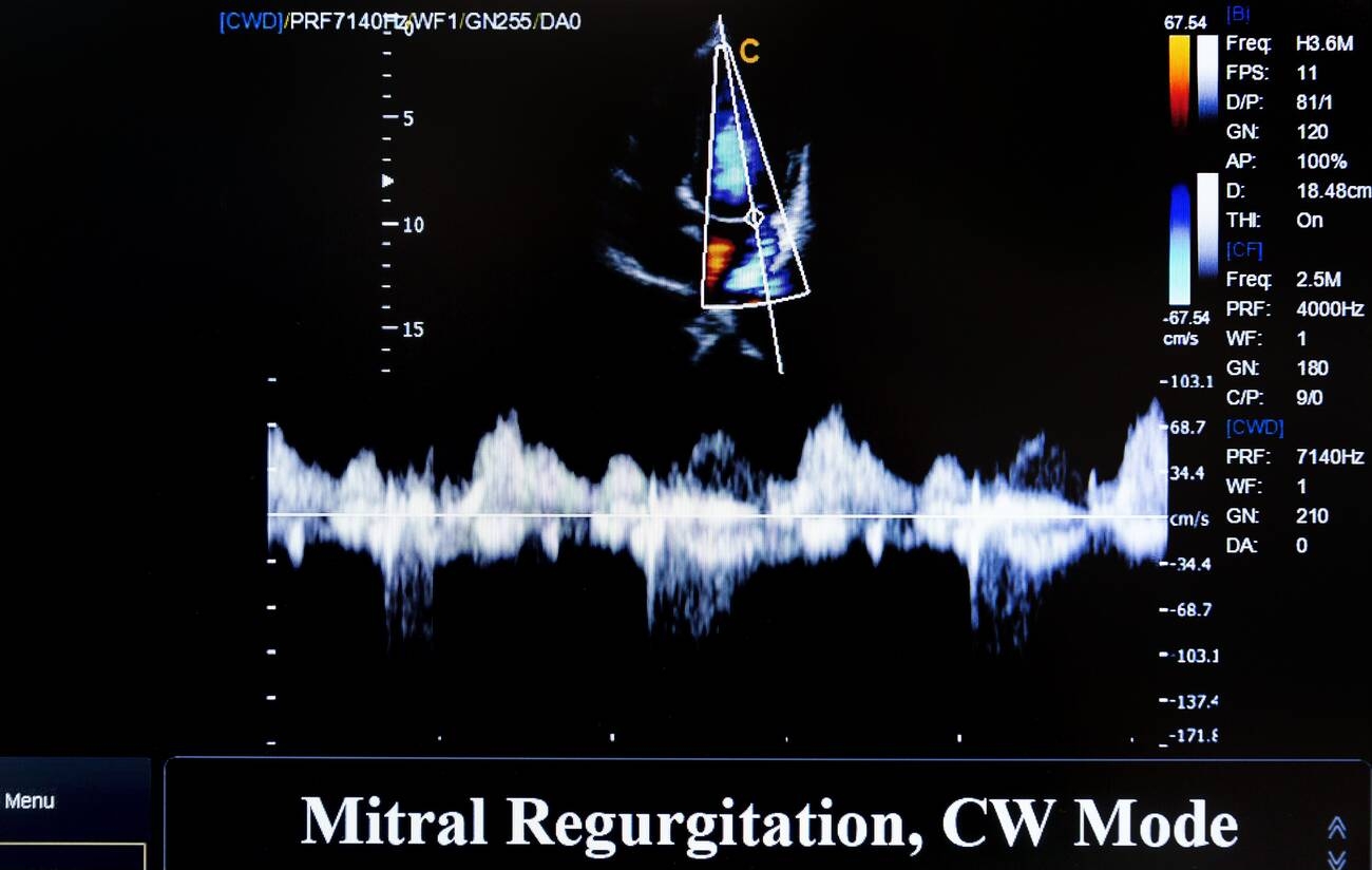
Continuous wave Doppler measures the speed of blood. It uses a beam of sound aimed through the heart. Blood moving toward or away from the beam shifts the sound’s pitch. The size of that shift gives the speed of the blood. The probe reads the shift and shows the speed on a trace. The shift is the Doppler effect at work. Sound bounced off moving blood comes back at a changed pitch. Faster blood changes the pitch more. The probe turns that pitch change into a speed.
The probe sends and listens at the same time. One crystal sends a steady beam of sound. Another listens to what comes back, without pause. This steady send and listen is what the name means. The beam is on continuously, end to end. The continuous beam is what gives the mode its reach. A steady listen catches the fastest moment of a jet. Nothing is missed between pulses. The mode hears the flow without a gap.
The reading is a speed, in metres per second. A normal flow runs around one metre per second. A tight valve drives blood far faster. The trace shows the speed climb as the jet speeds up. The number a clinician reads is the peak of that climb. The peak is the number that counts. A jet rises to a top speed each beat. The clinician reads that top. The rest of the climb leads up to it. A clinician sets the scale so the peak fits the screen. A jet that runs off the top reads no number. The scale is opened until the whole envelope shows. The peak then sits inside the trace, ready to mark.
No speed limit
Continuous wave Doppler has no speed limit. It reads any velocity, however high. A fast jet does not outrun it. The trace shows the true peak even at seven metres per second and beyond. A healthy heart rarely drives blood past one or two metres per second. A diseased valve can push it past five or six. The mode reads either with the same ease. No jet outpaces it.
This freedom from a speed limit is what sets the mode apart. A pulsed mode hits a ceiling and folds the signal over. Continuous wave never does. The ASE recommendations for Doppler quantification set out how the peak velocity is measured and turned into a gradient. A clinician reads the highest clean velocity on the trace. The number holds however fast the jet runs.
The high jets are the ones that matter. A narrowed valve speeds blood up sharply. A leaking valve fires a fast jet backward. These are the flows a clinician needs the speed of. Continuous wave reads them where a capped mode cannot. The fast jet is exactly the hard case. A clinician needs its speed to grade the valve. A mode that caps out loses the number right where it matters. Continuous wave holds it.
The speed it reads is the whole point of the mode. A clinician reaches for continuous wave when the flow is fast. The mode is built for the high-velocity jet. It reads the peak no other mode can hold. A clinician learns to switch to it for any fast flow. The colour and pulsed modes show the jet is there. Continuous wave measures how fast it runs. The reading is the number the grade is built on.
Range ambiguity, the trade
The speed comes at a price. Continuous wave listens along the whole beam at once. It cannot say where on the line the fast flow sits. The peak it reads could come from anywhere along the beam. This is called range ambiguity. The whole beam is one long microphone. It hears every speed along its length together. A fast spot anywhere on the line shows up on the trace. The mode cannot place that spot on its own. A clinician accepts the trade for the speed it buys. The fast jet is read whole, wherever it lies. The image fills in the missing where. The pair covers what neither does alone.
A clinician works around the ambiguity with the picture. The two-dimensional image shows where the jet is. The clinician lines the beam up through it. The trace then reads the velocity of that jet, placed by the eye on the image. The picture says where, the Doppler says how fast. The image and the trace work as a pair. A clinician finds the jet by eye on the picture. They lay the beam down it and read the speed. The two together place the velocity and measure it.
From velocity to pressure
A velocity is read for the number it gives. The number is the pressure gradient across the valve. A faster jet means a bigger pressure drop. The gradient grades how tight a valve is. A pressure gradient is a drop in pressure across a valve. Blood speeds up because the pressure behind it is higher. The faster it goes, the bigger that drop. The velocity is a window onto the pressure.
The velocity becomes a gradient through a simple sum. The pressure gradient is four times the velocity squared. A clinician reads the peak velocity and squares it, times four. The table below sets out the conversion. The rule is quick enough to do in the head. A jet at four metres per second squares to sixteen, times four is sixty-four. A clinician reads a gradient straight off a velocity. The machine shows both side by side.
| Peak jet velocity | Pressure gradient (4v²) | A rough read |
|---|---|---|
| 1 m/s | 4 mmHg | Normal or near-normal flow |
| 2 m/s | 16 mmHg | A mild jet |
| 3 m/s | 36 mmHg | A moderate jet |
| 4 m/s | 64 mmHg | Often a severe valve |
| 5 m/s | 100 mmHg | A tight, critical valve |
The gradient is the number that travels. A clinician records the gradient as the working number. The raw velocity is the step on the way to it. The gradient compares across patients and across visits. The conversion turns a speed into a pressure a whole team can read. A gradient in millimetres of mercury is a familiar number. A surgeon, a cardiologist, and a sonographer all read it the same. The conversion puts the Doppler reading into the language the team works in. The number leaves the echo lab and travels with the patient.
The highest velocity on the line
Continuous wave reads the fastest blood anywhere along its beam. The peak velocity is the number it gives.
The jets CW is for
Continuous wave is the mode for the fast jet. A healthy valve lets blood through at a gentle speed. A narrowed valve forces the same blood through a smaller hole. The blood speeds up to get through. The tighter the valve, the faster the jet. Continuous wave reads that speed and turns it into the pressure the heart is working against. A leaking valve makes a jet of its own. Blood fires backward through a valve that should be shut. The jet runs fast, driven by the pressure behind it. Continuous wave reads the leak the same way it reads a narrowing, by the speed of the jet. The aortic valve, the mitral valve, and the tricuspid valve each give their own jets. A clinician aims the beam through the one in question and reads its peak. The number grades the valve. The whole of valve assessment leans on the speed continuous wave can read.
The narrowed valve is read by the peak it drives. A clinician aims through the narrowing and reads the fastest flow. The peak velocity gives the gradient across the valve. The gradient says how hard the heart pushes to clear it. A clinician aims the beam from the apex up through the aortic valve. The jet runs away from the probe there. The trace reads its peak below the baseline. The number gives the gradient the left heart works against.
The leaking valve is read the same way. A clinician aims through the backward jet and reads its peak. The speed of the leak reads the pressure driving it. A regurgitant jet is graded by what continuous wave reads off it. A leak is a jet the wrong way through a shut valve. It runs fast because the chamber behind it is at high pressure. A clinician reads its peak the same as any jet. The speed grades how much pressure drives the leak.
The tricuspid jet reads the lung’s pressure. A small leak across the tricuspid valve is common and useful. Its peak velocity gives the pressure in the right heart. A clinician reads the lung’s pressure off that one jet. The tricuspid leak is small in many healthy hearts. A clinician still reads it for the right-heart pressure it carries. Its peak velocity plus a fixed number gives the pressure in the lung’s arteries. One small jet opens a window on the right side of the heart.
Every jet is read by its speed. A clinician finds the jet on the image. They aim the beam down its length. They read the peak off the trace. They turn it into a gradient. The same steps read every fast flow in the heart. The steps do not change from valve to valve. A clinician learns them once and runs them everywhere. The aortic, the mitral, and the tricuspid each take the same routine. The mode reads them all by the one method. A clinician builds the habit on the common valves first. The rarer jets follow the same drill. The method carries from one valve to the next. The reading is the same craft each time.
Reading the spectral trace
Continuous wave shows its reading as a spectral trace. The trace plots speed against time. A jet draws a filled envelope over each beat. The outer edge of the envelope is the peak velocity. A clinician reads the top of the envelope for the number. The trace runs left to right with time. Each beat draws a new envelope. A clinician watches a few beats and reads the tallest clean one. The peak of that envelope is the velocity.
The trace fills in because of the range ambiguity. Continuous wave hears every speed along the beam at once. The trace shows them all, stacked into a solid shape. A clinician reads the outer edge, the fastest flow, and lets the fill be. The peak is what the gradient needs. The solid fill is normal for the mode. A clinician does not read into the middle of it. The edge is the only line that matters. The fill is the price of hearing the whole beam.
A clean trace gives a clean peak. A well-aimed beam draws a crisp outer edge. A clinician traces that edge or marks its top. The machine reads the velocity and the gradient from the mark. A ragged trace is re-aimed until the edge comes clear. The machine can trace the edge by itself or by hand. A clinician checks the auto-trace and fixes it where it strays. The marked peak sets the velocity and the gradient. A clean edge makes the reading easy to trust.
The shape of the trace carries more than the peak. The rate of the rise, the time the jet takes to fall, and the shape of the envelope each tell the clinician something. A clinician reads the peak first and the shape after. The trace is the mode’s whole output, read top to bottom. A clinician reads the peak for the gradient first. The fall and the shape add to the picture after. The trace holds more than one number. The peak is the one the grade leans on. A clinician reads the trace across a few steady beats. An odd beat is skipped for a clean one. The peak is taken from a beat the clinician trusts. The reading rests on a good trace.
CW on a handheld
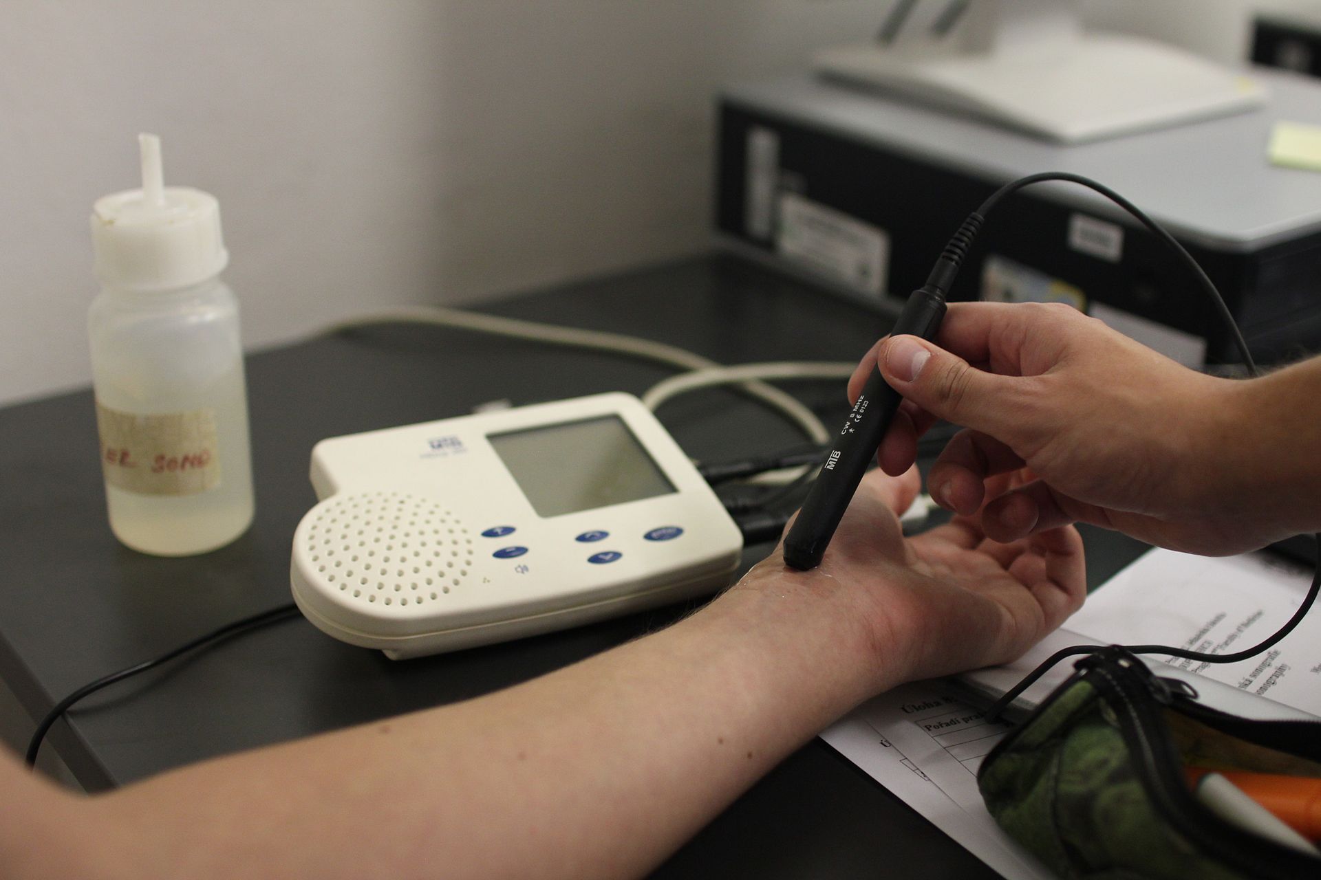
Continuous wave runs on a handheld probe. The mode asks for two crystals and the power to drive them. A handheld built for it carries the mode to the bedside. A clinician reads a valve gradient at the patient’s chair, on a probe in one hand. The bedside read is the mode’s home on a handheld. A clinician carries it to the ward or the clinic room. The valve is graded where the patient sits. No trip to the echo lab is needed for the number.
Few handhelds carry the mode. Continuous wave is harder to build into a small probe than the basic modes. A handheld that offers it brings valve assessment out of the echo lab. A clinician grades a valve at the bedside, on a machine that fits in a pocket. The mode on a handheld is what this page is about. A handheld with the mode does what a cart machine does for a valve. A clinician reads the same gradient on either one. The small probe brings the reading to places a cart cannot reach. The mode in the hand is what makes that possible. A clinician at a small clinic grades a valve on the spot. The handheld carries the mode to the patient. The reading is done where the patient already sits. No referral to the lab is needed for the gradient.
What CW leaves to other tools
Continuous wave reads fast flow along a line. It does not read where on the line the flow sits. A pulsed mode does that, at the cost of a speed limit. A clinician picks the mode the flow calls for. A clinician reaches for the pulsed mode to place a slow flow at a depth. They reach for continuous wave to measure a fast one. The choice turns on what the question needs.
The velocity-to-gradient sum has more to it than the simple form. The four-times-squared rule drops a term that sometimes matters. A clinician uses the simple form for everyday jets and the full one when it counts.
The pulse settings, the gain, and the scale each shape how the trace reads. A clinician tunes them to draw a clean edge. A clean trace is half the reading. The settings that draw it repay a clinician’s time. A well-set machine makes the peak easy to read.
Aligning the beam
The reading is only as true as the aim. Continuous wave reads the part of the speed that runs straight along the beam. A jet crossing the beam at an angle reads slower than it should. A clinician lines the beam up with the jet to read the true peak. The aim is the first thing the reading depends on. The beam reads only the speed along its own line. A jet square to the beam reads as almost nothing. A jet straight down the beam reads its full speed. A clinician aims to run the beam along the jet.
The angle is the main source of a wrong reading. A beam off the jet’s line reads a velocity too low. The gradient that follows reads too low as well. A clinician swings the probe until the peak reads highest. The highest peak is the best-aligned one. A clinician trusts the highest reading. A low one usually means a poor angle. A better aim gives a higher peak. The best aim gives the highest clean peak.
A clinician hunts for the true peak across several windows. The jet may read highest from one spot on the chest. A clinician tries more than one window and keeps the highest clean signal. The best window is the one the jet runs straight toward. The peak from there is the one to trust. A clinician does not settle for the first signal. They sweep the probe and watch the peak rise and fall. The window that reads highest is kept. The true peak hides until the beam finds its line.
The alignment is what separates a good reading from a poor one. A careless aim reads a jet too slow. The valve then grades kinder than it is. A careful aim reads the true peak. The number is only as good as the line it was read along.
The beam alignment is the skill the mode asks for. A clinician learns to find the jet and run the beam down it. The reading rewards a steady hand and a true aim. The mode gives a true number to the clinician who aims it true. The aim is the craft behind the gradient.
What CW Doppler gives the cardiac exam
Continuous wave gives the cardiac exam the speed of the fast jet. A clinician reads a velocity no capped mode can hold. The velocity becomes a gradient. The gradient grades the valve. The mode turns a fast jet into a number a clinician can act on. The grade flows from the speed the mode reads. A clinician turns a jet into a velocity, a velocity into a gradient, a gradient into a grade. Each step rests on the one before. The whole chain starts with the speed continuous wave can read.
The mode is what lets a handheld grade a valve. A clinician aims the beam through a jet and reads its peak. The peak gives the gradient. The gradient grades the valve. Continuous wave is the part of the exam that reads the high-velocity flow. It brings valve assessment to the probe in the hand. A clinician with the mode reads a valve without a referral. The jet is found, aimed, and read at the bedside. The gradient is in hand in a minute. The mode put valve grading where the patient is.
Common questions about continuous wave Doppler
What does continuous wave Doppler measure?
The speed of blood along the beam. It reads the fastest flow anywhere on the line, at any velocity, with no speed limit. A clinician aims the beam through a jet and reads its peak velocity. That velocity is turned into a pressure gradient by the four-times-squared rule. The gradient grades the valve.
Why does continuous wave have no aliasing?
Because it is not a pulsed mode. It sends and listens without pause, so it never has to wait for an echo to return. A pulsed mode samples in bursts and caps out at a speed limit. Continuous wave reads any velocity, however high. That is why it is used for the fast jets.
What is range ambiguity?
The trade for the speed. Continuous wave listens along the whole beam at once. It cannot say where on the line the fast flow sits. A clinician uses the image to see where the jet is and aims the beam through it. The picture says where; the Doppler says how fast.
How does velocity become a pressure gradient?
Through the simplified Bernoulli rule. The gradient is four times the peak velocity squared. A jet at three metres per second gives a gradient of thirty-six. A clinician reads the peak and the machine does the sum.
Can a handheld do continuous wave Doppler?
Yes, if it is built for it. The mode asks for the hardware to send and listen at once. A handheld that carries it grades a valve at the bedside. Few small probes offer the mode. The one that does brings valve assessment to the probe in the hand.

About the Author
Julien Mercier
Senior R&D Engineer · Medical Ultrasound Transducer Development
Senior R&D Engineer with an M.S. in Applied Physics and over 15 years of experience in medical ultrasound transducer development, specializing in the design verification and performance testing of high-frequency imaging transducers. Currently leading the development and verification of the company’s next-generation high-frequency linear-array transducer, responsible for imaging performance evaluation and reliability analysis in preclinical testing. Brings extensive hands-on experience in piezoelectric element tuning, beamforming parameter optimization, and system-level performance testing.