A fibroid is a benign growth of the muscle of the womb. The medical name is a leiomyoma. Ultrasound is the test that finds a fibroid and measures it. A scan shows where the fibroid sits in the wall of the womb. The place it sits tells a clinician what it is likely to cause. A scan reads the number, the size, and the position of every fibroid in one sitting.
What a fibroid is
A fibroid grows from the smooth muscle of the uterine wall. It builds a firm round lump of muscle and fibre. A womb can hold one fibroid or a dozen. A fibroid can stay the size of a pea for years. Another can grow to the size of a melon. The growth is benign. A fibroid turns to cancer in only the rarest of cases. A fibroid is made of muscle cells wound through with fibre. The mix gives it its firm feel. Estrogen feeds a fibroid through the menstrual years. A fibroid often grows in pregnancy, when those hormones run high. A fibroid commonly settles after the menopause, once the hormones fade. A fibroid sits in one of a few places in the womb. The wall holds the larger share of them. A reader tells a fibroid from a polyp by where it starts, since a polyp grows from the lining. Some women carry a higher risk of fibroids. The menstrual years raise it. A family history raises it, and a fibroid runs in some families more than others. Women of African heritage carry the highest risk of all. A fibroid can grow in the neck of the womb or out to the side. A cervical fibroid sits low and presses on the bladder. A fibroid out to the side reaches into the broad ligament. A reader maps these less common spots too. A fibroid carries receptors for the hormones of the cycle. Estrogen and progesterone both feed its growth. A drug that blocks those hormones can shrink a fibroid for a time. A clinician uses the plain word fibroid with a woman and the word leiomyoma in the report.
Fibroids are the commonest growth of the womb. More than seven in ten women grow at least one by the age of fifty. A fibroid often causes no trouble at all. A woman can carry one for years and never know. A scan for another reason finds many fibroids by chance. A small fibroid sits silent in the wall for years. A fibroid earns attention once it grows or once it bleeds. The number of fibroids in a womb ranges from one to many. A reader counts them all on the scan. Fibroids grow through the years a woman menstruates. They tend to appear from the thirties onward. A fibroid found in a young woman is watched across the years that follow. A fibroid grows at its own pace, and a scan across time reads how fast. One fibroid sits unchanged for years. The next doubles in a single year. A fibroid is one of the commonest reasons a woman is sent for a pelvic scan. Heavy periods bring her in. A scan reads the womb for the fibroids behind the bleeding. A fibroid earns its own line in the report, with its size, its place, and its FIGO type. A silent fibroid is left alone and watched. A fibroid shrinks after the menopause as a rule. Growth at that age breaks the rule and earns a closer look. A fibroid is a benign growth a woman can live with. A scan names it benign and a clinician reassures her. A fibroid earns care only when it causes trouble. A young woman with a small fibroid is reassured and watched across the years. A fibroid is measured in centimetres on the scan. A small one runs under a few centimetres. A large one can pass ten. The size and the symptoms together set whether a fibroid is treated. The size alone settles nothing. A scan reads the size of the whole womb beside the fibroids. A womb full of fibroids reads large for a woman who is not pregnant. A reader gives the womb size and the fibroid map together. A reader reads the count of the fibroids as well as the size, and both go into the report.
What it looks like on the scan
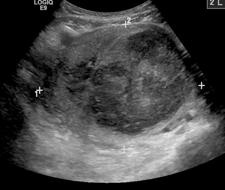
A large fibroid on ultrasound, measured with calipers across a rounded mass in the wall of the womb. It reads darker than the muscle around it.
A fibroid shows on ultrasound as a round mass in the wall of the womb. It reads darker than the muscle around it. Its edge is clear against the normal wall. A reader sees the mass stand apart from the tissue beside it. A reader reads a fibroid for its border first. A clear border is the mark a reader looks for. A fibroid can sit anywhere in the wall of the womb. A scan reads the front wall, the back wall, and the floor. The shape is round or oval. A whorled grain runs through some fibroids. A young fibroid reads as an even grey mass. An older fibroid breaks up inside as it outgrows its blood supply. The breakdown leaves dark pockets within the mass. A reader knows that change as degeneration. Degeneration takes a few forms in a fibroid. A quiet form leaves the mass a touch softer on the scan. A cystic form leaves dark fluid pockets inside. A fibroid in pregnancy can degenerate and turn painful as it outgrows its supply. A reader reads the pockets and the pattern for the kind of change. Red degeneration can strike a fibroid in pregnancy. The fibroid outgrows its blood and bleeds within itself. The change brings a sharp pain and a tender mass. A scan reads the fibroid for the dark change inside. The pain settles over days with rest and relief. A fibroid keeps a sharp edge a reader can trace. Adenomyosis has no such edge and blurs into the wall around it. A rare cancer of the muscle grows fast and breaks up inside. A scan cannot name that cancer for certain, and a fibroid that grows fast earns an MRI to look closer.
A fibroid bends sound in its own way. It casts thin shadows from its edges. A line of bright spots marks the calcium an old fibroid lays down. A reader knows that popcorn pattern as a fibroid grown old. Sound passes through it more slowly than through plain muscle.
Colour laid over a fibroid shows the blood that feeds it. A rim of flow runs around the edge of the mass. The centre carries less. A reader reads that ring of flow as the mark of a fibroid. The pattern sets it apart from the tangled flow of other growths. A fibroid in the wall can push the lining out of shape, and a reader reads the lining for the dent it leaves. A fibroid is sized to the millimetre, and the largest one sets the size of the whole womb. A reader holds the mass still and measures it in two views, down the womb and across, to catch its true size. A fibroid reads in shades of grey for the muscle, the fibre, and the calcium it holds. A dense fibroid throws a shadow that hides what sits behind it, and a reader tilts the probe to read around it. An old fibroid hardens with calcium at its rim, a bright line with a shadow behind.
A fibroid is measured in three directions. The reader records its width, its height, and its depth. The number, the size, and the place of each fibroid go into the report. A scan months later reads the new size against the old. A fibroid that grows fast earns a closer look. A reader maps a womb full of fibroids one by one. Each fibroid gets a size and a place in the report. A clinician reads that map before any treatment. The map of today reads against the map of the last scan. A submucosal fibroid can hide against a thick lining. A drop of salt water in the cavity lifts the walls apart and shows the fibroid clearly. This saline scan reads a fibroid that a plain scan left in doubt. A reader reaches for it when a fibroid sits near the cavity. A reader places a fibroid against the lining, the wall, and the outer surface. The exact place decides the FIGO type. A scan reads how much of a fibroid sits inside the wall and how much reaches the cavity.
Where it sits in the womb
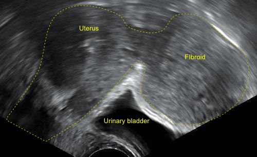
A subserosal fibroid bulging from the outer wall of the womb, above the bladder. The yellow dashed outlines and labels are added to mark the uterus, the fibroid, and the bladder.
The place a fibroid sits matters more than its size. A fibroid under the lining presses on the cavity. A fibroid in the wall sits in the muscle. A fibroid on the outside bulges into the pelvis. A reader maps each one to its place and marks it on a sketch of the womb for the surgeon.
Where a fibroid sits, and what it tends to cause
Where it sits
FIGO type
On ultrasound
Tends to cause
Submucosal (under the lining)
0–2
Bulges into the cavity
Heavy periods
Intramural (in the wall)
3–5
Sits in the muscle
Bleeding and bulk
Subserosal (on the outside)
6–7
Bulges from the surface
Pressure on the bladder or bowel
Pedunculated (on a stalk)
0 or 7
Hangs from a stalk
Pain if it twists
Cervical or broad-ligament
8
Sits apart, low or to the side
Pressure low down
Clinicians sort fibroids by a numbered system from FIGO. The table sets out the types and what each one tends to cause. A submucosal fibroid sits under the lining and reaches into the cavity. An intramural fibroid sits deep in the wall. A subserosal fibroid bulges from the outer surface. A pedunculated fibroid hangs from a stalk.
The map guides the care that follows. A fibroid under the lining drives heavy bleeding. A fibroid on the outside presses on the bladder or the bowel. A reader notes which fibroid sits where, so a clinician can match the treatment to the cause. The FIGO numbers run from zero to eight. A type zero fibroid sits wholly in the cavity on a stalk. A type one or two reaches in part into the cavity. A type three to five sits in the wall. A type six or seven bulges from the surface. A type eight sits apart, in the cervix or the ligament. A reader reads the number for where a fibroid sits. Ultrasound answers the question in the great run of wombs. A crowded womb of many fibroids can outrun a plain scan. An MRI maps such a womb before surgery, and it helps tell a fibroid from its rare cancer mimic. A three-dimensional scan builds a coronal view of the womb. The coronal view reads how far a submucosal fibroid reaches into the cavity.
What fibroids cause, and the map that guides care
Heavy bleeding is the commonest trouble a fibroid causes. A submucosal fibroid sits under the lining and pushes on the surface that sheds each month. The fibroid stretches that surface wider. The wider surface bleeds more with every period. A woman passes clots and soaks through her protection. The bleeding runs for more days than a normal period lasts. The steady loss drains the body of iron over the months. A woman grows tired and pale as the iron runs low. A blood test reads the iron alongside the scan. The iron is replaced where it runs low. A woman whose count runs low needs iron before anything else. A long heavy bleed can drop the count low enough to need a transfusion. Iron by mouth or by a drip builds the count back up over the weeks. A clinician treats the anaemia alongside the fibroid behind it. A heavy bleed shows on the scan as a thick lining over the fibroid. A reader reads the height of the lining beside the fibroids that push on it. A clot held in the cavity can show on the scan as well. The scan reads the source of the bleed and the toll it has taken in one pass. A clinician sets the urgency by the bleed and the count together. A heavy bleed with a low count brings a woman in soon. The scan and the blood test set the pace of the care that follows. A clinician weighs the size, the place, and the symptoms of a fibroid together. No one of these decides the care on its own. A submucosal fibroid is the one behind heavy bleeding more often than any other. A reader looks first at the lining and the fibroids that touch it. A fibroid deep in the wall can bleed too, once it reaches far enough to touch the cavity. A reader maps each fibroid by how close it comes to the lining. The scan names the fibroid behind the bleeding so a clinician can treat the right one. A fibroid that bleeds without let-up earns a quicker plan.
A large fibroid presses on what sits around it. It pushes on the bladder and a woman passes water often. It pushes on the bowel and leaves a fullness low in the belly. A big fibroid can swell the belly the way a pregnancy does. A scan measures the bulk and maps what it presses on. A fibroid pressing forward on the bladder lets a woman hold less water. She passes water by day and wakes for it at night. A fibroid pressing back on the bowel can leave her constipated. A fibroid low in the pelvis can press on a nerve and ache down a leg. A large fibroid can press on a vein and swell a leg. A fibroid on a stalk can twist on that stalk. A twisted fibroid cuts off its own blood and turns sharply painful. A scan reads the stalk and the flow to catch a twist. A fibroid low in the womb can block the path of a birth. A fibroid can ache when it grows large. A fibroid that outgrows its blood supply turns sharply painful as the tissue inside breaks down. A scan reads the fibroid behind a new pain. The flow and the inner pattern point to the cause.
A fibroid can stand in the way of a pregnancy. A submucosal fibroid crowds the cavity where an embryo would settle. The American College of Obstetricians and Gynecologists notes that ultrasound is the usual first test for fibroids, with more than seven in ten women growing at least one by the age of fifty. A scan reads whether a fibroid reaches the lining of the cavity. That reading shapes the advice on fertility. A fibroid in the cavity is often removed before a round of IVF, since it crowds the bed where an embryo would settle. A scan reads how close each fibroid comes to the cavity, and the closest ones are weighed for removal. A submucosal fibroid in the cavity can keep an embryo from settling, and it can raise the chance of a miscarriage. A fibroid can grow through a pregnancy under the rush of hormones. A reader maps the fibroids early in a pregnancy, and a scan later reads the new size against that baseline.
The map of the fibroids sets the treatment. A small fibroid that causes no trouble is left alone and watched on scans. A drug can shrink a fibroid or quiet the bleeding for a time. A fibroid in the cavity is shaved out through the neck of the womb. A fibroid in the wall is removed through a wider operation. A blocked blood supply starves a fibroid until it shrinks. A submucosal fibroid in the cavity is reached through the neck of the womb with a camera. The camera shaves the fibroid away with no cut on the belly. A fibroid deep in the wall is reached through keyhole surgery or an open one. Uterine artery embolization blocks the vessels that feed a fibroid, and the fibroid shrinks over the months that follow. A fibroid treated once is scanned again to confirm it has shrunk. The map after treatment reads against the map before. A clinician reads the map of the fibroids before the route is chosen. A womb crowded with fibroids can come out altogether. A single fibroid is shelled out on its own. A woman who wants a pregnancy keeps her womb where she can. A drug can hold a fibroid in check for the months before an operation. A beam of focused ultrasound can heat and kill a fibroid from outside the body. A scan maps the fibroid for that treatment. A large fibroid womb is sized in weeks, the way a pregnancy is.
A scan tracks a fibroid over time as well. A fibroid left alone is scanned again to watch its size. A fibroid that holds steady needs nothing. A fibroid that grows fast after the menopause earns a careful look, since growth at that age is out of the ordinary. A reader reads the trend across scans. A fibroid watched over years is read on the same handheld each time. The size on each scan builds a line a clinician reads. A fibroid that climbs fast on that line earns a closer look. A fibroid removed in surgery can grow back over the years. A scan after surgery reads the womb for new fibroids. A woman is watched on scans long after the first is gone. A fibroid with no symptom asks for no treatment at all. A scan once a year reads its size for any change. A scan steps in only when a symptom or a change calls for it. A fibroid found by chance with no symptom is noted and left, and the next scan years on reads it again.
Fibroid, or something else
A fibroid has look-alikes, and a reader tells it apart by its clear edge and its rim of flow.
Measuring fibroids on a handheld
A handheld scanner reads fibroids as well as a hospital cart. The endocavity probe sits close to the womb and draws the fibroids in fine detail. A scan through the belly maps a large fibroid the close probe cannot hold in one view. A reader uses the close probe for the small fibroids and the belly view for the large. The two together map a womb of every size. A clinic with a handheld maps a womb full of fibroids at the bedside, and a woman with heavy periods is read that day. A fibroid scan sweeps the ovaries and the lining in the same sitting. The answer comes without a wait for a hospital department.
The map of the fibroids travels with the woman to her care. A reader saves the size and the place of each one with the image. A fibroid scan is read in minutes, and a clinic gives the answer in one visit. A handheld scan ends with a clear answer, the size and the place of every fibroid named. A fibroid map built once is read again at every visit that follows, against the new scan each time. A handheld brings that reading within reach of any clinic, far from a hospital department. A handheld reads a womb of fibroids for a woman who would travel hours to a hospital.
Common questions about fibroids on ultrasound
What does a fibroid look like on ultrasound?
A fibroid shows as a round mass in the wall of the womb. It reads darker than the muscle around it, with a clear edge and often a whorled grain. An old fibroid lays down calcium that shows as bright spots with shadows. Colour Doppler shows a rim of blood flow around the edge.
Can ultrasound tell a fibroid from cancer?
Ultrasound names the great majority of fibroids with confidence. A benign fibroid has a clear edge, a rim of flow, and slow or no growth. A rare cancer of the muscle can grow fast and look irregular. A fibroid that grows quickly, especially after the menopause, is looked at more closely, sometimes with an MRI scan.
Why does the position of a fibroid matter?
The position drives the symptoms. A submucosal fibroid under the lining causes heavy bleeding and can affect fertility. A subserosal fibroid on the outside presses on the bladder or the bowel. Clinicians map each fibroid by the FIGO system, so the treatment matches the cause.
Is a transvaginal or an abdominal scan used for fibroids?
Both are used together. A transvaginal probe sits close to the womb and shows the fibroids in fine detail. An abdominal scan maps a large fibroid that reaches above the pelvis, beyond the close probe’s view. A handheld carries both, so a clinic can read the whole womb in one sitting.
Do fibroids always need treatment?
No. A fibroid that causes no symptom is often left alone and watched on scans. Treatment is reserved for fibroids that cause heavy bleeding, pressure, or trouble with fertility. A scan reads the size and the place of each fibroid to guide that decision.
About the Author
Julien Mercier
Senior R&D Engineer · Medical Ultrasound Transducer Development
Senior R&D Engineer with an M.S. in Applied Physics and over 15 years of experience in medical ultrasound transducer development, specializing in the design verification and performance testing of high-frequency imaging transducers. Currently leading the development and verification of the company’s next-generation high-frequency linear-array transducer, responsible for imaging performance evaluation and reliability analysis in preclinical testing. Brings extensive hands-on experience in piezoelectric element tuning, beamforming parameter optimization, and system-level performance testing.