Umbilical Artery Doppler Resistance Index Handheld Ultrasound
An umbilical artery Doppler measures how hard a baby’s blood has to push through the placenta. The probe listens to the blood moving in the cord and draws its speed as a waveform, a tall peak at each heartbeat above a dip in the lull between. The dip is the part that matters. A placenta easy to pour blood into keeps the flow high all through the lull, the dip staying well above the baseline. The waveform reads as a window onto the one organ a baby’s growth hangs on.
The flow in the cord
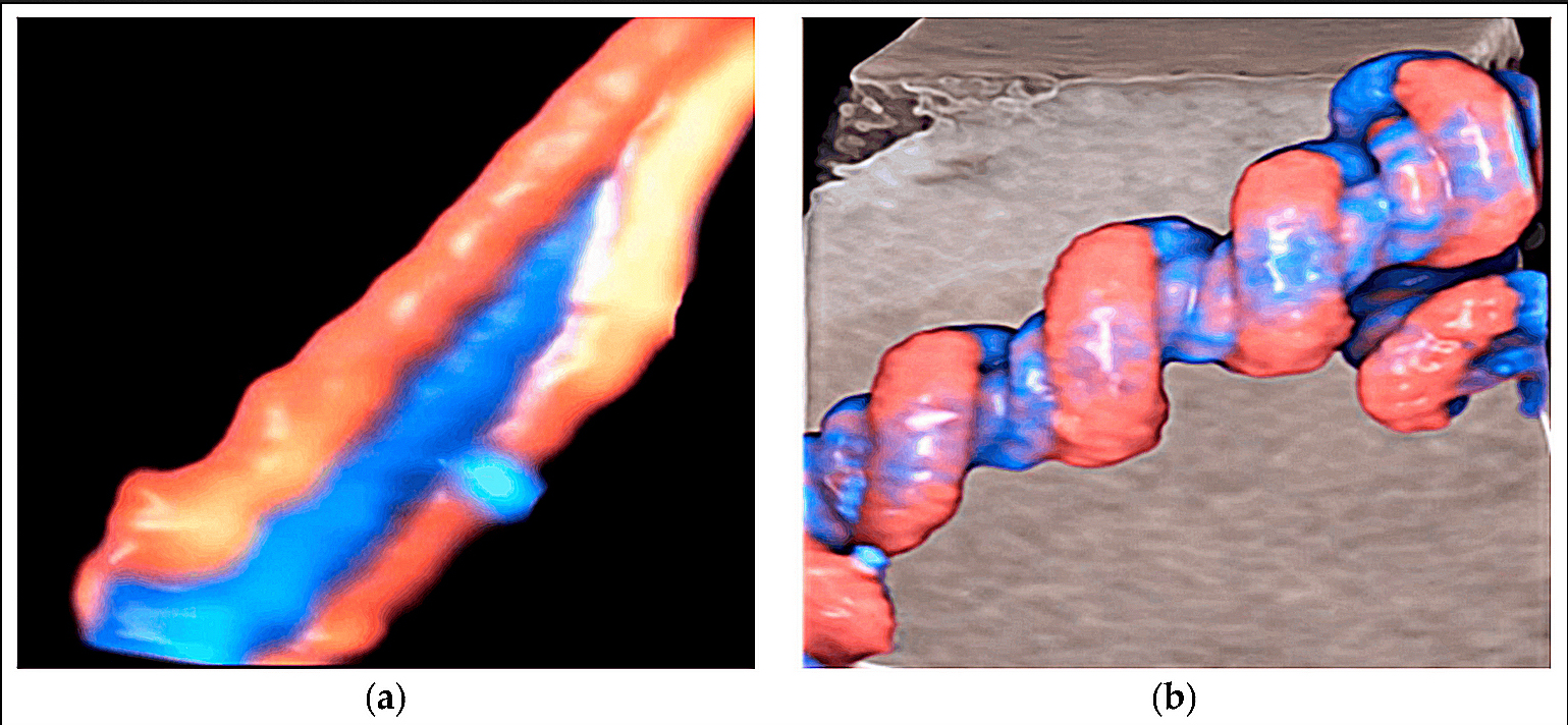
The umbilical cord in colour Doppler, the two arteries winding around the vein in a slow spiral. The red and blue are the two directions of flow in the looping vessels.
The cord carries two arteries and a vein, the arteries running the baby’s blood out to the placenta. The Doppler reads one of the arteries, the flow in it driven by the baby’s own heart. Some cords run only one artery, a variant a reader notes and reads the one it has. The cord’s vessels run in a soft jelly that keeps them from kinking, the artery held open for the probe to read. A reader drops a box of colour on the cord first, the red and blue of the flow showing the vessels before the gate reads their speed. The colour finds the artery in a tangle of loops. A clear stretch of free cord, floating in the fluid, gives the cleanest trace. The artery wraps the vein in a slow spiral along the cord, the colour on the screen showing the twist. The two arteries can read a touch apart, a reader taking the one with the cleaner trace. A reader steers the gate onto the artery and waits for a run of even beats. The cord shows at once on the screen, the harder part the reading of its flow. A reader samples a free loop in the middle of the cord, the figure steadiest there. The reader sets a small gate over the artery, the machine timing the blood that crosses it, a gate set too wide taking in the vein alongside. The machine can tilt its reading to the line of the flow, an angle correction that keeps the speeds true. The reader sets that angle along the vessel before the numbers are trusted.
The trace climbs to a peak with each squeeze of the heart, the systolic push. It settles to a low point in the rest between, the diastolic lull. The blood keeps moving through the lull in a healthy cord, carried on by a placenta that draws it through. The height of the lull, set against the peak, is what the reading turns on. The flow that runs on through the lull is the placenta’s doing, its open vessels pulling the blood through after the heartbeat has passed. A cord with strong lull flow speaks of a placenta wide open to the blood. A failing placenta has fewer working vessels in its bed, the blood held back and the cord reading high. The number stands as a measure of how much of that bed has gone. A healthy cord never empties its lull, blood moving forward through every beat of a working placenta.
The artery must be caught where the cord floats free, away from the baby and the wall. The reader keeps the beam near in line with the flow, an angle too steep flattening the trace. A few clean beats, alike in height, give a reading to trust. A fast fetal heart shortens the lull and bunches the trace, the indices holding their meaning across a range of rates. The machine reads the peak and the lull and prints the numbers. The machine traces the top edge of the flow, the fastest blood at each instant, the envelope a reader reads the peak and the lull from. A baby’s breathing movements ripple the trace, each breath nudging the indices, a reader waiting for a still spell to take the reading. The pulsed Doppler sends more energy than plain imaging, a reader holding the beam on the cord no longer than the reading needs.
The numbers
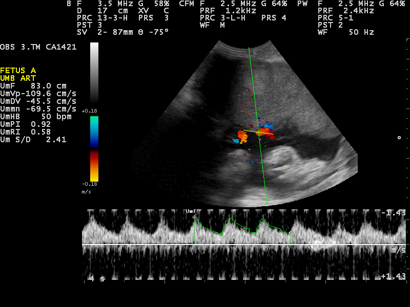
An umbilical artery spectral waveform along the foot of the image, a peak at each heartbeat above a lull that stays well clear of the baseline. The panel at the left reads the indices off it, a systolic-to-diastolic ratio of 2.41 and a resistance index of 0.58, the figures of a healthy cord. The colour box above marks the loop of cord the trace was taken from.
Three numbers come off the waveform, each a way to set the lull against the peak. The systolic-to-diastolic ratio divides the peak by the lull, a plain figure. A cord reading a peak of fifty over a lull of twenty gives a ratio near two and a half, a figure in the normal band near term. The resistance index and the pulsatility index do the same job in fuller ways. The resistance index takes the peak less the lull, divided by the peak, a figure between zero and one that reads near a third in a healthy cord by term. The pulsatility index takes the peak less the lull over the mean of the whole beat, a figure that still reads where the lull is flat or below the line. All three read higher the harder the placenta is to push blood through. The three share one strength, a freedom from the angle of the beam, the lull and the peak weighed as a ratio so the angle cancels out. A careless hand cannot spoil the figure as easily for it. The three figures move together, all rising as one when the placenta tightens.
The numbers are read against a chart for the weeks. A healthy placenta is easy to push blood through, its vessels wide and many by the second half of pregnancy. The figures sit low through those later weeks on a normal course. A number above the line for its week is the sign that the placenta is working too hard. The chart sets a line at the ninety-fifth centile for each week, a reading above it the one cord in twenty that runs high. A figure climbing across that line reads as a placenta tightening on the blood. The chart’s line falls from a ratio near four at twenty weeks toward two by term, the figure read against the week it was taken in. The baby’s own heart drives the flow the cord reads, a strong heart pushing blood through a stiffening placenta for a time.
The systolic-to-diastolic ratio is the one readers reach for first, quick to read off the trace. It sits around two to three in a healthy cord near term. A ratio past the chart for its week points to a placenta in trouble. The number is read again over days, its drift the thing that matters. The ratio asks no more than the peak and the lull, two heights off the trace, a reader reading it by eye before the machine prints it. The gap between peak and lull tells the story at a glance. A figure that holds steady across two scans reassures, the placenta holding its ground. A reader reads the cord across scans, the slope of its climb telling more than any single number.
The pulsatility index holds up best when the flow runs low. It keeps its footing where the simple ratio cannot be worked, the lull near flat. A reader leans on it in the worst cords, the ones where the diastolic flow has nearly gone. The pulsatility index reads on where the lull has emptied, its mean still a figure when the ratio is a division by nothing. The three numbers tell one story, read together. A reading is taken from a few clean beats and the worst of them kept, the cord judged on its hardest moments. The figure is logged with its week, the record built for the next reader to set against. A healthy cord waveform carries a high lull below its peak, the body of the trace full, a trained eye reading the shape before any number. The cadence of the watching follows the cord, a worse reading bringing the next scan sooner.
When resistance climbs
The flow fails in a known order.
The first sign is a lull that sinks, the diastolic flow low where the placenta has stiffened. The ratio rises above the line for the week, the earliest warning a reader catches. The flow still runs forward through the lull at this stage, only weaker than it should be. A placenta worse still lets the lull fall to nothing, the flow stopping dead between beats. This is absent end-diastolic flow, the blood moving out on the heartbeat and then halting until the next. It marks a placenta near the end of what it can do. The gravest sign comes when the lull turns backward, the blood running back toward the baby between beats. This is reversed end-diastolic flow, a placenta so stiff the blood rebounds off it. A baby with reversed flow is in real danger, its placenta failing fast. The order runs one way, from a high ratio to absent flow to reversed, each step a placenta further gone. Each step takes the baby closer to harm, the placenta passing less and less of what the baby needs. The reader’s response climbs with the danger, from closer watching at a rising ratio to action at once when the flow goes absent or backward. Early failing, before thirty-four weeks, shows in the cord first, the placenta the weak link in those early cases. A worsening cord tightens the watching, from twice-weekly scans to daily ones in hospital. The worse the flow runs, the sooner the baby must come out, a reversed cord the emergency that brooks no wait. The numbers behind the words, the ratio and the indices, put a figure on each step, a record a reader tracks from scan to scan. A cord can hold a high ratio for weeks before it worsens, the change slow in many. A cord can also fall from a high ratio to absent flow in days, a reason a reader watches it close once it climbs. A reversed-flow cord at thirty weeks or beyond is delivered without delay, the danger past what watching can hold. Below thirty weeks the call is harder, every day kept in the womb bought at a rising risk. A baby facing an early delivery for failing flow is given steroids first, a day or two to ready its lungs, the flow watched close across that wait. A baby due to come early is given magnesium as well, a guard for its brain through the birth. The Doppler sets the clock these treatments race.
Absent and reversed flow rarely come out of nowhere. They follow weeks of a placenta failing a small baby, the growth lagging and the fluid thinning first. A reader reading a high cord ratio on a small baby is watching a placenta on its way down. A cord turned bad sends a reader back over the whole pregnancy, the mother’s pressure and the baby’s build read for the cause behind the failing placenta. A placenta failing the cord often troubles the mother too, the same sick organ pushing her blood pressure up. The Doppler times the fall, the delivery set before the flow gives out. When the cord flow turns backward, a reader looks deeper still, at the vein that carries blood back to the baby’s heart. A wave running the wrong way there marks a baby close to its limit, the last sign before the heart itself begins to tire. A vein that should run smooth can take on a pulse late in the failing, the beat of a heart straining to take the blood back. A reader reads a pulsing vein as a grave sign. A baby with absent flow is brought into hospital, watched by monitors through the day, the delivery readied, the cord that has lost its lull never sent home. A reversed-flow cord rarely waits the two days steroids need, the baby out as soon as the trace allows no delay.
The reading guides the hardest call in obstetrics, when to bring a small baby out. The question is how long a baby can be left in a placenta that is giving out. A baby kept too long in a failing placenta can be lost before birth. The cord Doppler reads how close that placenta is to the edge, the flow the clock the timing runs on. A reader never reads a single high number into a delivery, the trend across scans the thing that decides. A reader reads the mother’s pressure beside the baby’s cord, the two failings of one placenta read as a pair. Cord Doppler was among the first uses of Doppler in pregnancy, the one with the firmest backing in the trials, a reader trusting it for the years of evidence behind it. An overdue baby has its cord read for a placenta thinning past its time, a rising ratio one of the signs the pregnancy has run long enough.
The flow is read beside the rest of the picture, the growth, the fluid, the baby’s moving. A high cord ratio weighs heaviest on a baby already small, the sign of a placenta in real decline. The Doppler is one reading among several, weighed for what it adds. A reader who reads it alone reads only half the story. The Doppler does not call the delivery on its own, the gestational age and the other signs weighed beside it. The cord flow is read at every growth scan on a small baby, a scan that skips it missing half of what it came for. Some babies are small and entirely well, their cord flow normal throughout, the Doppler pointing away from the placenta as the cause.
The cord and the brain
A failing placenta sets off a change a second Doppler can catch. The baby sends more of its blood to the brain, the head’s vessels opening wide to take it. A Doppler on the brain’s main artery reads the flow there running easy through the lull, the sign of a baby protecting its brain. This is brain-sparing. The head takes the share of blood the body gives up. The brain’s artery reads an easy flow there, the lull staying high. A reader reads that low resistance in the head as the baby’s own doing.
Read together, the two Dopplers show both the placenta’s failing and the baby’s answer to it, more than either tells alone. A reader sees how far the baby has had to shift its blood to cope. The ratio of the two flows, brain set against cord, makes a reading of its own, the cerebroplacental ratio. A low ratio marks a baby leaning hard on its brain to get by. Brain-sparing keeps a baby going for a time, the head kept fed at the body’s cost. It marks deepening trouble, the placenta still failing under it. A late, mild placenta failing can spare the brain before the cord flow ever climbs, the brain Doppler the earlier sign in those late cases. A reader late in pregnancy reads the brain as closely as the cord. The same brain artery serves a second reading, its peak speed a measure of a baby’s blood count, a high peak speed there the sign of an anaemic baby. In twins sharing a placenta, the two cords are read side by side, a wide gap between their flows the sign of one twin shortchanged. A cerebroplacental ratio below one turns a reader’s eye, the brain’s flow running easier than the cord’s. It catches a baby coping whom the cord flow alone would miss. A reader turns to the brain when the cord first climbs, the change in the head often the next step down.
What the flow cannot say
The Doppler reads the placenta’s resistance, never the whole of a baby’s health. A normal cord flow on a small baby calls for watching still, the other signs read alongside it. The angle of the beam and the baby’s breathing both nudge the reading, a careful reader taking several beats and the best of them. The cord tells how the placenta passes blood, the rest of the scan filling in around it. A cord wound round the baby’s neck can read a flow of its own, a reader untangling the trace from the loop and reading a free length in the fluid. Two readers reading the same cord land close, the indices steadier between hands than many a measure. A poor angle or a moving baby throws the trace, a reason a reader takes the cleanest beats and the best of them. The cord tells of the placenta, never of a knot or a cord too thin, the things a grey-scale look must find. The same ratio carries its meaning only with the week it was taken in, read always against the chart.
On a handheld
Cord Doppler asks more of a probe than plain imaging. The machine must read motion, the pulse of blood in a vessel a millimetre wide. Many handhelds carry the colour and the spectral Doppler the reading needs, the cord flow drawn on the same screen that sized the baby. A clinician finds the cord, drops the gate on an artery, and reads the trace in a minute or two. Reading the numbers needs a handheld with spectral Doppler. Some carry it, some only the colour that finds the vessel. The reading asks a trained hand, the gate and the angle set right and the trace read for what it means. A clinician learns the cord on a few dozen scans, the skill within reach of a clinic. The waveform saves to the record with its numbers, the trace ready for a specialist to read again. A reader who doubts a cord sends the clip on for a second eye.
The reading carries weight where it is hardest to get, in clinics far from a Doppler machine. A small baby found in such a place can have its cord flow read on the spot, the worst cases sent on early. A clinic that reads a reversed-flow cord sends the mother on the same day, the baby’s best chance a delivery where the care is. A cord Doppler read on the spot spares a mother a journey to a referral centre for one number, the flow read where she already sits. A clear reading settles the visit there and then. A district clinic with a Doppler handheld reads the one test that decides a small baby’s timing, the cases that need a hospital found early. The reading turns a guess into a number a team can act on. A normal cord flow reassures on a small baby, the placenta shown to be doing its work, time bought to watch on. A handheld puts the one test that times a failing pregnancy into the hands of a clinic that would otherwise have none.
Common questions about umbilical artery Doppler
What does umbilical artery Doppler measure?
It measures the resistance a baby’s blood meets flowing through the placenta. The probe reads the speed of blood in a cord artery as a waveform, with a peak at each heartbeat and a lull between. A sinking lull means a placenta growing hard to push blood through, where a healthy cord keeps its lull high.
What is a normal umbilical artery S/D ratio?
The systolic-to-diastolic ratio sits around two to three in a healthy cord near term. The ratio is read against a chart for the week of pregnancy, since a healthy placenta passes blood more easily by the second half of pregnancy. A ratio above the line for its week is the sign of a placenta working too hard.
What do absent and reversed end-diastolic flow mean?
Absent end-diastolic flow means the blood stops moving in the lull between heartbeats, a sign of a placenta near the end of what it can do. Worse still, reversed flow means the blood runs backward in the lull, the gravest sign of all. Both call for close watching and an early, planned delivery.
Why is umbilical artery Doppler used in growth restriction?
A small baby may owe its size to a failing placenta. The cord Doppler reads how hard that placenta has become and times the fall, so the baby can be delivered before the flow gives out. It guides the hardest call in obstetrics, when to bring a small baby out.
Can a handheld ultrasound read umbilical artery flow?
Yes, where it carries spectral Doppler. The clinician finds a free loop of cord, drops the gate on an artery, and reads the waveform in a minute or two. This brings the one test that times a failing pregnancy to clinics far from a Doppler machine, the one number that matters carried to where a mother lives. A cord read in a village clinic can move a mother to a city hospital in time, the number the reason for the journey.
About the Author
Julien Mercier
Senior R&D Engineer · Medical Ultrasound Transducer Development
Senior R&D Engineer with an M.S. in Applied Physics and over 15 years of experience in medical ultrasound transducer development, specializing in the design verification and performance testing of high-frequency imaging transducers. Currently leading the development and verification of the company’s next-generation high-frequency linear-array transducer, responsible for imaging performance evaluation and reliability analysis in preclinical testing. Brings extensive hands-on experience in piezoelectric element tuning, beamforming parameter optimization, and system-level performance testing.