





































Tricuspid Regurgitation TR Continuous Wave Doppler Pulmonary Pressure Handheld
Continuous wave Doppler turns a tricuspid leak into a pulmonary pressure at the bedside. The leak jets back into the right atrium in systole. Its speed reports the pressure the right ventricle works against. A clinician reads that speed, adds the pressure in the right atrium, and reaches a pulmonary pressure. A handheld carries the whole read to the chair, from the jet to the number.
What tricuspid regurgitation is
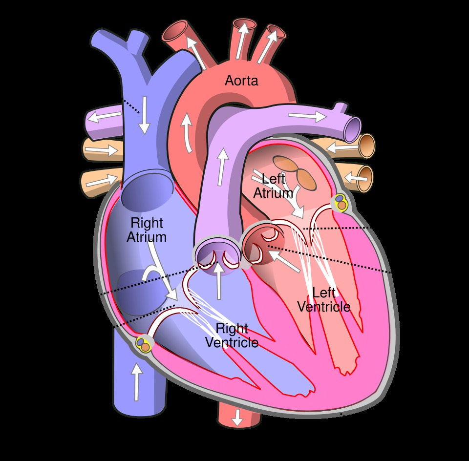
Tricuspid regurgitation is a backward leak through the valve between the right atrium and the right ventricle. The valve should seal in systole as the ventricle squeezes. A leak lets blood run back into the atrium with each beat. A small leak shows on the Doppler of many healthy hearts and means nothing. A larger leak loads the right atrium and the right ventricle with blood they pump twice. The valve has three leaflets, a ring, and cords to the right ventricle’s wall. A fault in any of these can break the seal. The right heart takes the load for years, often with no complaint. The leak earns attention when it grows large or when the pressure behind it climbs. The three leaflets sit at the ring as the septal, the anterior, and the posterior. A wide ring lets even healthy leaflets fail to meet. A trace of backward flow on colour is normal and needs no further thought.
A leak starts in the valve or in the ventricle that holds it. A stretched right ventricle widens the ring and pulls the leaflets apart, the common route to a leak. High pressure in the lungs drives that stretch, from left heart disease or lung disease. A pacemaker lead across the valve can hold a leaflet open. Infection on the valve, common in injected drug use, can tear a leaflet. Carcinoid disease and a valve born malformed each leak in their own way. A clinician notes the cause, since a leak from a stretched ring eases when the pressure falls. A valve born with its septal leaflet pulled down into the ventricle leaks from birth, the mark of Ebstein’s. Carcinoid tumour stiffens the leaflets into fixed boards that neither open nor close. A clinician reads the leaflets and their motion to name the cause.
The leak and its pressure run a clear path through the right heart. Blood should flow from the right atrium, through the tricuspid valve, into the right ventricle, and out to the lungs. A leak sends some of it back the way it came in systole. The right atrium swells to hold the returned blood. The pressure can back up into the veins that feed the atrium. A clinician sees the swollen neck veins, the large liver, and the leg swelling as the leak’s toll. The inferior vena cava feeds the atrium and shows that backed-up pressure. The path is the same one the pressure number traces, from the leak to the lungs. A clinician presses on the liver and watches the neck veins rise, a sign the right heart cannot take more. The liver can pulse with each beat under a large leak. Fluid gathers in the belly and the legs when the pressure holds high.
The right heart bears a large leak quietly for a long time. Symptoms come late, from the congestion behind the failing ventricle. A patient notices swollen legs, a bloated belly, and breathlessness. The murmur sits at the lower sternum and gets louder when the patient breathes in. A clinician hears it and turns to the Doppler to size the leak and read the pressure. The leak’s own jet carries the pressure a clinician needs to know. The bedside read gives the leak and the pulmonary pressure in one study. The louder murmur on a breath in sets the right-sided leak apart from a left-sided one. A clinician feels a heave at the lower sternum from the loaded right ventricle. An irregular pulse can mark the atrial fibrillation a swollen atrium breeds.
Why the TR jet matters for pressure
The tricuspid jet carries the pressure of the right ventricle. The ventricle drives the leak back at a speed set by the pressure gap between it and the atrium. A fast jet means a wide gap, which means a high pressure in the ventricle. A clinician takes the peak speed and turns it into that pressure gap. The gap plus the atrial pressure gives the pressure inside the right ventricle in systole. A clinician trusts the speed only from a complete envelope on a well-aligned beam. The number falls out of the jet a clinician can already see on colour. The gap the jet reads is the pressure at one instant, the peak of the beat. A clinician lines the beam along the jet, since an off-axis beam undershoots the speed. A small error in the speed grows fourfold in the pressure.
The pressure inside the right ventricle in systole is the systolic pressure of the lungs as well. The two match when the pulmonary valve opens without a block. Blood meets no obstacle between the ventricle and the lung arteries in systole. A clinician treats the right ventricular pressure as the pulmonary artery pressure on that basis, the method set out in the ASE guideline for the right heart. The tricuspid jet thus gives the pressure in the lungs from the bedside. The estimate holds in the common case, where the pulmonary valve is normal. A clinician confirms the pulmonary valve opens clean before trusting the link. The pulmonary pressure the jet gives is the systolic peak, the highest of the cycle. A clinician estimates the mean pressure from other signs when the case needs it.
A pulmonary pressure read from the jet answers a common bedside question. A clinician facing breathlessness wants to know whether the lung pressure is high. The tricuspid jet settles it in seconds on a handheld. A high pressure points toward the lungs or the left heart as the trouble. A normal pressure steers the search elsewhere. A clinician weighs the number alongside the size of the right ventricle and the leak. The pressure from the jet starts the workup of a breathless patient. A clinician reaches the pressure in the time it takes to find the jet. The number sorts a lung cause from a heart cause early in the visit. A clinician carries that fork into the rest of the study.
Reading the TR jet with continuous wave
A clinician finds the leak on colour flow first. The colour box at the tricuspid valve lights up the jet running back into the right atrium in systole. The jet marks the line for the continuous wave beam. A clinician lays the beam down the jet for its speed. The leak flies away from the probe in many windows, so the trace draws below the baseline. A clinician swings the probe to fill the envelope before reading the peak. A broken envelope reads the speed low. A clinician centres the jet in the colour box to aim the beam true. The beam should run down the length of the leak for the full speed. A clinician trusts the peak only once the envelope has filled.
Several windows open the tricuspid jet to the beam. The parasternal right ventricular inflow view lines up well with many jets. The apical four-chamber view sets the leak flying toward the probe. The subcostal view helps in a chest that hides the others. A clinician uses the colour jet to set the beam down the core of the leak. A clinician reads the jet from each window and keeps the highest clean peak. The window that fills the envelope and aligns the beam gives the truest speed. A clinician marks that window for the next study. A clinician tries each window in turn, since one often beats the rest for a given jet. The right ventricular inflow view catches a jet the apex misses. A clinician keeps the window that gives the fullest trace.
The read hangs on a complete envelope. A dense envelope traces the whole leak from start to finish. A clinician takes the peak from such an envelope with confidence. A faint signal misses the fastest blood and falls short of the true speed. A clinician who takes a weak signal at face value under-reads the pressure. A clinician keeps the gain fair, since too much gain fattens a thin signal into a false peak. A clinician swings and tilts the probe until the envelope fills and the edge sharpens. The complete envelope is the rule before any number comes off the trace. A clinician can sharpen a faint jet with a puff of agitated saline into a vein. The bubbles brighten the signal and fill a thin envelope. A clinician reads the strengthened trace for a peak the bare signal hid.
The peak velocity is the number the pressure read needs. A clinician marks the topmost tip of the envelope, the fastest moment of the leak. A normal jet peaks under three metres a second. A higher ventricular pressure drives a faster jet. A clinician records the peak in metres per second and keeps the same units across visits. The contour of the envelope rises and falls through systole, peaking in mid-beat. A clinician treats a clean envelope as a faithful trace of the leak. A clinician reads three or four beats and takes the highest clean peak. An irregular rhythm calls for an average across more beats. The peak holds steady across a settled run.
The same trace shows the size of the leak in its density. A dense signal as bright as the forward flow marks a large leak. A clinician judges the density for the size of the leak. A clinician reads the peak for the pressure. Both come off the one trace in the one study. A clinician grades the leak and reads the pressure from the one beam. The two readings need no second probe.
From velocity to pulmonary pressure
The peak velocity turns into a pressure gap by a simple rule. A clinician squares the peak speed and multiplies by four. A jet at three metres a second gives a gap near thirty-six millimetres of mercury. The gap is the pressure the ventricle builds over the atrium to drive the leak. A clinician reads that gap as the first half of the pulmonary pressure. The rule is the same modified Bernoulli that turns any jet speed into a pressure gap. A clinician keeps the units straight, since the squared speed swings the number fast. A clinician turns a jet of four metres a second into a gap near sixty-four millimetres of mercury. The squared term means a small rise in speed lifts the gap a long way. A clinician marks the peak with care for that reason.
The gap alone is not the pulmonary pressure. A clinician adds the pressure already in the right atrium to the gap. A jet of three metres a second over an atrial pressure of eight gives a pulmonary pressure near forty-four. The right atrial pressure comes from the inferior vena cava, read in the next step. A clinician reports the sum as the systolic pulmonary artery pressure. A clinician writes the velocity, the gap, and the atrial pressure in the notes for the next read. The whole estimate rests on the jet velocity and the atrial pressure together. A clinician treats the sum as the systolic pressure the lungs see. A pressure past about thirty-five to forty millimetres of mercury counts as raised. A clinician weighs a borderline number with the rest of the right-heart picture.
Estimating right atrial pressure
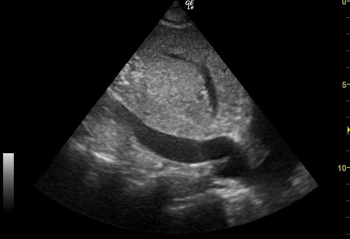
The right atrial pressure is the second half of the sum. A clinician reads it from the inferior vena cava in the subcostal view. The vein runs below the liver into the right atrium and mirrors the pressure there. A clinician measures its width and watches how far it collapses with a sniff. A clinician asks the patient to sniff and watches the vein narrow. The vein gives the pressure from the bedside. A clinician finds the vein from below the breastbone, with the probe aimed up toward the heart. The vein lies a little deep to the liver, easy to follow into the atrium. A clinician steadies the patient’s breathing before the measure.
| IVC diameter | Collapse with a sniff | Estimated RA pressure |
|---|---|---|
| 2.1 cm or less | More than 50% | Low, about 3 mmHg |
| 2.1 cm or less | Less than 50% | Intermediate, about 8 mmHg |
| Over 2.1 cm | More than 50% | Intermediate, about 8 mmHg |
| Over 2.1 cm | Less than 50% | High, about 15 mmHg |
A clinician matches the vein to the scale in the table. The width and the collapse together set the pressure. A clinician measures the width at end-expiration, a finger-width below the atrium. A clinician judges the collapse from a sharp sniff. The two together place the pressure on the scale. A clinician takes a dilated vein that does not collapse as a sign of a loaded right heart. A clinician takes that pressure into the sum for the pulmonary number. A clinician reads the width with an M-mode line across the vein for a steady trace. The measure sits about two centimetres from where the vein meets the atrium. A clinician times the read to a quiet breath and a sharp sniff.
The vein can mislead in a few settings. A young athlete can carry a wide vein at a normal pressure. A patient on a ventilator breathes against the machine, which clouds the collapse. A clinician reads the vein with the patient in mind. The sniff sharpens the read, since a forced collapse shows the vein’s true range. A clinician weighs a borderline vein against the rest of the picture. A clinician repeats the read when the first looks off. A clinician treats the vein as one piece of the picture, alongside the atrial size and the jet. A deep breath or a strain can fool the collapse for a moment. A clinician checks the vein at rest first, then with the sniff.
A clinician now has both halves of the pulmonary pressure. The jet velocity gives the gap across the tricuspid valve. The vein gives the pressure in the atrium. A clinician adds the two for the systolic pulmonary artery pressure. The number comes off one bedside study, with the jet and the vein read in turn. A clinician takes a high number as a flag for the lungs or the left heart. The handheld holds the velocity, the vein, and the sum in the one record. A clinician reads the two parts in turn, the jet then the vein, and adds them. The sum gives the pulmonary pressure a clinician can act on. A clinician dates the number to track it across the visits.
Pitfalls and limits
The pressure read carries a few traps. A broken envelope reads the velocity low and undercalls the pressure. A clinician fills the envelope before trusting the peak. A massive leak can drop the velocity even with a high pressure, since the wide-open valve lets the pressures equalise. A clinician reads a torrential leak by its size and the clinical picture. A narrowed pulmonary valve breaks the link between the ventricle and the lung pressures, so the jet then reads the ventricle alone. A clinician checks the pulmonary valve before calling the number a lung pressure. The read holds best when the envelope is full and the pulmonary valve is clear. A clinician reads a low velocity in a sick patient with care, since the equalised pressures of a torrent can hide the load. A direct pressure tracing in the catheter lab settles a doubtful case. A clinician sends the unclear number on for that check. A clinician reads the jet number as a strong bedside clue, with the catheter as the final check.
What the number guides
The pulmonary pressure from the jet steers the next step. A high number sends a clinician looking for its cause in the lungs or the left heart. A clinician reads the left-sided valves and the ventricle, since left heart disease drives the bulk of high lung pressures. A clear left heart with a high pressure points toward the lung arteries themselves. The handheld screens for the high pressure and starts the hunt for its source. A rising pressure over visits marks a disease on the move. A clinician sorts the high pressure into a left-heart cause, a lung cause, or a vessel cause. The left heart drives the common case, so a clinician reads it first. A clinician sends a high pressure with a clear left heart on for a fuller workup.
The size of the leak guides the heart’s own care. A clinician grades the leak from the jet density, the vein width, and the right heart’s size. A large leak swells the right atrium and the right ventricle over time. A clinician watches the right ventricle’s function, since it can fail under a long-standing heavy leak. A failing right ventricle and a severe leak together move a patient toward a valve repair. A clinician measures the right ventricle’s size beside the leak each time. The handheld tracks the leak and the right ventricle at each visit. A clinician measures the right ventricle’s long-axis motion as a quick read of its function. A ring stretched past about four centimetres marks a valve likely to keep leaking. A clinician weighs the leak, the ring, and the ventricle as one before any referral.
Treatment follows the cause as much as the leak. A secondary leak eases when the pressure behind it falls, so a clinician treats the lungs or the left heart first. A leak that stays severe on full therapy can earn a valve repair. A clinician refers a severe leak with a struggling right ventricle in good time. The handheld reads the leak and the pressure before and after each change in care. A clinician carries the whole right-heart read in a pocket probe to every visit. A surgeon often repairs the tricuspid valve during an operation on the left heart. A clinician flags a severe leak before that surgery, since it is easier to fix in the same sitting. A clinician checks the leak again after the left-sided repair to see what remains.
Common questions about TR and pulmonary pressure
How does continuous wave Doppler estimate pulmonary pressure?
Continuous wave Doppler reads the speed of the tricuspid leak in systole. A clinician squares that speed and multiplies by four for the pressure gap across the valve. A clinician adds the right atrial pressure to the gap for the systolic pulmonary artery pressure. The whole estimate comes from one bedside study.
What tricuspid jet velocity is normal, and what is high?
A normal tricuspid jet peaks under three metres a second. A jet past about 2.8 to 2.9 metres a second points to a raised pulmonary pressure. A clinician reads the peak from a complete envelope, since a thin signal reads the speed low. The peak feeds straight into the pressure sum.
Can a handheld scanner measure pulmonary pressure?
Yes. A handheld with continuous wave Doppler reads the tricuspid jet at the bedside. A clinician finds the leak on colour, lays the beam down it, and reads the peak speed. The handheld also images the inferior vena cava for the right atrial pressure. A clinician reaches a pulmonary pressure from one bedside study.
How is right atrial pressure estimated?
A clinician reads the right atrial pressure from the inferior vena cava: its width and its collapse with a sniff. The reading runs from about three millimetres of mercury for a small vein to about fifteen for a wide one. A clinician adds the collapse to place the vein on that scale. The number then joins the jet gap for the pulmonary pressure.
Why can severe tricuspid regurgitation underestimate the pressure?
A massive leak can read a low velocity even with a high pressure. The wide-open valve lets the right ventricle and the right atrium equalise their pressures fast. The small gap that remains drives the leak at a low speed. A clinician reads such a torrential leak by its size and the clinical picture, since the velocity alone misleads.

About the Author
Julien Mercier
Senior R&D Engineer · Medical Ultrasound Transducer Development
Senior R&D Engineer with an M.S. in Applied Physics and over 15 years of experience in medical ultrasound transducer development, specializing in the design verification and performance testing of high-frequency imaging transducers. Currently leading the development and verification of the company’s next-generation high-frequency linear-array transducer, responsible for imaging performance evaluation and reliability analysis in preclinical testing. Brings extensive hands-on experience in piezoelectric element tuning, beamforming parameter optimization, and system-level performance testing.