Subcutaneous Mass Ultrasound Scanning Handheld Linear Probe
A subcutaneous mass is a lump in the soft tissue under the skin. A probe held over it reads which layer it lives in and what fills it. It matches the lump to the short list of things a soft tissue lump turns out to be, and names the harmless ones on the spot. The rare lump that fits no friendly pattern is the one the scan is built to catch.
Name it, or refer it
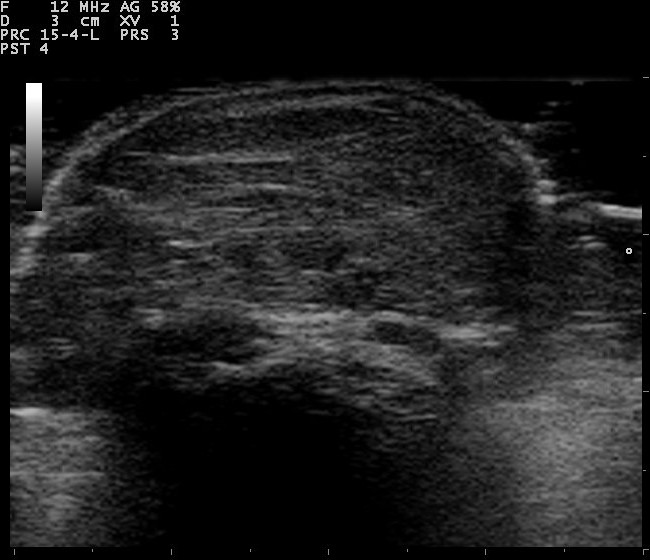
A lipoma on ultrasound. The mass is a flat oval, its long axis lying along the skin, with fine bright lines running through it like the grain in the fat. It reads close to the shade of the fat around it. A soft striped oval like this, up in the fat layer, is the look the scan names as a lipoma. Image: Nevit Dilmen, CC BY-SA 3.0.
Nearly all lumps under the skin are harmless. A few are the kind the scan can name outright, by a look it knows. A lump of fat in the fat layer, soft and striped, is a lipoma. A round pocket in the skin with a little tail to the surface is a cyst. A clear sac of fluid by a wrist joint is a ganglion. A tangle that fills with color on Doppler is a vessel lump. The scan that names one of these can set the worry aside, and spare a person a cut for a lump that needed none. The naming is half the work. Guarding against the rare bad lump is the other half. The two tasks run together on every lump the probe meets. A rare lump in the soft tissue is a cancer, a sarcoma, and the one mistake that matters is to shell it out as though it were a lipoma. A sarcoma that is cut out by surprise is far harder to treat than one that was known before the knife. So the scan reads every lump for the marks of that rare bad one. The deep fascia, the sheet that wraps the muscle, is the line. The fascia shows on the scan as a bright band under the fat. A probe pressed over the lump shows whether it lifts off that band or pushes through it. A lump above it, in the fat, is the common harmless kind. A lump that sits below the fascia, in or under the muscle, is read with a raised guard. Size is the next mark. A lump past about five centimeters calls for a careful look, the more so as it grows. Growth is the loudest mark. A lump that is getting bigger, week on week, is taken seriously whatever its size. A lump that has come back after a past removal is read with that history in mind. The look inside carries weight too. An even quiet lump reads gently. A jumbled lump, with a busy chaotic flow running through it, reads as one to send on. The scan sorts the many harmless lumps from the rare dangerous one by these marks: the layer, the size, the growth, and the flow. The job is to name the lump where it can, and to refer the lump it cannot name with confidence, before a blind cut turns a treatable cancer into a hard one.
The layers under the skin
A lump is placed by the layer it sits in. The scan reads the soft tissue as a stack of layers. The skin and the dermis lie on top, a thin bright band. The fat lies under that, a darker layer threaded with bright lines. The deep fascia runs below the fat, a bright sheet over the muscle. The muscle lies deepest, with its own feathered grain.
The layer sets the first read of a lump. A lump in the skin is a lump of the dermis, a cyst or a skin nodule. A lump in the fat is the home ground of the lipoma and the common cyst. A lump that sits against or below the deep fascia is the one read with care.
The deep fascia is the line that counts. Above it lie the lumps that are nearly all harmless. A lump that pushes below it, into or under the muscle, joins the group that needs an MRI and a specialist eye. The probe finds the bright fascial line and reads the lump against it.
The probe is laid with a light hand. A high-frequency linear probe, with plenty of gel, reads the shallow layers in fine detail. A thick layer of gel, or a soft pad, brings a lump right at the skin into focus. The lump is read along its length and across, and pressed to test how it gives. A lump that slides freely on the layers under it reads as a loose, friendly one. The probe checks that glide on every lump.
The lipoma
The lipoma is the commonest lump of all. It is a soft growth of fat, sitting in the fat layer under the skin. The scan knows it by a steady look.
It lies as an oval, its long axis along the skin. It rarely stands up tall. It reads close to the shade of the fat around it, a touch brighter or the same. Fine bright lines run through it, parallel to the skin, like the grain in the fat. The grain runs the way the fat runs, a sign the lump is fat and nothing stranger.
It gives under the probe. A press flattens a lipoma with ease. The soft squeeze is part of its friendly look.
It carries little flow. Color Doppler laid on a lipoma shows next to nothing, a quiet lump with maybe a single vessel crossing it. The quiet flow fits the soft fatty mass.
A plain lipoma in the fat needs no more than a name. The scan that calls it can reassure the person and close the matter. A lipoma that grows large, dives below the fascia, or fills with chaotic flow loses its plain label, and goes on for an MRI. A lipoma can run a few millimetres or fill a whole forearm. It can sit alone, or in a scatter of several across the body, a pattern that runs in some families. A lump of fat with a pink tint of flow is an angiolipoma, a benign cousin that can ache.
The cyst in the skin
An epidermoid cyst is a common round lump in the skin. It grows from the skin’s own lining, filling with soft keratin. The scan reads it with a set of marks.
It sits as a round or oval pocket, well bounded, in the skin or the fat right under it. It reads dark, with fine internal echoes that swirl like the inside of a testis. The sound passes through it and brightens the tissue behind. That brightening behind a round pocket is a steady sign of fluid or soft contents within.
It often keeps a link to the surface. A fine tract, or a small dark dot of a punctum, runs from the cyst to the skin. That link is a clue to its kind.
A cyst can rupture and turn angry. A cyst that leaks its keratin into the tissue swells, reddens, and fills the area around it with flow. An inflamed cyst reads as a messy lump with a bright rim, settling back to a quiet pocket once the flare passes.
The scan names the plain cyst and the inflamed one alike. A quiet round pocket with a tract to the skin is a cyst at rest. Hot and haloed with flow, it is the same cyst inflamed. Both are benign. A cyst on the scalp grows from the hair root, a pilar cyst, often firm and sometimes in a cluster. The scan reads it the same way, a round pocket with through-sound and a quiet inside.
The lump beside a joint
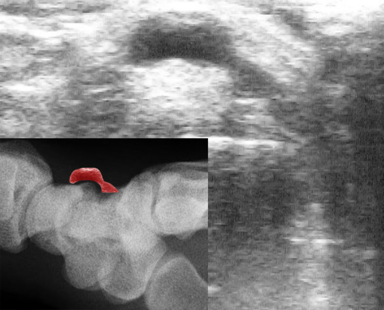
A ganglion at the wrist on ultrasound. The main image shows the ganglion as a dark, clear space over the wrist bones, the sound passing straight through it. The inset, lower left, is an X-ray of the same wrist, with the ganglion’s place shaded red over the carpal bones. A clear sac like this, off a joint, is a ganglion. Image: J. Lengerke, public domain.
Some lumps grow from a joint or a tendon. The scan reads the lump against the structures it sits near.
A ganglion is a sac of clear jelly off a joint or a tendon sheath. It shows as a smooth dark space, often lobed, by the back of the wrist or the top of the foot. The sound passes straight through it. A fine stalk can be traced from the sac to the joint it springs from. No flow runs inside it.
A lump on a tendon sheath of the hand can be solid. A giant cell tumor of the tendon sheath sits as a firm dark nodule hugging a finger tendon. It carries flow on Doppler. It moves with the tendon when the finger bends.
The place of the lump tells much here. A clear sac off a joint is a ganglion, named and left. A solid nodule on a tendon is read further, measured and watched, sampled if it grows. Behind the knee, a sac of joint fluid can bulge into a lump, a popliteal cyst. The scan traces its neck back to the knee joint, and reads the fluid inside for the debris of a worn joint. A swollen bursa over a kneecap or an elbow makes a flat fluid lump the same way.
The vascular lump
A lump can be made of vessels. The scan reads these with color Doppler, and they give themselves away.
A lump that fills with color is a vascular one. A hemangioma, or a venous malformation, reads as a soft tangle that floods with flow when the Doppler is laid on it. It can squash under the probe and fill again when the press lifts.
Hard bright specks can sit within it. A phlebolith, a small stone in a sluggish vessel, shows as a bright dot with a shadow below. A few of these in a soft lump point to a vessel malformation. The bright stones and the soft channels together make a picture the scan can call with confidence.
The flow sets the pace. A slow venous lump fills gently and drains slow. A fast lump, fed by an artery, runs a brisk noisy flow. The scan reads the speed of the flow, a guide to the kind of vessel lump and the way it is treated. A small vascular lump under a nail, or in the skin, can be sharply painful, a glomus tumor, tender out of all proportion to its tiny size. The scan finds it as a dark nodule with a brisk flow, in the spot the person points to.
The lump on a nerve
A lump can grow on a nerve. The scan reads how a lump sits with the nerve beside it.
A nerve sheath tumor is a smooth dark oval, lying along the line of a nerve. The nerve can be seen running into one end of it and out of the other. That tail of nerve at each pole is the giveaway. A press on such a lump can shoot a pins-and-needles down the nerve. The tingle and the tail of nerve together name the lump before a needle is near it.
The scan that finds a nerve entering a lump treats it with care. A nerve sheath tumor is nearly always benign. The aim is to spare the nerve, so the lump is mapped against the nerve for the surgeon to lift it cleanly. A painful lump between the bones of the forefoot can be a nerve grown thick, a Morton neuroma. The scan reads it as a dark teardrop, pressed out from between the bones when the foot is squeezed.
The hot, tender lump
A lump that is hot and sore reads a different way. The scan looks for a pocket of pus or a buried thorn.
An abscess is a pocket of pus in the tissue. It shows as a dark collection, full of fine floating debris, with no flow inside the pus. A rim of bright flow rings it. A push on the probe sets the contents swirling. A swirling, rimmed collection in a hot red lump is an abscess for draining. A cooler pocket of clear fluid, gathered after surgery or a knock, is a seroma or an old bruise, drained only if it presses or lingers.
The tissue around it can light up. An infection spreads the fat into a bright cobblestone of swollen lobules. The flow runs high through the inflamed tissue. The scan reads the spread of the heat around the pocket.
A buried foreign body can sit at the heart of it. A splinter or a shard shows as a bright line with a shadow or a comet trail behind. A ring of dark inflamed tissue often wraps it. The scan that finds the thorn guides its way out.
The lump that comes and goes
Some lumps are not there all the time. A lump that swells on standing or straining, and softens on lying flat, behaves like a hernia or a vein. The scan reads the lump as the person moves and strains, a thing a still picture cannot do.
A hernia pushes a plug of fat or a loop of bowel through a weak point in the wall. It bulges out on a cough, and slides back when the strain lets go. A muscle can bulge the same way, pushing through a gap in its own fascia as it tightens. The scan watches the lump fill and empty in real time, and traces it to the gap it comes through.
The lump that has not changed
A lump that has sat the same for years carries its own comfort. A soft lump, the same size and feel as it was, behaves like the benign thing it almost always is. The scan reads its quiet look, and with the long still history beside it can rest the matter.
The history is read with the picture. A photograph or a note from a year back, set against today’s scan, shows whether a lump has held still or crept up. A lump that has held its size and its soft feel is the easiest of all to leave alone.
The features that send a lump onward
A handful of marks turn a lump from a name into a referral. The scan reads every lump for them. They are the marks of a soft tissue sarcoma, the rare cancer of the soft parts.
Depth is the first mark. A lump that sits below the deep fascia, in or under the muscle, is read as a sarcoma until a specialist says otherwise. The fascia is the line that sorts the common lump from the one that needs a plan. A lump that is hard to the touch, fixed and unmoving on the layers under it, joins the depth as a mark to heed.
Size is the next. A lump past about five centimeters, the width of a golf ball, is large for a soft tissue lump. The bigger a lump, the harder the scan looks at it.
Growth is the loudest. A lump that grows month on month, that the person watched get bigger, is taken seriously at any size. A lump that keeps growing has lost the patience the scan gives a quiet one. A lump that has grown back where one was cut out before carries the same alarm, and goes the same way.
The inside speaks as well. A lump that is jumbled inside, with a chaotic flow that springs from no clear center, reads as one to send on. A hard fixed lump that will not slide on the tissue under it joins the list.
A lump that wears these marks goes whole to a sarcoma unit, for an MRI and a planned needle, before any knife is set to it. The whole point of the read is to catch this lump before a blind cut does.
Measuring the lump for the report
The scan ends in a few clear numbers and words. A surgeon, or a sarcoma unit, reads the report to plan the next step. The scan gives them what they need to act.
The size is taken in three planes. The lump is measured along its length, its width, and its depth. A growing lump is judged against these numbers on the next scan.
The depth is named against the layers. The report says whether the lump sits in the skin, in the fat, or below the deep fascia. That one word, above or below the fascia, steers the whole plan.
The lump is mapped to what lies around it. Its place against a nerve, a vessel, a joint, or a tendon is noted. A picture saved from the scan shows the surgeon where the lump sits before the first cut. The same numbers, read again in a few months, show at a glance whether the lump has held still or grown.
Leaving it, or acting on it
The scan ends with a plain plan for the lump. The plan follows from what the lump turned out to be.
A named benign lump can be left in peace. A lipoma, a plain cyst, a ganglion, a vessel lump: each can be named and let be, watched only if it troubles the person or grows. The scan spares these the knife.
A lump that troubles the person can still come out. A cyst that keeps flaring, a lipoma that catches on a belt, a ganglion that aches: any of these can be removed for comfort, once the scan has shown it is benign.
A lump with the marks of a sarcoma takes a set path. It goes to a sarcoma unit. An MRI maps it. A core needle, set along a planned line, reads its tissue. The wide removal that follows is planned around clean margins. The path guards against the cut that seeds a cancer.
The needle has its place in the read. A doubtful lump, or a pocket of fluid, can be sampled or drained under the probe. The scan sets the needle in the part that gives the truest answer, clear of the vessels. A cyst or a collection drained under the probe can be sent for a culture or a cell count, and the scan checks that the pocket has emptied. A lump the scan cannot place is watched on a short repeat, a fresh look in a few weeks telling growth from rest.
What the lump is made of
A lump under the skin is a question with a short list of answers. The scan runs down the list. A soft striped oval in the fat is a lipoma. A round pocket with a tail to the skin is a cyst. A clear sac by a joint is a ganglion. A tangle that fills with color is a vessel. A swirling pocket in a hot lump is an abscess. Each of these the scan can name. A named lump loses its fear.
The list has one entry that the scan must never miss. A lump deep under the fascia, large, growing, jumbled inside, can be a sarcoma. The scan reads every lump against that one, and sends the suspect on whole, before a blind cut makes a hard cancer of a treatable one.
A person finds a lump and fears the worst. The probe, in a few minutes, names the lump for what it nearly always is, a harmless thing of fat or fluid. For the rare lump that does not fit, the same probe raises the flag in time. Naming the many, and catching the few: that is the whole of the soft tissue scan.
Common questions about ultrasound for a lump under the skin
Which lumps under the skin can ultrasound name?
Many of them. A soft striped oval in the fat is a lipoma. A round pocket with a tail to the skin is a cyst. A clear sac by a joint is a ganglion. A swirling pocket in a hot lump is an abscess. The scan reads the layer and the make of each, and checks whether the lump sits above the deep fascia or below it.
Can ultrasound tell if a lump is cancer?
It can name the common harmless lumps with confidence, and it can flag the marks of a sarcoma. A lump deep below the fascia, larger than about five centimeters, growing, or jumbled with chaotic flow, is sent on for an MRI and a biopsy. A named lipoma or cyst is cleared. The certain word on a doubtful lump comes from the tissue under the microscope.
Is a lipoma dangerous?
A lipoma is a benign growth of fat. The common one sits in the fat under the skin, soft and striped, and stays harmless for years. A lipoma that grows large, sits deep below the fascia, or fills with busy flow is checked more closely, to be sure of its kind. A plain lipoma needs no more than a name.
Why does a cyst sometimes hurt?
A cyst hurts when it leaks. A skin cyst that ruptures spills its keratin into the tissue around it. The tissue swells, reddens, and fills with flow. The cyst turns into a hot, sore lump for a time. It settles back to a quiet pocket once the flare passes, and stays benign throughout.
What does a lump on a nerve feel like?
A lump on a nerve can shoot a tingle when it is pressed. The pins-and-needles run down the path of the nerve. On ultrasound the lump is a smooth oval with the nerve running into each end. A nerve sheath tumor is nearly always benign. The scan maps it against the nerve, so a surgeon can lift it and spare the nerve.
Should a lump under the skin be removed?
Not always. A lipoma, a plain cyst, or a ganglion that causes no trouble can be left alone, once the scan has named it. A lump that aches, catches, or keeps flaring can be taken out for comfort. A lump with the marks of a sarcoma is sent to a specialist unit, for imaging and a planned biopsy before any wide removal. The scan sorts which lump goes which way.
About the Author
Julien Mercier
Senior R&D Engineer · Medical Ultrasound Transducer Development
Senior R&D Engineer with an M.S. in Applied Physics and over 15 years of experience in medical ultrasound transducer development, specializing in the design verification and performance testing of high-frequency imaging transducers. Currently leading the development and verification of the company’s next-generation high-frequency linear-array transducer, responsible for imaging performance evaluation and reliability analysis in preclinical testing. Brings extensive hands-on experience in piezoelectric element tuning, beamforming parameter optimization, and system-level performance testing.