Sports Medicine Injury Assessment With Handheld Ultrasound Probe
Handheld ultrasound lets a sports injury be read at the side of the pitch, in the minutes after it happens, on the muscle or joint that was hurt. The probe shows the tear, the strain, or the break on the spot, and answers the one question that cannot wait: can the athlete go on. The reading that once waited for a clinic and a scan booked days out is now made on the touchline. The first call of a sports injury is made where the injury happens.
The probe at the side of the pitch
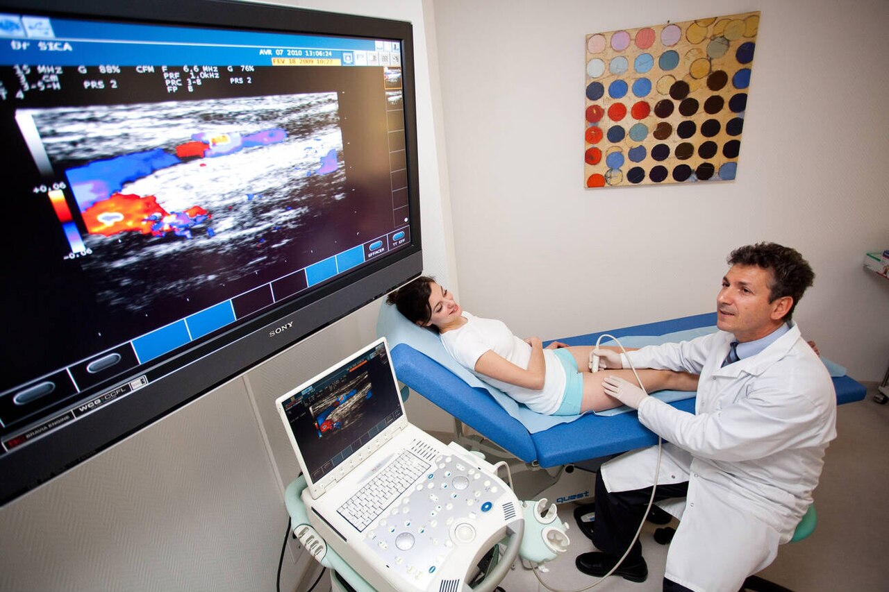
A portable ultrasound read in real time, the probe in one hand and the screen watched at a glance. This example is a vascular study in a clinic; a scan at the side of the pitch uses the same probe and the same live reading, turned on the injury. Photo: Goleisureintl, CC BY 4.0.
Sports injuries happen far from the imaging department. They happen on the field, on the track, in the gym, at the moment of a sprint or a tackle or a landing. The team’s doctor is there with the athlete. A handheld probe in the kit bag is the only imaging within reach, the machine now small enough to travel with the team.
The probe meets the injury at its freshest. A muscle tear is at its clearest in the first hours, the torn ends apart and the blood still pooling, before swelling and healing blur the picture. A scan taken at the side of the pitch catches the injury at a stage a scan days later cannot reach.
What the doctor needs in that moment is not a full report. It is an answer to a narrow question: is this the kind of injury that ends the game, ends the season, or lets the athlete run on. The probe is aimed straight at that question, and the rest can wait for the clinic.
The setting asks nothing fancy of the probe. A bench, a treatment table, or the turf itself does for a couch. The athlete is scanned sitting, lying, or standing, in the kit they were hurt in. The scan fits itself to the field. The picture it gives is the one a clinic couch would give an hour later, only sooner and where it counts.
What one probe reads
A single linear probe reads nearly all of what a sports injury damages. Muscle, tendon, ligament, the lining of a joint, the fluid inside it, and the surface of a bone all lie within a few centimeters of the skin, in the range the probe shows best. One tool covers the soft tissues that sport tears.
Each tissue has its own look on the screen. Muscle is a dark mass feathered with bright lines. A tendon is a tight, bright band of parallel fibers. A ligament is a shorter band bridging two bones. The lining of a joint is a thin film. Fluid within it reads as a dark space. The surface of a bone reads as a bright, unbroken line.
An injury shows as a break in that normal look: a torn muscle losing its feathered order, a torn tendon its bright band, a sprained ligament swollen and frayed, a joint filled with dark fluid, a cracked bone stepped where its line should run straight. The scan learns the normal first so the break stands out.
The breadth is the point. The doctor at the field does not know in advance what the athlete has hurt. One probe answers for the muscle, the tendon, the ligament, the joint, and the bone, in the order the injury calls for. The same scan that clears one part rules in another.
No other tool at the field reads across so many tissues.
The call at the side of the pitch
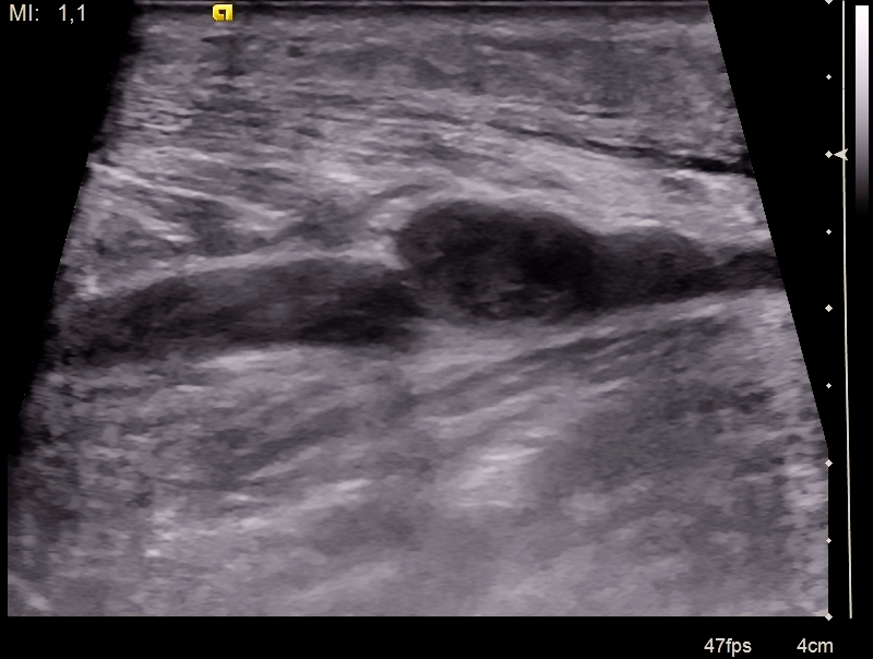
A torn calf muscle on ultrasound. The dark pool in the middle is the gap where the muscle has torn, filled with blood. A strain ruffles the fibers without parting them. A tear like this one opens a space the probe can measure. Image: Nevit Dilmen, CC BY-SA 3.0.
The scan is built around one decision. An athlete goes down, and a crowd of people wait on a single question: can the player carry on, or is the game over for them today. A handheld probe answers it at the side of the pitch, in real time, with the injury minutes old. The doctor lays the probe on the hurt part and reads it live. A muscle felt as a painful knot shows its true state on the screen, a strain that will settle or a tear with a gap that ends the match. A joint that has given way shows whether it holds fluid and blood or sits dry. A bone struck hard is run along for a step in its bright line. The reading is dynamic, which is the heart of it. The athlete is asked to tense the muscle, to bend the joint, to take the weight, and the probe watches the injured part work under load. A muscle that contracts and draws its torn ends together behaves unlike one whose ends pull apart and gape. A ligament stressed under the probe opens at the tear or holds firm. This live test, the injured part working under the doctor’s eye, is what no still picture and no test off the field can give. A call made on the field can go wrong in two ways. A torn muscle sent back into play tears worse. A sound one held back loses the athlete a game for nothing. The live reading guards against both, the true state of the injury seen before the choice is made. The decision turns on what the probe shows. A minor strain, the fibers ruffled and whole, can let a player return with care. A torn muscle with a wide gap, a ruptured tendon, or a joint full of blood takes the player off at once and sets the road to recovery. A break in a bone sends the athlete straight to hospital. The probe sorts these on the spot, in the minutes that matter, as the player, the coach, and the medical team wait on the answer. The largest call of a sports injury, whether to keep playing, is made at the side of the pitch with the probe in hand. Everything that follows, the scan in the clinic, the imaging in the department, the weeks of recovery, builds on that first reading taken where the injury fell.
Muscle, the commonest tear
Muscle is the tissue sport tears more than any other. A sprint strains a hamstring, a push-off tears a calf, a kick pulls a thigh. The probe reads the muscle as a dark mass combed through with bright lines, the sheets of fiber and their coverings. A tear breaks that comb. The break is what the scan looks for.
A minor strain ruffles the fibers without parting them, a little swelling and a darker patch where the muscle is hurt. A partial tear opens a gap that fills with blood, a dark pool sitting in the torn muscle. A complete tear parts the muscle across, the ends drawing back and the gap wide. The size of the tear and the gap grade the injury and set the time off.
The probe measures the tear and follows it as the muscle is tensed, the torn ends seen to move or to gape. The reading at the field is an overview. The full grading of a serious tear in an elite athlete still goes to MRI. The scan’s job at the pitch is to tell a strain that plays on from a tear that does not.
The time the scan sets matters as much as the name it gives. A small tear may keep a sprinter out for a week, a large one for two months. The gap the probe measures, read again as the weeks pass, guides the athlete back to the track at the right time. A muscle returned to sport before the gap has filled tears again more easily, so the healing tissue itself sets the safe day to come back.
The bleed in the muscle
A torn muscle bleeds. The blood pools in the gap as a dark, shifting collection. A small bleed is left to settle on its own. A large, tense one, painful and swelling fast, can be drained under the probe, the needle laid into the pool and clear of the sound muscle around it. The probe finds the blood, measures it, and guides the needle that lets it out.
An old bleed left in the muscle can harden, a fleck of bone forming where blood should have cleared. The probe catches this early as a bright patch in the healing muscle, a warning to rest the part and let it settle. A bleed found early and managed at the field settles without leaving that hard fleck behind.
Tendons and ligaments
Tendons take the next share of sports injury. A strained tendon reads thickened and dulled, its bright fibers swollen. A torn tendon shows a gap in the band, the ends drawn apart. The Achilles is the one that snaps outright at a push-off, read in full on its own page. The probe at the field confirms the gap and watches the ends move as the ankle bends.
Ligaments hold the joints together. A twist tears them. The lateral ankle ligaments go in the classic rolled ankle. The thumb’s side ligament tears in a fall onto a pole or a ski stick. The probe shows the torn ligament swollen and slack. A gentle stress under the probe opens a tear that a still joint hides.
The stress test is the probe’s strength here. The joint is pushed gently to the side that loads the hurt ligament, and the probe watches the gap. A ligament that opens under the load is torn. A ligament that holds firm under it is strained, its fibers stretched and sound. The live test sorts the two. A still image leaves them looking alike.
Overuse wears a tendon down with no single tear, the strain of training week on week. The probe shows the worn tendon thickened and dull, its bright fibers blurred, with new vessels lit on color flow where the wear runs deepest. A patellar tendon in a jumper, an Achilles in a runner, read early and watched over a season, can be eased before it gives way outright. The scan gives the worn tendon a measured thickness to watch, season on season.
Joints, fluid, and blood
A struck or twisted joint fills with fluid. The probe reads the fill. A clear fluid is a simple effusion. A dark, mottled fluid is blood, a hemarthrosis, the mark of a tear inside the joint that bleeds. The knee is the joint that fills more than any other in sport, with a page of its own.
Blood in a joint within an hour of the injury points to something torn that bleeds fast, a ligament or a bone surface. It raises the stakes of the injury. The probe finds the blood at the field and marks the joint for a closer look. A swollen knee that is dry tells a different story from one full of blood.
The same probe drains the joint when it needs draining. A tense, painful effusion can be tapped under sight at the side of the pitch, the needle laid into the fluid and clear of the cartilage. The scan that finds the fluid guides the needle that lets it out. The relief is felt at once.
The joint is tested as well as drained. The probe watches the joint move and its surfaces meet. A fleck of bone or cartilage knocked loose can be caught drifting in the fluid, a fragment a blind tap would never find. A joint read live at the field gives up more of its story than a joint felt from the outside.
What comes off the pitch
Some injuries come off the pitch again and again. The probe knows them well. The hamstring tears in a sprint, high at the back of the thigh, the gap and the blood plain on the screen. It is the injury that keeps a runner out the longest. The scan that grades the tear shapes the weeks ahead.
The calf tears low and inward, where the inner head of the calf muscle meets its sheet of tendon, the injury an older player calls a tennis leg. The probe shows the torn edge and a slick of fluid between the muscle and the sheet below it. The find is quick. The picture is clear.
The groin strains where the inner thigh pulls on the pelvis, a nagging tear that dogs footballers. The quadriceps tears at the front of the thigh from a kick or a block. The probe reads each at the field, a strain told from a tear, a player’s day decided.
The ankle rolls and its outer ligaments give, the commonest sprain in sport. The probe shows the torn band. A gentle stress opens the gap. The tip of the shoulder injures in a fall, the joint between collarbone and shoulder blade lifted and sore, set out on a page of its own.
A handful of injuries make up the bulk of a season’s work. The probe carries a picture of each. The doctor who has read a hundred hamstrings reads the next one fast. The team has its answer before the stretcher is called.
The break the X-ray might miss
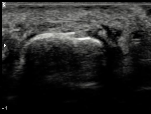
A fractured foot bone on ultrasound. The bright curved line is the surface of the bone. The step and notch in it is the break. The probe reads only this near surface, not the depth of the bone. Image: Mme Mim, CC BY-SA 4.0.
Bone turns the sound back as a bright, smooth line. The probe reads only its surface. A fracture shows as a step or a break in that line, with a small dark gap and sometimes a lift of the lining over it. The probe cannot see into the bone or behind it. It reads the cortex where the cortex faces the skin.
This narrow view still earns its keep. A rib cracked in a tackle is often missed on an X-ray. The probe can find it, the break felt for where it hurts. A stress fracture that an early X-ray misses can show as a lift of the lining and a fleck of new bone. A finger or a foot bone can be checked at the field before a trip for an X-ray is weighed.
The probe does not replace the X-ray. A normal-looking cortex does not clear a bone, since the probe sees only the near surface and nothing of the depth. A break found on the probe is useful. A break not seen is not ruled out. The bone goes for an X-ray when the injury or the pain calls for it.
Following the injury back to play
The injury is scanned again as it heals. A muscle tear is followed over the weeks, the gap seen to fill and the fibers to knit. A tendon and a ligament are watched the same way, the swelling settling and the band firming. The same probe that read the injury reads the repair.
The scan helps time the return to play. A tear that has knit, with a muscle that contracts cleanly under the probe, argues for a safe return. A gap still open, or pain still felt where the probe presses, argues for more rest. The decision rests on more than the scan. The scan adds a look at the tissue itself.
A return made on a tear not fully healed risks the worst outcome in sport, the same muscle torn again, worse than the first. The scan that shows a repair still short can save an athlete a second, longer spell out. The picture of the healing tissue weighs in the timing of the return.
The stakes of that timing run high. A torn muscle is far likelier to tear again in the weeks after it knits. The second tear runs worse and heals slower than the first. A scan that holds an athlete back a week can spare a season lost to a tear that came back. The look at the tissue is one more guard on a decision that has to be got right.
What the probe misses
The probe has its blind spots. It reads the near surface and a few centimeters of depth. It cannot see into a bone or behind one. A deep tear, a joint surface, a bruise in the bone itself lie beyond its reach. The probe is read for what it shows. What it cannot reach is left to other imaging.
The reading leans on the operator. A tilted probe darkens a sound tendon into a false tear. A finding is checked from more than one angle before it is called. The skill of the hand and the eye sets how far the scan can be trusted.
Magnetic resonance imaging is the fuller test for many sports injuries, the one that maps a deep tear, a bone bruise, and the inside of a joint. The X-ray remains the test for a fracture. The probe at the field does not replace them. It brings a first answer to the place and the moment the others cannot reach. It points the way to them when they are needed. It tells the doctor which of the two to send the athlete for, and when.
Sports ultrasound on a handheld unit
The handheld scan and the sports injury are made for each other. The injured tissues lie shallow, in easy reach of a linear probe. The probe travels in a bag to the field, the track, the court. The imaging comes to the athlete, in the place and the minute of the injury.
The scan is quick, painless, and repeatable. It can be run on the pitch, run again at half-time, and run a third time the next day, at no cost beyond the minutes it takes. It carries no radiation, so it bears repeating as often as the injury asks. The healthy limb is a moment away for a comparison on the same athlete.
The probe also acts on what it finds. It guides a needle to drain a tense joint or to place an injection. It steers a treatment as well as it reads an injury. The same tool that makes the call carries out part of the care.
The whole team gathers around the screen. The doctor, the physio, the coach, and the athlete see the same picture at once. A player shown the gap in their own muscle takes the rest the recovery needs. The probe turns a hidden injury into something the people who must act on it can see for themselves.
What the handheld scan asks for is a trained hand and a clear head: the right tissue found, read square, and tested under load, with the question of return held in mind throughout. Used that way, a probe in a kit bag reads a sports injury at the side of the pitch as surely as a department machine reads it days later.
Common questions about sports ultrasound
What can handheld ultrasound assess in a sports injury?
It reads muscle, tendon, ligament, joint linings and their fluid, and the surface of bone, all of which lie shallow under the skin. It shows a muscle tear, a tendon or ligament injury, a joint effusion, and some fractures, and it tests the injured part as it moves. One probe covers nearly all of what a sports injury damages.
How does ultrasound help the return-to-play decision?
It reads the injury at the side of the pitch, in real time, as the athlete tenses the muscle or bends the joint. A minor strain with whole fibers can let a player return with care. A wide tear, a ruptured tendon, or a joint full of blood takes the player off. The scan is repeated as the injury heals to help time a safe return.
Can ultrasound find a fracture?
It can show some fractures as a step or break in the bright surface of the bone, which is useful for a rib or a stress fracture that an X-ray may miss. It cannot see into or behind the bone, so a normal scan does not clear a fracture. A bone goes for an X-ray when the injury or the pain calls for it.
Does ultrasound replace MRI in sport?
No. Magnetic resonance imaging maps the deep tears, bone bruises, and joint surfaces that ultrasound cannot reach, and it remains the fuller test for grading a serious injury. Ultrasound brings a first answer at the field and points the way to MRI when it is needed.
Why scan a sports injury in the first minutes at the field?
A fresh injury reads at its clearest, the torn ends apart and the bleeding still pooling before swelling sets in. A scan at the field also answers the immediate question of whether the athlete can carry on. Both the picture and the decision are best served in the minutes after the injury.
Can a handheld probe do this at the pitch-side?
Yes. The injured tissues are shallow and within reach of a linear probe, and the machine is small enough to carry in a kit bag. It is quick, painless, and free of radiation, so it can be repeated as often as needed. It also guides a needle to drain a joint or place an injection on the spot.
About the Author
Julien Mercier
Senior R&D Engineer · Medical Ultrasound Transducer Development
Senior R&D Engineer with an M.S. in Applied Physics and over 15 years of experience in medical ultrasound transducer development, specializing in the design verification and performance testing of high-frequency imaging transducers. Currently leading the development and verification of the company’s next-generation high-frequency linear-array transducer, responsible for imaging performance evaluation and reliability analysis in preclinical testing. Brings extensive hands-on experience in piezoelectric element tuning, beamforming parameter optimization, and system-level performance testing.