Second Trimester Anomaly Scan Handheld Ultrasound Protocol
A second trimester anomaly scan, run at around twenty weeks, checks the baby’s body part by part for anything built wrong. It works head to toe through a fixed list, the brain, the face, the spine, the heart, the belly, the kidneys, the limbs, reading each against how it should look at this age. A handheld probe runs the same survey at the bedside, the detailed scan that maps a whole fetus done in one careful pass. It is the one scan in pregnancy built to read the baby’s structure.
When the scan is done
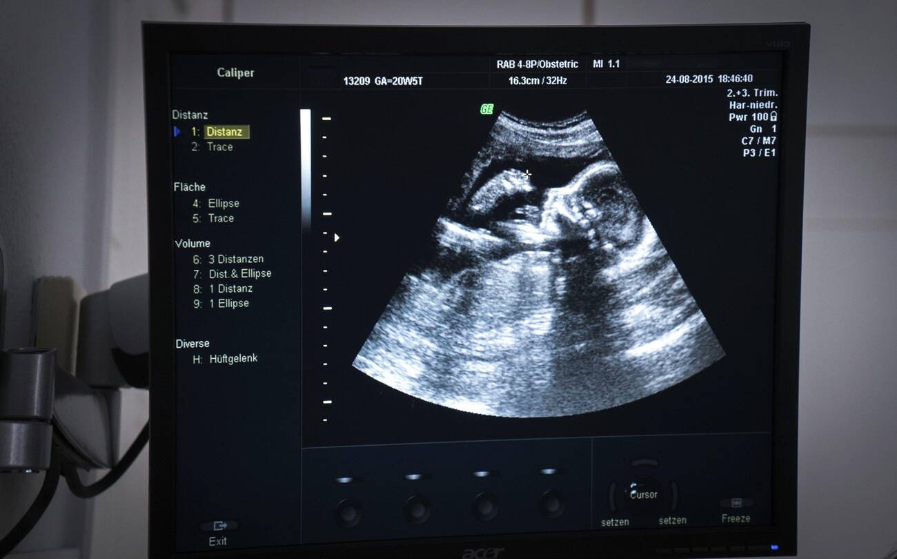
A twenty-week anatomy scan on the machine, the fetus in view at twenty weeks and five days. The menu down the side holds the measuring tools, the photograph taken of the scanner’s own screen.
The scan is timed to a window around eighteen to twenty-two weeks. The body is built enough to read by then, the fetus large enough to see, with room still to shift it for a better view. The weeks around twenty are the sweet spot, late enough that the organs have formed and early enough that the baby moves freely under the probe. The window is set by the baby itself, the middle weeks the time the body is built and the room to read it is still there, the organs large enough and the baby free enough to turn. The eighteen-to-twenty-two window is the same the world over, set by what the fetus shows. A clinic that scans too early calls more women back for the views a small fetus hides, the right week the one that reads the fullest in a single pass.
Twenty weeks and on reads more completely than the weeks just before. A scan done a little later catches the views a smaller fetus hides, fewer of them needing a repeat. The body keeps growing into the window, every organ clearer at twenty-two than at eighteen. A view the probe cannot get on the day, a baby lying the wrong way, a hand across the face, is the commonest reason a woman is asked back. The right timing cuts those callbacks down. A baby that lies face down, or curls away from the probe, hides the views that need a clear line to it. A gentle push, a walk, a wait, often turns the baby into a better lie, the reader patient where a view will not come.
The scan is the one full look at the baby’s structure the pregnancy gives. It comes once, takes its time, and works through every system in turn. A scan rushed or cut short misses what only a careful pass can find. The reader gives it the minutes it needs. The scan carries a weight beyond reassurance, what it finds able to change the care of the whole pregnancy, the plan for the birth, the place it should happen. A serious problem caught at twenty weeks gives months to prepare, a team and a plan ready before the baby arrives. The weeks around twenty give the one clean window the whole pregnancy offers for this depth of look, the scan rewarding the reader who works the list to its end. A dating scan in the first weeks has usually fixed the age before this one, the anomaly scan freed to read structure with the date already set. The months the scan buys are its quiet gift, a heart problem named early letting a birth be planned at a centre with the team to meet it.
The head-to-toe sweep
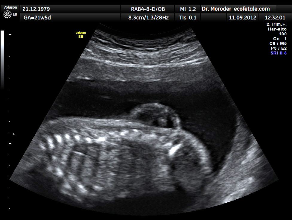
A fetal spine in long section at twenty-one weeks, the bright bones running along the back. The break in their line low down is a lumbar spina bifida, the open spine the anomaly scan is built to catch.
The scan follows the same path through the baby every time.
It starts at the head and works down, the order fixed, no part skipped. The skull is read first for its shape and its bones, a smooth oval the sign of a normal vault, the brain inside checked for the fluid spaces that should sit where they do, the cerebellum and the hollow behind it read at the back of the head. The face comes next, the probe turned to read the upper lip for the gap of a cleft, the profile for the set of the nose and the chin. The head holds more than the skull, the reader reading the midline of the brain, the two halves matched across it, the fluid spaces measured for a width that should stay small. A space grown wide is the sign of a brain under pressure, read and measured and watched. The fluid spaces of the brain are measured at a fixed point, the width read against a number that should stay under ten. A space wider than that is followed, the cause hunted, the brain watched across later scans. The spine is run from the neck to the tail in two ways, along its length for the line of the bones and across it for the ring each should make, a break or a bulge in that line the mark of an open spine. The chest is read for the lungs filling it evenly and the heart sitting where it should, pointing the way it should. The belly is read for the stomach as a dark pocket on the left, the bowel packed neat without a swelling, the wall closed around the cord where it enters. The kidneys are found either side of the spine and the bladder as a small dark pocket low down, the fluid around the baby proof that the kidneys are passing water. The limbs are counted and read, two arms and two legs, each with its long bones measured, the hands and feet checked for their place and their count. Every system has its views. The reader takes them all in the same order each time, the routine itself the guard against a part left unread. The whole baby is read in a set order, a reader who keeps to it reading the same scan every time, sure and complete. A view caught and kept is a view that can be read again, by the same eye or another, the saved picture the proof the scan was done.
The brain and the spine are read with special care, the defects there among the gravest, and among the easiest to catch. An open spine shows as a break in the run of the bones, often with a tell-tale pull on the shape of the skull and the brain behind it. The scan that reads the back cleanly, bone by bone, is the one that catches the open spine early. A skull pulled into a lemon shape, or a brain drawn back into a curve, points to an open spine even before the back is read. The reader who catches those head signs reads the spine that bit harder. The skull gives its own clue to the spine below it. A reader who knows the head signs reads the back with the spine already half-suspected. The two are read as a pair, the head and the spine each telling on the other. The lips and the palate are read for the gap of a cleft, a common difference the scan often catches in profile and head-on.
The belly and the limbs round out the survey. A wall that has not closed lets the bowel float outside it, a defect the scan catches almost every time it is there. The long bones are measured for their length and read for their shape, a bone too short or bent a clue to a problem with the way the skeleton forms. The count of fingers and toes is read where the hands open. The belly holds the early signs of trouble in the gut, a stomach not seen where it should be, a loop of bowel swollen with what cannot pass. A wall left open at the cord lets the organs sit outside the body, the clearest defect the scan reads and near always caught. The bladder seen and the stomach seen, both dark pockets where they belong, tell that the gut and the kidneys have plumbed themselves right. A stomach not found, scan after scan, is a sign chased, the tube to it sometimes blocked or astray. The long bones are read for length and for the clean straight line a normal bone holds. The kidneys read as two bean-shaped organs hugging the spine, each with its own dark centre where the urine gathers. A kidney swollen with backed-up water, or one missing, is read for what it means.
The whole sweep takes its time and skips nothing. A reader who runs the list the same way every scan builds the habit that catches the rare wrong thing among the many right ones. A reader who freelances, reading what catches the eye and skipping what does not, is the reader who misses the quiet defect. The list run whole is what makes the scan one to trust. The reader names each view as it is caught, building the report view by view, a record of what was seen and what was clear. A view not got is written as not got, the honest report naming its own gaps. Completeness is the measure of a good anomaly scan, well ahead of speed.
The heart, the hardest part
The heart is the hardest organ to read, the one a scan misses more than any other. It is small, it beats fast, and its defects can hide in a view that looks nearly normal. The scan reads it in steps, the four chambers first, a clean cross of two pumps and two filling rooms, then the great vessels leaving it, checked for their cross and their size. The heart is read live, its beat the thing that lets it be read at all. A reader watches the four chambers fill and empty, the walls moving evenly, the valves opening clean, the rhythm read as steady. The four-chamber view is the heart of the heart scan, a single picture showing the two sides matched, the walls whole between them, the valves set right. A reader holds that view and reads it slow, the one picture that clears the commonest serious defects in a stroke, the first heart view a reader learns and the one leaned on hardest. The heart sits at an angle in the chest, its apex pointing down and to the left, a tilt read as part of the check, a heart pointing the wrong way a sign before the chambers are even counted. The reader who reads a hundred normal hearts reads the odd one by how it breaks the look, the trained eye the best tool the heart scan has. The heart is the part a reader trains longest on, the rest of the survey learned faster than this one organ.
A heart that shows four equal chambers and two vessels crossing as they leave is a heart largely cleared. The defects that slip past are the subtle ones, a narrowed vessel or a small hole, caught at rates far lower than the obvious wrongs elsewhere. A reader spends longer on the heart than on any other organ. The doubtful one is sent to a specialist scan built for it. A clean heart on this scan is a quiet kind of good news, the organ that worries families read and cleared. The outflow tracts are the views that catch what the four chambers miss, two great vessels leaving the heart and crossing one another as they go, a cross read for on every scan. A vessel that runs parallel where it should cross, or one too narrow, is the defect a careful outflow view catches. A heart cleared on the four chambers and the outflows is a heart through the screen well. The defects that still slip past are the ones that change little at twenty weeks, sometimes clearer only on a later scan, the heart never read once and forgotten.
The rest of the picture
The scan measures the baby as well as reads it. Four lengths are taken, across the head, around the head, around the belly, and along the thigh bone. Together they plot the baby’s size against its age. A baby measuring on track is a baby growing well, the numbers a baseline every later scan is read against. The four lengths fall each on a line for its age, the four together telling a baby grown evenly. A head large for the belly, or a belly small for the dates, is a shape read for what it means. The measurements are taken on set views, the head on a flat round slice, the belly on a circle at the stomach, the thigh along the bone. A length read off the right view is a length to trust, the chart turning it into a place for the baby on the growth curve. The growth read here is a starting line, the baby measured again later to see how far it has come along since.
The placenta is found and its position read. One lying low, over or near the neck of the womb, is noted and checked again later. The cord is traced to where it joins the placenta and the baby. The low placenta is the one the scan flags to read again. Many that sit low at twenty weeks have climbed clear by the third trimester, the lower womb stretching up beneath them. The few that stay low are the ones a later scan watches, the position that matters for the birth read in good time. The placenta is read for more than its place, its look checked for the even grain of a healthy one, its edge for where it sits against the neck of the womb. A placenta low and covering the way out is the one that changes how a baby must be born, flagged early and watched to the end.
The fluid around the baby is read for its amount, too little or too much each a sign to chase. The cord is checked for its three vessels, two arteries and a vein, a count that hints at how the baby has formed. These are the surrounds the baby grows in, read alongside the baby itself. The fluid is the baby’s room and its drink, made by the kidneys and swallowed and made again. A pocket too small, or too deep, is the sign the reader chases, the cause hunted from there. The cord usually holds three vessels, a cord with two noted and the baby read with extra care. The bladder filling and emptying across the scan is its own quiet sign, the proof the kidneys are at work. The fluid and the placenta and the cord are read as the baby’s world, the things it lives in read with the thing itself.
The scan looks too for the small signs that raise the odds of a chromosome problem. A bright spot in the heart, a touch of fluid in a kidney, a slightly short bone, each on its own means little. The markers are weighed together, and against the blood tests, before any of them counts for much. A single soft marker on a low-risk pregnancy rarely changes anything. A soft marker is a hint read for the company it keeps, no diagnosis on its own. Several together, or one beside a worrying blood test, is read more seriously, the markers weighed as a set. The soft markers were named when scans were the main test for chromosome trouble. The blood tests of today carry much of that weight now, the markers a second voice read beside them, a clean blood test and a single marker together rarely raising the odds enough to act on.
What the scan can and cannot find
A normal anomaly scan is a strong reassurance, never a full guarantee. The scan finds many structural problems, near all of some kinds and only a share of others, the subtle heart defect the hardest of all to catch. Some problems show only later, some never on a scan at all. A reader reads the scan for what it finds well and names plainly the limit on the rest. A clear scan is read as good news held with honest care. The scan reads structure, the working of an organ and the months ahead beyond its sight. A heart built right can still fail to work right, a brain formed well can still grow wrong, the scan reading the baby as it is built on the day, no more and no less. A problem in how a gene is written, a deafness, a difference in how a brain will work, leaves no mark on the picture at twenty weeks. The scan reads structure well and claims no more, the unseen left plainly unclaimed, a clear result named as a good sign held with care. A scan names what it sees and holds its tongue on what it cannot, the honest reading the safe one for the woman who trusts it. The scan gives what it can give, honestly and in full, the rest left to the tests built for it.
On a handheld
The anomaly scan asks a lot of a probe. A good handheld answers the call. The views are detailed, the structures small, the heart a test of any machine, the better handhelds reading them as a cart once did. A machine that reads a clean four-chamber view and a crossing pair of vessels reads enough to run the protocol, its small screen no bar to the fine detail it shows. A clinician trained in the protocol runs the full survey at the bedside, the whole map of the baby read in one sitting. A clinician who runs the protocol day in and day out reads a handheld as surely as a cart, the skill living in the hands and the eye, the machine’s size counting for little. The probe that travels turns a scan from a journey a woman makes into a service that comes to her, the care carried the last mile.
The reach carries the scan to where the care is thin. A pregnancy that would never reach a scanning centre is read where the woman lives, the major problems caught early enough to plan for. The image saves to the phone, the views stored, the heart filmed, ready for a specialist to read from afar. A woman in a village, a camp, a clinic with no scanner of its own, carries a baby whose problems can be found by a probe that travels to her. The scan that reaches her early gives the same months to prepare a city mother takes for granted. A baby read early, wherever the mother is, is a baby whose problems are met with a plan made in good time. The twenty-week check travels to clinics far from a hospital on a probe one clinician carries.
Common questions about the anomaly scan
When is the anomaly scan done?
Between about eighteen and twenty-two weeks, with twenty to twenty-two the preferred window. By then the organs are formed and the baby is large enough to read in full, with room still to shift it for a clear view. A scan at this age reads more completely than one done a week or two earlier.
What does the anomaly scan check?
The baby’s whole structure, head to toe: the brain and skull, the face and lip, the spine, the heart, the stomach and bowel, the kidneys and bladder, and the limbs. It also measures the baby’s growth, finds the placenta, reads the fluid around the baby, and counts the vessels in the cord.
Can the scan find every problem?
No. It finds many structural problems, near all of some kinds and only a share of others. The subtle heart defects are the hardest to catch, and some problems show only later or never on a scan. A normal scan lowers the odds of a serious problem a great deal, without ruling every one out.
Why is the heart looked at so carefully?
The heart holds the structural problems hardest to see and easiest to miss. It is small and fast, its subtle defects able to hide in a view that looks almost normal. The scan reads the four chambers and the great vessels in steps. A doubtful heart is sent on to a specialist fetal heart scan.
Can a handheld ultrasound do an anomaly scan?
Yes, in trained hands. The anomaly scan is detailed work. A good handheld reads the small structures a cart once did. A clinician who knows the protocol can run the full head-to-toe survey at the bedside, which brings the scan to women who would otherwise never reach a scanning centre.
About the Author
Julien Mercier
Senior R&D Engineer · Medical Ultrasound Transducer Development
Senior R&D Engineer with an M.S. in Applied Physics and over 15 years of experience in medical ultrasound transducer development, specializing in the design verification and performance testing of high-frequency imaging transducers. Currently leading the development and verification of the company’s next-generation high-frequency linear-array transducer, responsible for imaging performance evaluation and reliability analysis in preclinical testing. Brings extensive hands-on experience in piezoelectric element tuning, beamforming parameter optimization, and system-level performance testing.