A nerve block puts local anaesthetic around a nerve to numb the part that nerve serves. Ultrasound guidance does the placing under a live picture, the needle and the spreading drug both watched on screen. The probe finds the target, follows the needle in to it, and shows the anaesthetic pooling where it belongs. A handheld version carries that whole sequence to the bedside, the probe and the screen shrunk to a pair of objects that travel in a coat pocket.
What the live picture gives
The whole value of ultrasound here is that the drug is placed under sight. The operator watches the needle tip travel to the target and stop short of the nerve. The anaesthetic is seen to spread around the nerve, a dark cuff growing where it is wanted. A spread that pools in the wrong place shows at once, before a second millilitre follows. The needle, the tip, the first bead of anaesthetic, and the cuff that grows from it all sit on the one screen, nothing of the block left to a sense the operator cannot check. The eye confirms every step the old landmark method left to feel and hope. A nerve that runs deep, or a plexus packed against a vessel, comes into plain view on the screen. The depth of the target reads off the picture, so the needle is set at the right angle from the skin. The picture turns a block once judged by surface marks and a reported tingle into one read off a live image. Every move answers to that picture, the operator never an inch ahead of what the screen shows.
The record bears the value out. A block placed under ultrasound takes hold more often and faster than one set by feel, the drug landing where it works on the first pass. Fewer repeat needles mean fewer chances to stray. This reliability is what made the guided block a standard within a generation.
The patient gains a great deal from the sight. A limb numbed for surgery lets a patient stay awake and breathing on their own, clear-headed soon after. A block carries its own pain relief into the night, sparing the heavier drugs a general anaesthetic would call for. The picture serves the recovery as much as the surgery.
That sight buys two things at once: a block that works and a block that is safe. A drug laid right against the nerve takes a fast, full hold. A tip kept out of the vessel beside it, and out of the nerve itself, spares the patient the harms that a blind needle courts. Smaller volumes do the job when the drug is steered to the spot, which lowers the dose a patient carries. A block placed under sight takes hold in a known time, the operator watching the cuff form on the screen. A patient spared a general anaesthetic keeps their own breathing and wakes without its grogginess, a gain the guided block makes safe enough to offer. A failed first pass does less harm under sight, a needle seen to miss pulled back along its own track and sent again on a clear view.
The steps every block shares
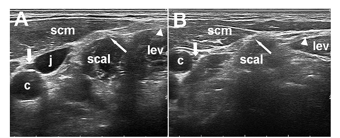
A nerve seen in cross section on ultrasound, here the vagus in the neck beside the carotid (c) and the jugular (j). The probe picks the nerve out from the vessels around it, the first task of any block. The letters and arrows are the authors’. Cropped from a figure by Uwe Walter and colleagues, CC BY 4.0.
Three moves run through every ultrasound block.
The first is to find the target and know it for certain. A nerve shows as a bundle of dark dots in a bright sheath, a honeycomb the probe learns to pick out from the tendon and the vessel around it. A fascial plane shows as a bright line between two muscles, the seam the drug will open. The operator slides and tilts the probe until the structure sits clear in the middle of the screen, named beyond doubt before any needle moves. Reading these targets is a craft of its own, set out in full on its own page. The whole block rests on this first certainty, since a needle aimed at a misread shape carries the drug to the wrong place. A probe held square and steady turns a confusing grey field into a map with the target marked. Only once the target is sure does the needle earn its entry. The honeycomb of a nerve has a look the eye comes to trust, a cluster of dark pockets in a bright frame that holds its shape when the limb moves. A tendon nearby slides and a vessel pulses, each giving itself away under the probe. Colour over the spot lights any vessel hiding by the nerve, marking the ground the needle must skirt. The colour is run over the target as a matter of habit before the needle goes in, any vessel in the path lit before it can be struck. A target that hides behind bone or air is coaxed into view by a tilt of the probe or a shift of the patient, the picture worked until the nerve sits clear. A gentle press folds a vein flat, sorting it from the artery beside it before a needle is drawn. The operator builds a small map of the target and its neighbours before committing, every structure named, every danger marked. This patient reading is the part an expert never skips. The target is chased into the clearest window the body allows before a needle is ever drawn. A nerve named in one plane is turned to read in the other, the honeycomb seen across and the cable seen along its length. A structure that holds its look in both views is a structure named beyond argument. The patient is positioned to bring the target up toward the skin, the head turned or the arm laid out, so the nerve sits in the easy reach of the probe. The reading is unhurried, the few seconds it costs repaid many times over by a needle that goes to the right place the first time.
The second move is to bring the needle in along the beam, its whole length in view. The probe runs in line with the needle. The shaft and the tip then show as one bright line from skin to target. The tip is watched every millimetre, never pushed into tissue the eye has not cleared. This in-plane discipline is the safety of the whole field, the tip stopped a hair short of the nerve and held clear of it. A tip kept in sight is a tip kept out of the artery, the vein, the lung, and the nerve. The needle is chosen for the screen as much as for the skin, a textured shaft catching the beam even at a steep angle. Small jiggles and a breath of fluid betray a tip gone faint, the same tricks a guided line leans on. The whole craft of seeing a needle carries straight across from the vessels to the nerves. The needle for a block is often a short, blunt bevel, shaped to slide a nerve aside on contact. The blunt tip gives a feel as it crosses each layer, a second sense backing up the eye. The angle of entry is set from the depth read off the screen, the needle laid flat for a shallow plane and pitched steeper only when the target sits deep. The needle enters far enough from the probe to lie along the beam, its whole length lit on the way to the nerve.
The third move is to inject and read the spread. A test of a millilitre tells the story before the full dose follows. Anaesthetic that opens a dark ring around the nerve, the donut the operators look for, says the drug sits where it works. A bulge that swells the nerve itself, or one that vanishes into a vessel, calls a halt. The spread is the proof the block will take, read live, millilitre by millilitre. Watching it turns a hopeful injection into a confirmed one. The needle is nudged a touch and the drug redirected when the first spread sits wrong, the dose steered until the cuff closes clean. A block read this way rarely fails for want of placement, the proof gathered before the full dose commits. A clean cuff seen to close is a block the operator can trust before the patient ever reports it. A spread read wrong is caught early. Drug that tracks the wrong way, or gathers in a single blob, tells the operator to stop and move the tip before the dose is spent. The first millilitre is the scout for the rest. The volume needed shows in the spread itself, a cuff that closes clean around a nerve asking no more drug than that. The operator reads the dose off the picture itself.
The three moves run as one motion in a practised block. The target is found, the needle walked in, the spread read, in a single unbroken sequence that takes a minute or two. The eye moves from target to tip to cuff without a pause, each move checked before the next is begun. The same three steps underlie every block on every page that follows, learned once and carried through the whole field.
Two families of block
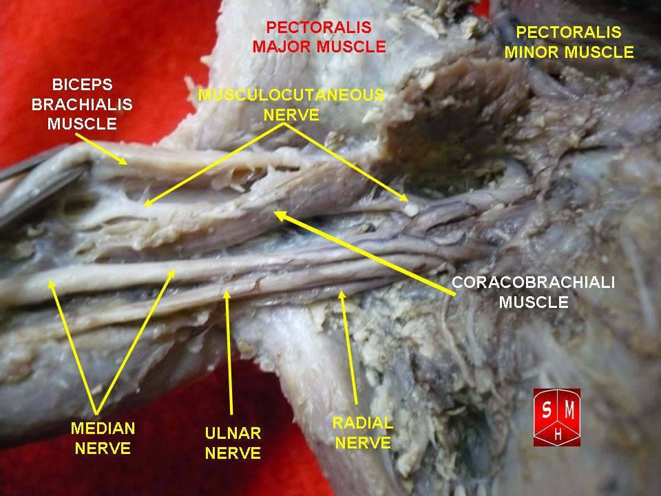
A dissection of the arm’s nerves where they branch from the brachial plexus, the median, ulnar, and radial nerves a perineural block bathes in anaesthetic. The yellow arrows and labels were added to the photograph. Image: Anatomist90, CC BY-SA 3.0.
The blocks fall into two kinds by what the needle aims at. A perineural block lays the drug around a named nerve or a plexus, the bundle that feeds a limb. The anaesthetic cuffs the nerve directly. The numbing follows the map of that nerve. The blocks at the neck and the shoulder work this way, the brachial plexus bathed where it passes. A perineural block numbs the whole limb it serves, enough to carry a patient through surgery on the arm awake and comfortable. The drug sits close to a bundle that also runs beside vessels and, at the neck, beside the lung, so the placing asks a steady hand and a clear picture. The numbing of a perineural block reaches deep and lasts for hours, the whole territory of the nerve going quiet. It asks a great deal of the operator, the target small and the dangers close.
A fascial plane block lays the drug into the sheet of tissue between two muscles, where the small nerves of a region run. The anaesthetic spreads along the plane and reaches the nerves where they travel, a wider and shallower numbing than a single nerve gives. The block of the abdominal wall works this way, a pool opened in a plane the nerves cross. The drug finds the small nerves by spreading along the seam to reach them, so a fascial block leans on volume to carry the anaesthetic the length of the plane. The target is a broad bright line, easier to reach than a fine bundle, which makes the plane block a friendly one to learn. The plane block works at a safe distance from the named nerves and their vessels, the needle laid into an open seam well clear of trouble. The wide field it gives suits the long incisions of the trunk, where one pool covers many small nerves at once.
The plane block is the gentler of the two to place, its target a broad seam the width of a muscle border. It gives a wide, shallow field of numbing across the wall it covers. Both families share the one screen and the same needle skill, the discipline of seeing carried from block to block. Each family has its place. The choice follows the surgery the block is meant to cover, the operation on the table deciding which the patient needs. A third path threads a fine catheter in beside the nerve and leaves it there, the numbing topped up over days for the worst of the pain.
The drug and its limits
The anaesthetic is the active part of the whole exercise. A local agent blocks the nerve from carrying its signal. The part goes numb for the hours the drug lasts. A short-acting agent covers a brief procedure. A longer one carries the numbing well past the end of surgery into the first sore hours of recovery. A small dose of adrenaline mixed into the drug slows its uptake into the blood and stretches the block, a marker as well that warns if the needle has slipped into a vessel. The choice of agent and mix follows the length of the surgery and the pain that comes after it. The longer-acting agents hold a block well past the day of surgery, the numbing measured in many hours. A clinician picks the agent for the job, the duration matched to the recovery the patient faces. The block is one piece of a plan, set inside an anaesthetic built for the patient and the surgery. A dilute solution numbs the feeling and spares much of the movement, letting a patient keep some use of the limb. The block can be set before the surgery or at its end, the numbing timed to cover the cut and the hours after it.
Volume and dose are watched with care. Every block has a ceiling on how much drug a patient can take, set by weight and by the agent. A dose pushed past that ceiling can reach the blood and harm the heart and the brain, the danger named local anaesthetic systemic toxicity. The sight ultrasound gives lowers the volume a block needs, which buys a margin against that ceiling. The toxic dose is a hard line to respect. A patient’s weight sets the ceiling. The operator adds up every millilitre against it across a long case or a second block on the same day, the ceiling shared across them all. The running total stays in the operator’s head through the case, the safe limit a number never far from mind.
Aspiration before each push guards the same line. A gentle pull on the syringe that draws back blood warns of a needle in a vessel, the place the drug must never go. The live picture and the empty syringe together keep the anaesthetic out of the bloodstream. Safe practice leans on both at once. The signs of the drug reaching the blood run from a ringing in the ears and a metal taste through to a seizure and a failing heart. A theatre that does these blocks keeps the antidote, a fat emulsion given into a vein, within arm’s reach. The smaller volume a guided block needs is the first guard against ever reaching that point.
What a block cannot promise
A block is not a sure thing. Even placed well, a block can come in patchy, a stretch of skin left with feeling where a nerve ran a course of its own. A second top-up, or a switch to a general anaesthetic, covers the gap. The careful operator plans for the block that does not fully take. The numbing wears off on its own schedule. A patient is warned to guard a limb that cannot yet feel a burn or a knock.
Some patients cannot have a block. A patient on blood thinners bleeds where the needle passes. A deep block near the spine carries the gravest version of that risk. An infection over the site, or a patient who cannot lie still, rules the block out, and the choice then weighs the block against the general it would replace.
The skill takes time to build. Reading a target, holding a needle in plane, and judging a spread are each a craft. Together they are the work of many supervised blocks. A handheld lowers the cost of the kit. The cost of the learning stays the same, and the field rewards the operator who has put in the hours. A block also takes time the clock may not allow. Finding the target, placing the needle, and waiting for the numbing to set can run past the minutes a crash theatre has. The block is the tool of the planned case more than the dire emergency.
The blocks in detail
Each block has its own page in full. The craft of reading the target underlies them all and is set out in how to locate a nerve with ultrasound. At the neck, the interscalene brachial plexus block covers the shoulder, with its own trade-off at the breathing nerve. Lower in the neck, the supraclavicular brachial plexus block covers the whole arm, the lung sitting close below. On the trunk, the transversus abdominis plane block numbs the abdominal wall through a fascial plane. Beside the spine, the paravertebral block reaches the nerves where they leave the backbone. Each builds on the three shared steps above, the pages running from the single nerve through the wide plane to the deep paravertebral space.
On a handheld
A pocket probe suits regional anaesthesia as well as any field. The targets sit shallow on the common blocks, within the reach of a small linear head. The operator holds the probe in one hand and the needle in the other, a balance a light wireless probe makes easier than a heavy cabled one. The screen on a phone sits in the line of sight, the needle and the spread read without a glance away to a cart. The block is a craft of the hand, the machine a pair of eyes the operator points where it is needed. A scanner who has the targets and the needle drilled gets the same block from a pocket probe as from a cart. The whole exam asks less of luck than a block placed by feel ever did.
The reach of the small probe carries the block to where the patient is. A block under a pocket probe reaches a patient a department could never have served in time. A block placed in a clinic, an emergency room, or a field hospital spares a patient the wait and the risk of a general anaesthetic. A block that once needed a fixed machine and a procedure room travels now to the bedside in a coat pocket. A clip of the spread saved to the phone shows the next clinician that the drug reached its mark, a proof a written note cannot carry. The block goes to the patient now, in place of the patient going to the block. The probe wiped down moves from one block to the next, the same head serving a shoulder in the morning and an abdomen in the afternoon. The whole of a regional service rides in a bag: the probe, the phone, the needles, and the drug, ready wherever a patient needs numbing. A trainee learns the blocks on the same probe a consultant carries, the picture on the phone shared between the two.
Common questions about ultrasound nerve blocks
What is an ultrasound-guided nerve block?
It is the placing of local anaesthetic around a nerve under a live ultrasound picture. The probe finds the nerve, the needle is watched to it, and the spreading drug is seen to cuff the nerve. The numbing follows the part that nerve serves, for the hours the drug lasts.
Why use ultrasound instead of landmarks?
The live picture shows the needle tip and the spreading drug in motion. The tip is kept clear of the vessel, the nerve, and the lung. The drug is seen to pool in the right place. This makes the block both safer and more reliable than a needle placed by feel alone.
What is the donut sign?
It is the dark ring of anaesthetic that opens around a nerve when the drug is injected. The ring shows that the drug sits in the right plane, cuffing the nerve without swelling it. A spread that fails to form this ring warns that the needle tip needs moving before more drug follows.
What is the main danger of a nerve block?
The chief risk is the drug reaching the bloodstream, which can harm the heart and brain, a state called local anaesthetic systemic toxicity. The dose is capped by the patient’s weight, aspiration checks for a needle in a vessel, and the smaller volume ultrasound allows lowers the risk.
Can a handheld ultrasound guide a nerve block?
Yes. The targets sit shallow on the common blocks, within reach of a small linear probe. The exam leans on the hand more than the machine. A light wireless probe run from a phone frees one hand for the needle and brings the block to the bedside or the field.
About the Author
Julien Mercier
Senior R&D Engineer · Medical Ultrasound Transducer Development
Senior R&D Engineer with an M.S. in Applied Physics and over 15 years of experience in medical ultrasound transducer development, specializing in the design verification and performance testing of high-frequency imaging transducers. Currently leading the development and verification of the company’s next-generation high-frequency linear-array transducer, responsible for imaging performance evaluation and reliability analysis in preclinical testing. Brings extensive hands-on experience in piezoelectric element tuning, beamforming parameter optimization, and system-level performance testing.