Routine IV lines go in by sight and touch, no scanner needed. Ultrasound comes in only when the veins cannot be seen or felt, in the patient stuck again and again for access that is not there on the surface. The probe finds a vein the hand misses, usually a deeper one, then guides the cannula in under view.
What difficult intravenous access is
Difficult intravenous access is the term for a patient whose veins are hard to cannulate. The veins may run deep under fat or scar from old lines. They may be collapsed by dehydration, or small and rolling under the skin. The arm gives no vein to see and the finger feels none to aim at.
The cost of difficult access falls on the patient. A nurse may try a stick three or four times before a vein is found, each one a fresh needle and a fresh bruise. In the general run of patients, the first peripheral stick fails between 12 and 26 percent of the time. The rate climbs far higher in the difficult group.
Some patients are difficult by their build or their history. Obesity buries the veins under fat. Repeated lines from chemotherapy or long illness scar and harden them. Dehydration and low blood pressure flatten them. Small children are difficult by their size alone. For all of these, the surface offers little and the depth offers more.
These are the patients who used to go to a central line for want of a vein. A person who needs fluids or antibiotics, with no peripheral vein to take them, would be sent for a deeper and riskier central catheter. Ultrasound offers another way, by finding a peripheral vein that the eye and hand missed.
Why ultrasound helps the difficult stick
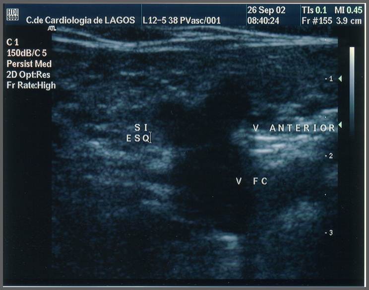
A transverse vascular scan, here at the groin. A deep vein shows as a dark, compressible circle, the kind of target the operator looks for in a difficult arm.
Ultrasound helps because it sees the veins the surface hides. A vein a centimeter or two deep, invisible to the eye and beyond the finger, shows up clearly on the screen as a dark, compressible circle. The operator can pick a vein, read its depth, then watch the needle enter it, none of which the blind method allows. The numbers from difficult-access patients are striking. One cohort study reported a first-attempt success of 84 percent with ultrasound against 51 percent by the landmark method, with the cannulation taking under 7 minutes against over 11, and fewer needle passes along the way. Reviews that pooled many trials put ultrasound success above 90 percent in difficult access, against 25 to 35 percent for the traditional method in the same hard group. The first-stick gain matters more than comfort. Each failed stick is a delay in the fluids or antibiotics the patient came for. A patient stuck four times is a patient whose treatment has waited. The scan also changes the choice in front of the operator. The blind method offers only the veins that can be felt, a short list in a difficult patient. Ultrasound shows several veins at once, the felt and the unfelt, then the operator takes the best of them. A vein is sized up before the stick, picked for being wide enough for the cannula and clear of nearby arteries, a judgment the blind method cannot make. The depth reading sets the angle before the needle moves, so blind probing is not needed. The same scan rules out the wrong targets. An artery, pulsing and round, is set aside. A nerve, a bright bundle beside some veins, is given a wide berth. A tourniquet higher on the arm swells the deep veins as it does the surface ones, making the target larger before the scan begins. A vein that rolls away from the needle is steadied by the probe’s pressure or by a thumb beside it. Watching the needle the entire way is the core of it, since a vein lost from view mid-stick is a vein the needle can pass through unseen. The probe also confirms the stick worked, the tip sitting in the lumen, before the line is trusted. The minute or two the scan adds before the stick is paid back when the first pass lands, sparing the repeated tries a blind stick would need. Little of this is possible by feel alone.
The deeper veins the probe reaches
The veins ultrasound opens up are the deep ones in the upper arm. The basilic vein runs along the inner arm, large and straight, the favorite once it can be seen. Deeper still are the brachial veins beside the artery. The cephalic vein sits on the outer arm as another option. All lie too deep for a blind stick, reachable only because the probe shows them.
Depth is the reason these veins were off the table before. A vein two or three centimeters down gives nothing to see or feel at the skin. The blind method never had them. Ultrasound brings them into play, adding a deep set of veins to the surface few an operator could otherwise reach in a difficult arm.
Choosing among the deep veins takes judgment. The basilic is often best, away from the artery and the nerve that run with the brachial veins. A vein sitting right beside an artery is passed over, since a needle could stray into it. The probe shows what lies around each vein, so the safe one is picked.
Depth and width both guide the choice. A deeper vein needs a longer catheter and a steeper angle of approach. A wider vein is the easier target and holds the cannula better. The probe reads both before the operator settles on a vein. Both arms can be scanned, since a vein buried on one side may sit clear on the other. The better of the two is taken.
The catheter has to be long enough
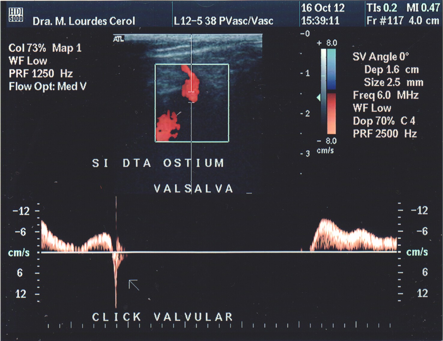
A vein on ultrasound with colour and spectral Doppler. Flow and the response to a Valsalva breath confirm a vessel is a vein before a line goes in.
Reaching a deep vein creates a problem of its own. A standard IV cannula is short, made for a vein near the skin. Pushed toward a deep vein, only its tip reaches the lumen, with much of the cannula still in the tissue above. A line held by its tip alone slips out easily, with the movement of the arm or the vein.
The numbers show the catch. Short cannulas placed in deep veins under ultrasound have failed at high rates, reported between 45 and 56 percent in some series, against 19 to 25 percent for ordinary surface IVs. The first stick succeeds, then the line fails hours or days later, pulled out of a vein it barely reached.
The fix is a longer catheter. A catheter of around 6 centimeters, against the standard of under 5, reaches far enough into a deep vein that a solid length sits inside the lumen. Long catheters placed under ultrasound have held for a wide range of dwell times, with one series reporting treatment finished on a single catheter in 84 percent of patients. The lesson is that ultrasound and a long catheter belong together, since the depth that ultrasound opens up is the same depth that a short catheter cannot hold.
The width of the vein matters here too. A good rule keeps the catheter well under the vein’s width, so blood still flows around it. A catheter that fills the vein slows the flow and invites a clot. The deep veins ultrasound reaches are wide enough to take a long catheter and still leave that room, which is part of why they suit the job.
How long the deep line lasts
A line placed well can stay for days, where a surface IV in a difficult patient might fail within hours. The deep vein holds the long catheter steady. The wide vessel leaves room around it. Dwell times for these lines run far longer than the short surface ones they replace. For a patient facing weeks of treatment, that durability spares a string of repeat sticks.
Lasting still depends on care. A tip that ends too shallow works loose. A catheter packed against the vein wall risks a clot. The same probe that placed the line can check it later, looking for a clot around the catheter or fluid leaking into the tissue.
Guiding the cannula in
With a vein chosen, the cannula goes in under live view. The operator watches the vein on the screen and brings the needle toward it, looking for the tip to enter the lumen. The way the needle is aligned with the probe, across the beam or along it, is its own technique, set out on its own pages.
The sign of success is a flash of blood in the cannula, with the tip seen inside the vein at that moment. The cannula is then advanced off the needle into the vein. The needle is withdrawn. With a deep vein the operator threads a good length of the cannula in before pulling the needle, so the line sits secure.
The angle is dropped once the vein is entered, since a steep needle that crossed the skin would push out through the far wall if kept high. Lowering the needle and easing it a little further slides the cannula into the lumen before it is threaded.
A short pull on the syringe draws blood back when the tip is in the vein. No blood, or bright blood that pulses, sends the needle back for another look before the cannula goes anywhere.
What ultrasound access saves
The reach of ultrasound at the bedside saves more than a few extra sticks. The patient who once needed a central line for lack of a peripheral vein can often keep a peripheral one. In one observational study, ultrasound in the emergency department let 85 percent of difficult-access patients avoid a central catheter they would otherwise have had.
Avoiding a central line is worth a great deal. A central catheter carries its own placement risks, a punctured lung or artery, along with a higher chance of a bloodstream infection over its life. A peripheral line, even a deep one placed under ultrasound, carries none of that placement risk and less of the infection risk.
The deep peripheral line also fills a gap between the plain surface IV and the central line. For treatments lasting a week or two, a longer peripheral catheter or a midline can carry what once called for a central catheter or a PICC. The bigger lines are kept for the cases that need them, the longest courses and the harshest drugs.
This shifts where the line is decided. A difficult arm no longer forces the step up to a central line. The question becomes whether ultrasound can find a peripheral vein first. The answer is often yes, which keeps the patient on the simpler and safer line.
The shift has a cost side worth naming. A deep ultrasound line takes longer to place than a quick surface stick. It ties up a trained operator and a machine. For an easy vein it is not needed. The extra minutes pay off only where the vein is genuinely hard to reach, the setting this technique was built for.
Who places the deep line
The deep peripheral line is commonly a nurse’s job. Trained nurses and vascular access teams place many of them on the ward and in the emergency department.
The skill takes teaching.
Holding the probe in one hand and the needle in the other is a coordination learned with practice. Keeping the tip in view and reading the depth come with it, on phantoms and under supervision. A trained operator places a deep line as a routine task. That is what brings the technique to the bedside.
Placing a line on a handheld unit
A handheld unit puts this within reach of any ward or clinic. The same probe scans the arm, finds a deep vein, then guides the cannula in at the bedside. No move to a procedure room is needed. A difficult stick is settled where the patient lies, often on the first try.
Common questions about ultrasound-guided peripheral IVs
What is ultrasound-guided peripheral IV access?
It is placing a peripheral IV while the operator watches the vein and the needle on an ultrasound screen. It is used when veins cannot be seen or felt, letting the operator reach a vein deeper than a blind stick can find.
Who needs an ultrasound-guided peripheral IV?
Patients with difficult intravenous access, whose veins are deep, scarred, collapsed, or small. In these patients the first blind stick often fails. Ultrasound finds a usable vein, frequently a deep one in the upper arm.
How much does ultrasound improve the success rate?
One cohort study reported first-attempt success rising from 51 percent by the blind method to 84 percent with ultrasound in difficult patients. Reviews put ultrasound success above 90 percent in difficult access, against 25 to 35 percent for the traditional method.
Why does an ultrasound-guided IV need a longer catheter?
A deep vein is far from the skin, so a standard short cannula reaches it by only its tip and slips out easily. A longer catheter, around 6 centimeters, keeps a solid length inside the vein, which holds the line in place far better.
Can ultrasound-guided peripheral IV avoid a central line?
Often, yes. In one observational study, ultrasound let 85 percent of difficult-access patients avoid a central catheter. A peripheral line carries none of the placement risk of a central line and a lower infection risk.
Can a handheld ultrasound place a peripheral IV?
Yes. A handheld probe scans the arm, shows a deep vein, then guides the cannula in at the bedside. The stick is done where the patient lies, with no move to a procedure room.
About the Author
Julien Mercier
Senior R&D Engineer · Medical Ultrasound Transducer Development
Senior R&D Engineer with an M.S. in Applied Physics and over 15 years of experience in medical ultrasound transducer development, specializing in the design verification and performance testing of high-frequency imaging transducers. Currently leading the development and verification of the company’s next-generation high-frequency linear-array transducer, responsible for imaging performance evaluation and reliability analysis in preclinical testing. Brings extensive hands-on experience in piezoelectric element tuning, beamforming parameter optimization, and system-level performance testing.