Pelvic Ultrasound Tool Selection Handheld Probe for Gynecology
A pelvic ultrasound for gynaecology is done in one of two ways. A scan can read the pelvis through the belly. A scan can read it from inside the vagina. Each way uses its own probe at its own frequency. The choice of tool sets what the scan can show. A clinician picks the route and the probe to fit the question and the woman.
The two ways into the pelvis
A transabdominal scan reads the pelvis through the wall of the belly. The probe rests on the skin below the navel. It looks down through a full bladder to the womb and the ovaries. A transabdominal scan gives a wide view of the whole pelvis. A transabdominal scan is the first scan a woman often meets. It is quick to set up and easy to start. A reader places the probe and sweeps the lower belly to open the view wide. A pelvic scan often uses both ways in one sitting. A reader starts with one route and adds the other as the case needs, the two views answering together what neither answers alone. The two routes read the same pelvis from two distances, and a reader chooses the distance the case needs. A pelvic scan is named for the route it takes: transabdominal or transvaginal. A reader names the route in every report. A reader meets both routes in training and uses both in practice, as a scan calls for one, the other, or both. A reader reads which the case needs. A reader makes the choice of route many times a day, the first decision of every pelvic scan. A reader picks before the probe touches the woman. A reader chooses the route, the probe, and the frequency as one set, all following from the question and the woman.
Transabdominal and transvaginal scanning for a gynaecology pelvic ultrasound
Feature
Transabdominal
Transvaginal
Probe
Curved, low frequency
Endocavity, high frequency
Bladder
Full
Empty
Reach
Deep, wide field
Close, narrow field
Best for
Large masses, overview
Fine detail, early pregnancy
When
The close probe cannot reach
The first choice for routine work
A transvaginal scan reads the pelvis from inside. A slim probe sits in the vagina, close to the organs. It draws the womb and the ovaries in fine detail. A transvaginal scan is the closer of the two views. The transvaginal scan came later, with the rise of the slim endocavity probe. It changed what a pelvic scan could see. The fine detail follows from bringing the crystal to within a finger’s width of the womb. A woman who wants no internal scan keeps to the belly view, and a reader reads as much as that view gives. The choice of route respects the woman as well as the question.
Through the belly
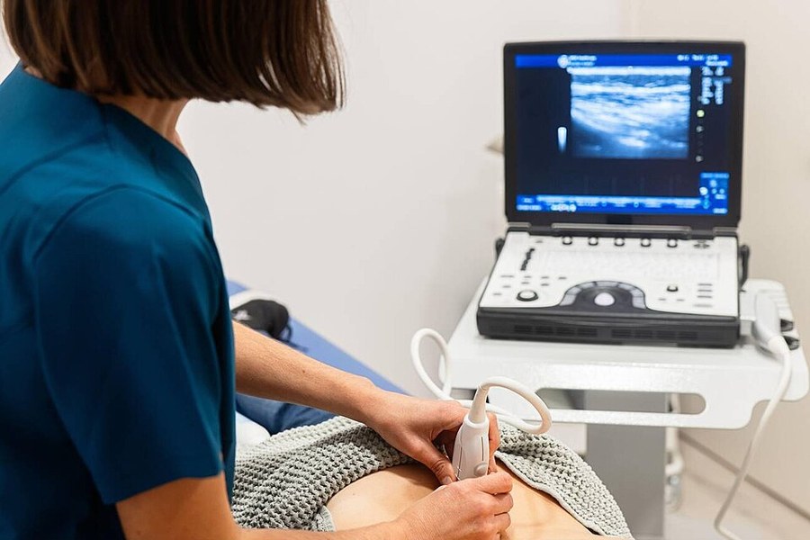
A transabdominal scan in progress, a curved probe sweeping the lower belly with the machine alongside. This is the through-the-belly route to the pelvis.
The transabdominal probe is a curved one with a wide face and a low frequency. The low frequency reaches deep into the pelvis. The wide face sweeps a broad field in one view. A curved probe is the tool for the overview. The curved probe spreads its beams in a fan that opens wide with depth. A reader reads the womb, both ovaries, and the spaces between in that one fan. The wide field is the strength of the belly view. The belly probe reads through gel on the skin, which carries the sound across to the body. A reader sweeps the probe over the lower belly in long passes. The whole pelvis comes into view in a minute. A belly scan needs no special room and no chaperone, and a reader scans the lower belly in the open clinic. The scan is quick to set up and quick to read. A reader sweeps the curved probe in lines across the lower belly, down the long axis of the womb and then across it. The two passes build the full shape of the pelvis in planes. A belly scan reads a pelvis no probe has to enter, and a woman keeps her clothes and lies back for it. A reader reads through the gel on her skin, and the scan moves from the broad view to the fine.
A full bladder is the key to a transabdominal scan. The bladder fills with urine and rises over the womb. The fluid pushes the bowel aside and opens a window. A reader scans the pelvis through that clear window. A woman drinks water and waits an hour or so for the bladder to fill before the scan. A reader checks the bladder is full before starting. An empty bladder leaves the pelvis hidden behind the bowel. A transabdominal scan gives a fast first look in an emergency. A woman in pain gets a quick belly sweep before anything else, and the wide view finds free fluid or a large mass at once. A reader reads the bladder itself along the way, its wall and any stone or growth, as both the window to the pelvis and an organ in its own right.
A transabdominal scan suits a large mass. A fibroid womb or a big cyst can reach above the pelvis, beyond the close probe. A transabdominal scan maps the whole of it. A reader reaches for it when a mass is large. A transabdominal scan has its limits. A thick belly wall blunts the view. Gas in the bowel scatters the sound. A womb tipped far back can sit beyond the clearest part of the field. A reader reads what the belly view allows and turns to the close probe for the rest. The sound weakens for the extra layer of a heavy belly wall, and a reader presses gently and lowers the frequency for the reach. A belly scan reads a pregnancy grown past the early weeks, a womb risen out of the pelvis and read through the belly. The wide view holds the whole of a later pregnancy. A belly scan reads around the gas of the bowel, which throws a shadow that hides what sits behind. A reader shifts the probe and presses to clear it, and the window opens where the gas moves aside.
A transabdominal scan suits a woman who cannot have the close probe. A young girl or a woman who has never had sex is scanned through the belly. A woman who cannot take the vaginal probe is scanned the same way. A reader reads the pelvis through the belly for them. A belly scan reads a child’s pelvis with no internal probe, and a reader reads her ovaries and womb from outside. The belly route serves where the close one cannot go. A belly scan is open to any woman, whatever her age or her history, asking only that she lie back with a full bladder.
A transabdominal scan reads the pelvis as a whole first. It places the womb, the ovaries, and any mass in the round. A reader starts wide to see the lie of the land. The close probe then fills in the detail. The belly view sets the scene for the whole scan. It places a mass against the womb and the bladder. It shows how big a thing has grown and what it presses on. A reader reads the lie of the pelvis before the close look.
The empty bladder
A transvaginal scan works best with the bladder emptied first, the probe then sitting close to the organs.
From close inside
The transvaginal probe is a slim wand with the crystal at its tip. It slides into the vagina to sit a finger’s width from the womb. Its high frequency draws the organs in fine detail. A transvaginal scan is the sharpest view of the pelvis. The transvaginal probe is covered and gelled before it goes in. A clinician explains the scan and gains consent, with a chaperone standing by. The woman or the clinician can guide the probe in.
A transvaginal scan reads the small and the early. It reads the lining of the womb to the millimetre. It reads a small ovarian cyst or an early pregnancy. A reader reaches for it when the detail matters. Nothing sits between the close probe and the organs. No bladder and no bowel lie in the way. The high-frequency beam reaches the womb and the ovaries direct, for the sharpest picture a pelvic scan can give. A close scan reads what a smear cannot, the womb and the ovaries the body hides, through the wall of the vagina. A close scan needs no full bladder and no wait, since a woman empties her bladder and the scan begins. A reader reads at once, with no hour of drinking water first. A close scan suits a woman who can take the probe and wants the detail, reading her in a few quiet minutes.
The close probe reads the tubes and the ovaries best, sitting near the adnexa where so much gynaecology happens. It reads an ovarian mass or an ectopic in the detail the close view gives, and it separates the ovary from the tube beside it. It reads the follicles on an ovary one by one and counts them for a fertility plan. It is the tool for early pregnancy, dating a sac to the day when it is only millimetres across, then finding the yolk and the heartbeat. It measures the lining of the womb to the millimetre, and a drop of salt water in the cavity lifts the walls for a clearer look still. It reads the lining for a polyp and reads a coil and its place in the cavity. A three-dimensional close scan builds a face-on view of the womb, showing a cavity that is divided or oddly shaped. Colour on the close probe fills a small vessel for a torsion or an ectopic. The close probe even reads the deep pelvis a belly scan strains to reach in a heavy woman, sitting below the fat and near the organs, down to a short cervix or a low-lying mass. It is gentle used with care, causing little more than the pressure of a smear, and the scan takes only a few minutes. A transperineal scan reads the cervix and the pelvic floor from the skin between the legs, filling the gap when the close probe cannot go in and the belly view sits too far. A transrectal scan serves a woman who needs the close view without the vaginal probe, reading the pelvis from the rectum nearby. The close probe is cleaned to a high standard between women, with a fresh cover and a disinfection after each scan, and a reader follows the steps that keep it safe.
Frequency against depth
Frequency sets the trade in every ultrasound probe. The higher the frequency, the finer the detail and the shorter its reach into the body. A modern probe runs a broad band, and a reader picks within it for the depth in front of the probe. A high frequency holds its fine detail for a few centimetres of reach. The machine sets the band to the depth on the screen and can sharpen the picture with harmonics on top. A reader places the focus on the organ it reads, so the picture is sharpest where the reader looks. Resolution and depth pull against each other, and the frequency sets where on that line a scan sits. A reader sets the focal zone to the depth of the target, where the beam is narrowest and the picture sharpest. The detail follows the focus down the field. A high band draws the fine grain near the probe, and a reader reads the small things on it. A close scan reads the lining and the cavity a belly scan can only suggest, the fine grain caught from a finger’s width away.
The route sets the frequency a probe runs. A scan from inside has a short reach, so it runs the highest frequency and the finest detail. A scan through the belly runs lower, for the depth it has to cross. A reader trades a little detail for the reach to see a deep target. A reader sets the machine to the woman and the question, a near and small target calling for the highest band. The frequency follows the depth and the detail the case needs.
One handheld, both probes
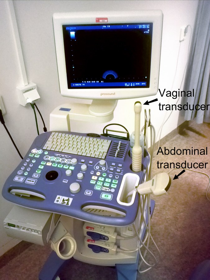
An ultrasound machine carrying both probes a pelvic scan can call on: an abdominal transducer and a vaginal one. The two labels are added to the photo to mark each probe.
A handheld scanner carries both probes a pelvic scan needs. A curved probe and an endocavity probe each plug into the same unit, and a clinician clips on the one a scan needs and switches between them. The device holds the whole pelvic toolkit in one hand. Some handhelds run a single probe for both jobs, where a wand made for the vagina can also read a slim woman through the belly. A two-probe handheld covers the full range.
The two probes cover the whole pelvic exam on a handheld. A reader starts with the belly probe for the overview. A reader changes to the close probe for the detail. One device reads the pelvis from both routes. A handheld costs a fraction of a cart and needs no fixed room, so a small clinic can own one and read its own pelvic scans. A reader works where a cart could never go, the tool reaching the patient where she is. A handheld runs on a battery for a day of scanning, charged overnight and carried through the rounds. It is light enough to rest on the woman and read for as long as the scan takes. A reader learns both routes to read the whole pelvis, the belly view first in training and the close view after, with its own feel and its own care. A reader trained in both reaches for the right one.
The handheld adds colour to either probe. Colour shows the blood that flows in a mass or a tube. A reader reads the flow for a torsion, an ectopic, or a tumour. The same device carries the colour to the bedside. A three-dimensional sweep builds a fuller picture of the womb on the same handheld. A reader calls on the colour and the volume from the one device. The extra tools ride along with the probe. A handheld reads the pelvis to the standard a cart sets, since the probe and the picture are the same. A trained reader gets the same answer on either, and the device changes the place of the scan and holds the quality. A handheld stores its scans and sends them on, so a reader shares a scan with a specialist far away. A second eye reads the pelvis from the saved images. A handheld brings both probes to one pair of hands, with no second machine and no second room. It reads a pelvis in the time a cart takes to warm up, ready when the reader is. A handheld runs the newer tools a cart runs, a live three-dimensional scan moving the womb in real time. The machine can flag a measurement for the reader, holding the large toolkit in the small device. The choice a big machine holds, the small one holds too, and a reader carries it to the woman. A handheld puts the whole pelvic kit where a cart cannot reach, and a reader picks the route once and reads the pelvis whole.
A handheld carries the pelvic scan up a ward and out to a home, reaching a woman who cannot come to a department. A reader picks the probe and reads the answer in minutes, the choice of tool travelling with the device. A wireless handheld sends its picture to a phone or a tablet, so a reader scans with the probe in one hand and reads the screen in the other. The handheld saves the pelvic scan to the woman’s record, and a reader reads the next scan against the saved one.
Choosing for the case
A reader picks the tool to fit the woman and the question. The American Institute of Ultrasound in Medicine holds that the vaginal approach should read all the structures it can, with a scan through the belly added when the close probe cannot reach a high mass or a woman cannot take the vaginal one. A reader reads much of gynaecology from close, and reaches over the belly for the rest. The close probe goes first in the usual case, for the detail of the womb and the ovaries. The belly probe comes in for a mass that reaches high or a woman who needs it. The two routes meet in one report. The question in hand points to a route, and an early pregnancy or pelvic pain in a young woman sends a reader to the close probe first. A reader reads the simplest route that works, going no further than the question needs. A clear answer from the belly view ends the scan with no internal one. The clinical question steers the choice in a few set ways. Bleeding after the menopause goes to the close probe for the lining of the womb. Pelvic pain in a young woman goes to the close probe for the ovaries and the tubes. A large mass found on the belly view stays there for its full reach. A lost coil goes to the close probe to be found. Infertility goes to the close probe for the ovaries and the follicles. A reader matches the route to the question each time, and the two routes between them answer the run of gynaecology. The same pelvis reads the same on whichever route the case sets, the answer holding whoever takes the scan.
The woman shapes the choice as much as the question. A large body can blunt the belly view, and a reader turns to the close probe for it. A reader reads the woman and picks the route that serves her. A woman has a say in the route, and a clinician offers the belly scan first and explains why the close one helps. A reader reads the woman, the question, and the body as one before the choice. A reader reaches for the belly view when the close probe cannot be used, since a blocked vagina or a woman’s refusal sends a reader outside. A reader always has a way to read the pelvis. A reader reads the route into the plan for the next scan, keeping to the route that answered before. The plan names the tool as well as the question. A reader writes the route, the probe, and the frequency into the report, so the next reader knows how the scan was taken. A scan read one way can be matched the next time.
The two routes work as one exam. A reader reads the belly view for the lie of the pelvis, then the close view for the detail, and writes both into a single report. The woman gets the whole picture from one sitting. Bodies that set the standard call for both routes in a full scan.
Common questions about choosing a pelvic ultrasound tool
Transabdominal or transvaginal for a gynaecology scan?
Both have a place, and many scans use them together. The transvaginal route reads the womb and the ovaries in fine detail from close range. The transabdominal route gives a wide overview and reaches a large mass above the pelvis. A clinician picks the route to fit the woman and the question.
Why does a transabdominal scan need a full bladder?
The full bladder lifts the bowel out of the way and opens a clear window to the womb. The fluid carries the sound down to the pelvic organs. Without it, gas in the bowel blocks the view. A transvaginal scan is done instead with the bladder empty, the probe sitting close to the organs.
What frequency is used for a pelvic ultrasound?
A transabdominal scan runs a lower frequency, around 3.5 megahertz or higher, for the depth it must reach. A transvaginal scan runs 5 megahertz or higher for fine detail at close range. A reader uses the highest frequency the depth allows, since higher frequency trades reach for detail.
When is a transvaginal scan not used?
It is not used in a girl or a woman who has never had sex, or in a woman who cannot take the probe. A large mass that reaches high above the pelvis can also outrun the close probe. A transabdominal scan covers these through the belly.
Can a handheld do both transabdominal and transvaginal scans?
Yes. A handheld carries a curved probe for the belly and an endocavity probe for the close view. A clinician switches between them on one device and reads the whole pelvis at the bedside. The same device adds colour Doppler for blood flow.
About the Author
Julien Mercier
Senior R&D Engineer · Medical Ultrasound Transducer Development
Senior R&D Engineer with an M.S. in Applied Physics and over 15 years of experience in medical ultrasound transducer development, specializing in the design verification and performance testing of high-frequency imaging transducers. Currently leading the development and verification of the company’s next-generation high-frequency linear-array transducer, responsible for imaging performance evaluation and reliability analysis in preclinical testing. Brings extensive hands-on experience in piezoelectric element tuning, beamforming parameter optimization, and system-level performance testing.