An ovarian cyst is a fluid-filled sac on or in the ovary, one of the commonest findings a pelvic scan turns up. Ultrasound is how a cyst is read, the scan telling a harmless one from the rare cyst that needs a closer look. The reading rests on a handful of features, the look of the fluid, the wall, and what sits inside. A scan settles in minutes whether a cyst is one to watch or one to leave alone.
A common thing, almost always benign
Ovarian cysts come and go with the cycle in women of childbearing age. An egg grows in a sac each month, the sac sometimes filling with fluid and lingering as a cyst. These functional cysts are the commonest of all, a normal part of the ovary’s month read on a scan. The reader names a simple cyst before the woman is off the couch. The answer is that quick.
The great majority of cysts are benign, harmless sacs that need no more than a name. The Royal College of Obstetricians and Gynaecologists puts the chance of a symptomatic cyst being a cancer at around one in a thousand in a woman before the menopause, a figure that frames every scan that follows. A reader reads a cyst expecting the benign and watching for the rare exception. A cyst can announce itself with a dull ache low on one side, a fullness, a pain with a period, or it can sit silent and be found by chance on a scan for something else. The scan reads a cyst for its nature and gives it a name, the name carrying the plan that follows. Cysts come in kinds beyond the functional, the simple sac that grew too big, the cyst lined with cells that make fluid, the cyst grown from tissue out of place, the scan reading the look and the name following from it. A cyst after the menopause carries more weight, the ovary quiet by then and a new sac more cause for a look, the scan reading the same features whatever the age. A cyst comes to a scan found because it hurts or found by chance on a scan for something else, the reader reading it the same either way and the look of the cyst all that matters. A cyst is named, measured, and sorted in a single scan, a woman often reassured before she has left the room. The ovary is a small almond of an organ, an inch or so long, low in the pelvis to one side of the womb, a cyst on it able to swell it to several times that size. A cyst is graded in the reading from the plainly benign to the plainly worrying, the great run of them sitting at the safe end. A functional cyst is part of the monthly cycle. It comes with the egg and goes again after it.
What the scan looks at
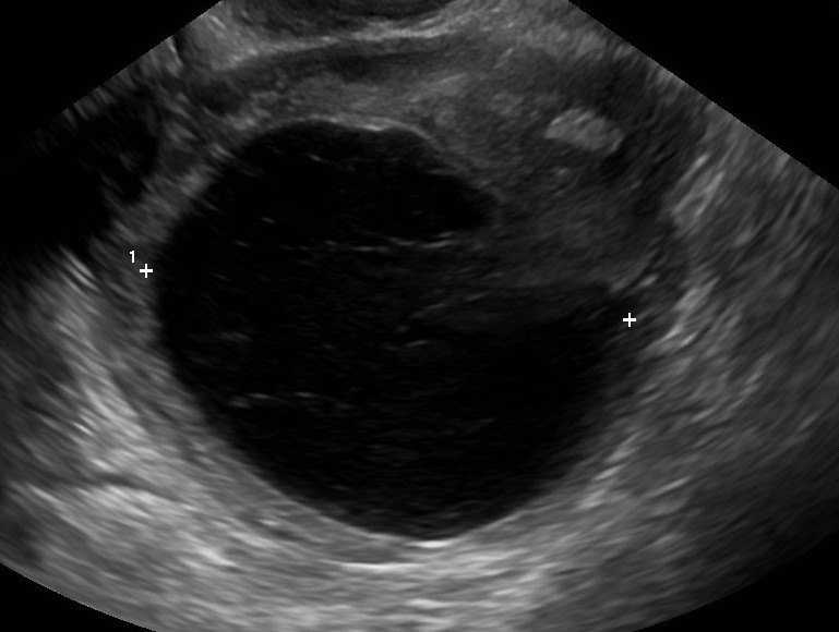
A large ovarian cyst on ultrasound, the dark fluid filling nearly all of it and the two caliper marks sizing it. The faint strands within mark it a haemorrhagic cyst, blood that has settled into the sac and clears on its own.
A cyst is read by a few features, each telling something about what it is. The scan asks what the cyst holds, how its wall is built, what lies inside it, and how blood moves through it. The answers sort a cyst into the simple and the rest. A cyst is measured across its widest, the size set down with its look, the reader logging the number and the features both for the next scan to read against. A cyst is read against any scan that came before, the same sac smaller or larger telling its story, a cyst unchanged across two scans a cyst at rest. A reader who reads a cyst well saves a woman both a needless fright and a needless operation, the right read counting for as much as the right treatment. The size goes into the report with a date. The next scan reads the new size against it.
How a scan tells a simple ovarian cyst from one that needs a closer look
Feature
Simple, reassuring
Concerning
Contents
Clear black, anechoic
Solid areas, debris
Wall
Thin-walled
Thick or irregular
Dividers (septa)
None, or thin
Thick, with blood flow
Projections
None
Buds growing into the fluid
Blood flow (Doppler)
None or little
Brisk flow in solid parts
Size
Under 5 cm, stable
Large, or growing
A simple cyst is the picture of harmlessness. It holds clear black fluid, the screen dark all through with no speck inside. Its wall is a thin line around the dark. Nothing solid sits within it, no divider crosses it, no bud grows from its lining. A touch of colour finds little or no flow in its walls. The black of the fluid brightens the tissue behind it, a glow the reader reads as proof the cyst is fluid all the way through. Each feature is read in turn, the fluid first, then the wall, then the inside, then the flow. A cyst that passes all four is a cyst a reader can call benign with confidence. The simple cyst is the commonest the scan finds, the ovary forever making and shedding them through the years a woman cycles. A reader who knows the look of a simple cyst names it without a second thought. The whole picture, dark and clean and still, is one a reader learns to trust and let be. The features that make a cyst simple are the features that make it safe. The clearer the fluid reads, the surer the cyst is benign, the cleanest black the safest sign of all. A reader builds the picture feature by feature, no single sign enough on its own, the fluid clear, the wall thin, the inside empty, the colour quiet, all four together naming a cyst benign. The bright glow behind a cyst, the posterior enhancement, is the sound passing easily through fluid and lighting the tissue beyond, a sign the cyst is fluid through and through. A simple cyst is read and dismissed in the same breath, the eye trained to see harmlessness in a black, quiet sac.
A cyst that strays from that picture asks for more thought. Solid tissue inside it, a wall gone thick or ragged, a divider running across it, a bud of tissue poking into the fluid, each is a feature that lifts a cyst out of the simple. Blood flowing briskly through a solid part is the feature a reader watches for hardest. A divider inside a cyst, a septum, is read for how thick it is and whether blood runs in it, a thick septum carrying flow the one that asks for a second look. A bud of tissue growing from the wall into the fluid, a papillary projection, is the feature a reader fears the deepest, the bud that carries its own blood supply the strongest single sign a cyst is more than benign. Colour laid over a cyst shows where blood flows, the solid part where the colour lights up the one a reader reads hardest. A reader reads the colour gently, too much power painting flow where there is none, a whisper of colour over a simple cyst showing the quiet a reader expects. A cyst is measured in three directions and its volume read from them, a growing cyst the one the numbers catch. A wall is read for its evenness and its thickness, the thin wall of a benign cyst the one a reader hopes to see. Echoes inside a cyst are read for their pattern, a solid lump among them the finding a reader marks.
These features have been gathered into simple rules a reader can follow. A set of benign features and a set of worrying ones each have their list, a cyst read against both. A cyst that shows the benign features and none of the worrying is called benign with confidence, the rest sent on for more. The rules read a cyst right the great part of the time, missing few of the cancers and frightening few of the well. A cyst that does not fit either set, neither clearly benign nor clearly worrying, is the one a specialist eye is asked to settle. A blood marker, the CA125, is read beside the scan in an older woman, a raised level adding to the worry a cyst already carries, the number raised by many a benign thing and weighed with care.
Size is read alongside the look. A cyst is kept under an eye once it passes a few centimetres or once it starts to grow, the change scan on scan a thing the reader weighs. A small cyst that holds still is the least of a reader’s worries, read and let be. A worrying feature sends even a small cyst on its way, size and look read together. The cyst that turns a reader cold has a solid heart, a tangle of new vessels, a wall that will not stay thin, a size that climbs, the picture they make together the thing that sends it on.
The simple cyst that needs nothing
A simple cyst in a woman who still has periods is the ovary doing its ordinary work. A follicle that holds onto its egg swells into a follicular cyst, the commonest of all. The sac left behind after the egg goes can fill with fluid or blood, a corpus luteum cyst that fades with the cycle. A cyst is common in early pregnancy too, the corpus luteum that holds it often a cyst of its own, read as a normal sac that is gone once the placenta takes over. A functional cyst is the ovary’s normal work caught mid-task, time the test it passes and the next cycle or two clearing it away. A cyst can grow on the pill or with treatment for fertility, the ovary stirred to make more than its usual one, a known effect read and set aside. A simple cyst asks for patience, the scan in a few weeks doing the work a knife once might have.
How a simple cyst is handled turns on its size. A small one, under about five centimetres, is left alone, the guidance plain that it needs no repeat scan at all. A larger one is checked again in a few months to watch it settle or grow. The biggest are looked at harder, a size beyond which the scan alone cannot give full reassurance. A simple cyst rarely grows large. One that keeps growing is read again as something more than functional. Many small sacs ringing an ovary is the look of polycystic ovaries, a pattern a reader names at a glance. A cyst comes out only when it must, a size too big to leave, a pain it will not stop causing, a feature that cannot be called benign, the scan reading the reasons and the knife held until they are clear. A simple cyst between five and seven centimetres is scanned again each year, watched for a change that rarely comes, a cyst above that read by a closer look or a different machine called in. A woman told her small cyst is a simple one is reassured on solid ground, the scan having earned that by reading every feature that would mean otherwise. A scan that names a cyst benign lifts a fear a woman may have carried for weeks, the fear gone in the few minutes the scan takes.
A simple cyst rarely causes trouble. It can ache when it stretches the ovary, or pass without a sign at all. A cyst found by chance on a scan for something else is the commonest way they come to light, a quiet thing read and named. A simple cyst found one month is scanned again the next to watch it go, the proof of its nature in its fading and a cyst cleared by the repeat scan never anything to fear. A simple cyst is given time to do what it does, fade, a scan in six or twelve weeks reading it gone or shrinking and the watching the whole of the treatment for the commonest cyst. A simple cyst needs no treatment at all in the great run of cases, a woman spared an operation by a scan that reads the cyst right. A cyst that fades on its own needs no more than the scan that watched it go, the watching the treatment and the fading the cure. A simple cyst is the ovary’s own handiwork. The body clears it without any help.
The cyst that needs a closer look
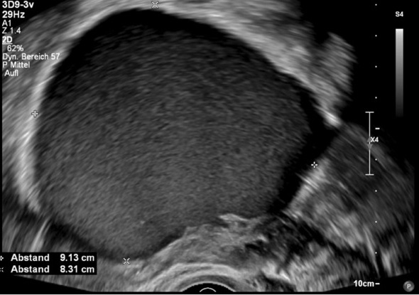
An endometrioma on ultrasound, the cyst filled with an even, dim “ground-glass” haze, the look of old blood walled in the ovary. The calipers and measurements (about nine centimetres here) size it. A reader names the pattern on sight.
Some cysts have a look all their own, neither simple nor sinister, that a reader learns to know. A dermoid grows the body’s own tissue in the wrong place, fat and hair and even a fleck of tooth or bone, casting a bright tangle with a shadow behind that a reader names on sight. Its weight of fat and bone makes it prone to twist the ovary, a torsion risk a reader notes whenever one is found. An endometrioma is old blood walled in the ovary, the lining’s tissue bleeding each month into a sac it cannot leave, reading as an even dim grey like ground glass. It marks endometriosis, a reader who finds one reading the pelvis for the disease behind it. A cystadenoma is a cyst lined with cells that pour out fluid, a benign growth that can reach a large size, its thin walls and clear fluid the marks of a harmless thing. A solid mass on the ovary, a fibroma or a growth from its own tissue, reads firm and grey with no fluid in it, told from a cyst by the simple fact of the fluid.
A cyst that bled into itself reads busy, fine strands crossing it like a net and a clot settling to one side. It is a hemorrhagic cyst, the kind that clears on its own and a repeat scan proves harmless. A reader knows the busy net of it for the harmless thing it is. A cyst can twist the ovary on its stalk, an ovarian torsion that cuts the blood and brings sudden, sharp pain, the scan reading a swollen ovary and looking for flow, a torsion an emergency that asks for surgery within hours. A cyst can burst, spilling its fluid and bringing a stab of pain, free fluid the scan finds pooled in the pelvis, a ruptured simple cyst settling on its own with the scan watching only for the rare one that bleeds on.
A cyst with solid tissue, brisk flow within it, or a thick divider is the one a reader sends on, each feature raising the chance the cyst is something more than fluid. A cyst handed to a specialist is read again with more tools, a scan that scores its risk and a team that decides, the first scan there to flag the cyst for the team that settles it. The few cancers caught this way are the reason the features are read at all. A flagged cyst goes to a gynaecology team. They read it with a fuller set of tools.
Cancer of the ovary is uncommon, rarer still before the menopause, the scan reading the same worrying features whatever the age. A solid, vascular, growing mass is the gravest picture of all. A blood test and a scan together sort the risk, the cyst that scores high sent to the team that handles it. A cyst unchanged for months is read as settled and left be. The hardest cyst to call is the one simple in every way save a single odd feature, a small solid area or a thick septum on an otherwise clear sac, the reader who cannot settle it sending it on with the guess left unmade. Between the plainly benign and the plainly cancerous sit a few cysts a scan cannot fully place, borderline growths the scan flags for the doubt they raise and the answer found only in the tissue itself. A simple cyst after the menopause earns a closer watch, the quiet ovary a place a new sac is read with more care, a small simple one often still left with the scan and a marker watched together.
The one rule
A simple black-walled cyst is almost always nothing to fear.
The endocavity probe and the handheld
The ovary is read best from close range. An endocavity probe, a slim wand with the crystal at its tip, is slid into the vagina to sit a finger’s width from the ovary, its high frequency drawing the cyst in fine detail from that close range. The wall, the inside, and the flow all read sharper from there than through the belly, the high note a match for a small, deep organ. A scan through the belly needs a full bladder to lift the bowel and open a window to the ovary. The same probe reads the womb and the lining in the same sitting, the whole pelvis swept in the one scan around the cyst.
A scan through the belly has its place for a large cyst that reaches up out of the pelvis, read whole from above where the close probe sits too near. A reader uses whichever view the cyst calls for, the answer the same from either once the cyst is fully seen. The same scan is run in a clinic or a ward. The cyst reads the same in each.
A handheld carries both views in one probe, the same class a hospital uses with the endocavity option built in, a clinic with one reading a cyst to the standard of a cart. A scan often starts from the belly to read the lie of the land, the close probe going in after for the detail, the two views building one picture. The inside scan is done with care and consent and a chaperone present, a woman who would rather not have it read from above where the answer is often there either way. Reading an ovary takes a trained eye, the cyst found, measured, and read for its features in a few minutes, a clinician learning the look of a simple cyst over a few dozen scans. A clinic far from a scanner can read a cyst, name it simple, and reassure a woman the same day, a cyst followed for its size read again on the same probe and closed off the day it fades. A woman with pain low in the belly can have her ovary read on the spot, a cyst found or ruled out before she leaves, the scan answering at the bedside a question that once meant a wait for a department. A handheld in an emergency room reads the ovary of a woman in sudden pain, a torsion or a bled cyst found in the minutes that matter and a twisted ovary sent to surgery without delay. A cyst read on a handheld is filmed and saved, its size and features set down for the next scan, the record travelling with the woman wherever she is seen. The cyst that would have meant a wait and a worry is read where she sits. A pocket probe holds the whole pelvic scan. The ovary is one stop along it.
Common questions about ovarian cysts on ultrasound
Are ovarian cysts usually dangerous?
No. The great majority of ovarian cysts are benign, and in a woman before the menopause the chance of a symptomatic cyst being a cancer is around one in a thousand. Many are functional cysts that come and go with the cycle and clear on their own within a month or two.
What does a simple ovarian cyst look like on ultrasound?
A simple cyst holds clear black fluid with a thin wall, nothing solid inside, no dividers, and little or no blood flow. The fluid brightens the tissue behind it. A cyst that reads this way is benign in all but the rarest case, and a reader names it and moves to its size.
When does an ovarian cyst need a closer look?
A cyst with solid tissue, a thick or irregular wall, a divider with blood flowing through it, or a bud growing into the fluid is read as complex. These features lift a cyst out of the simple and send it to a specialist team. Brisk blood flow in a solid part weighs heaviest of all.
Does a simple cyst under 5 cm need follow-up?
Usually not. Guidance holds that an asymptomatic simple cyst under about five centimetres in a premenopausal woman needs no repeat scan. A larger one is checked again in a few months to watch it settle or grow.
Why is a transvaginal probe used for ovarian cysts?
An endocavity probe sits a finger’s width from the ovary and draws the cyst in fine detail, sharper than a scan through the belly. A scan from above is added for a large cyst that reaches up out of the pelvis. A handheld carries both, so a clinic can read a cyst at the bedside.
About the Author
Julien Mercier
Senior R&D Engineer · Medical Ultrasound Transducer Development
Senior R&D Engineer with an M.S. in Applied Physics and over 15 years of experience in medical ultrasound transducer development, specializing in the design verification and performance testing of high-frequency imaging transducers. Currently leading the development and verification of the company’s next-generation high-frequency linear-array transducer, responsible for imaging performance evaluation and reliability analysis in preclinical testing. Brings extensive hands-on experience in piezoelectric element tuning, beamforming parameter optimization, and system-level performance testing.