The upper abdomen holds four organs a single scan must cover: the liver, the gallbladder, the pancreas, and the spleen. Each one is read through its own set of views, taken in a fixed order. A probe that runs the four in turn, with a full set caught at each, leaves the upper belly fully seen. Get this sequence right, and the rest of the abdominal scan follows from it.
A full set of views for each organ
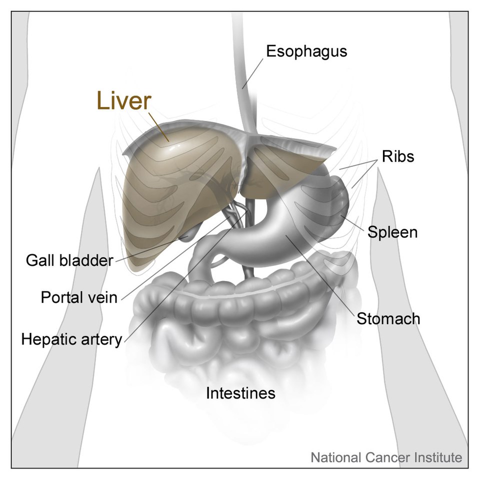
The organs of the upper abdomen. The liver fills the right side, the gallbladder tucked under it; the spleen sits high on the left behind the ribs; the stomach lies in the middle. The pancreas, hidden behind the stomach, is not drawn here, which is much how the scan finds it. Illustration: Don Bliss, National Cancer Institute, public domain.
Each upper-abdomen organ has a short set of views that proves it was wholly seen. That set measures whether a scan was complete. Skip a view, and the organ goes down only half read. The eye that knows the set looks first for the missing view.
The liver carries the longest set. Its right lobe is swept from under the ribs and between them, its left lobe from below the breastbone. The veins are traced, the dome is reached, the far edge is cleared. The gallbladder asks for two clean planes, its long axis and its short, with the wall measured and the inside searched for stones. The patient is rolled to roll the stones, so none hides in a corner. The pancreas asks for its three parts, the head, the body, and the tail, read across the line of the splenic vein behind it. The spleen asks for its full length down the left flank, with its tip and its top both reached.
The order runs the same way each time: the liver first, the gallbladder next, the pancreas across the midline, the spleen last on the far side. Run them in a fixed order, and no organ slips by unread. A scan run loosely, organ to organ as they catch the eye, leaves gaps that a busy day will not fill. The set for each organ, run in the same order, turns a glance into an examination.
A set is short. The liver needs a handful of sweeps, the gallbladder two planes, the pancreas three parts, the spleen one long view. The whole of it runs in a minute or two each organ. Each set is the same on every patient, learned once and run for life.
What the sequence guards against is the quiet miss, the lesion in the part of an organ the probe never reached. A liver read without its dome, a gallbladder read in one plane, a pancreas read without its tail: each leaves a blind spot where trouble can sit. The set is built around exactly these blind spots. It is the cheapest insurance a scan carries. The full set for each, every time, is the habit that closes those blind spots. The four organs, each with its set caught, are the whole of the upper-abdomen scan. A reader who runs the set can say at the end that the whole upper belly was truly seen.
Before the probe: the empty stomach
The scan begins before the gel goes on. A patient fasted for six hours or more gives the cleanest pictures. The fast fills the gallbladder, so its wall and its stones show clear. It empties the stomach and quiets the bowel, so less gas stands between the probe and the organs behind. Water can be drunk at the table to fill the stomach for the pancreas.
The patient lies on the back to start. The arm is raised above the head to open the rib spaces. The patient’s breath does as much work as the probe. A deep breath in pushes the liver and the spleen down below the ribs, into reach. The patient breathes to order through the scan, holding still where a clear view is caught. A patient full of gas, or unable to hold a breath, gives a harder scan, met with patience and a change of position.
The convex probe and its windows
The upper abdomen calls for a convex probe. Its low frequency reaches deep, down to the back wall of the belly. Its wide curved face fans the beam over a broad field, taking in a whole organ in one view.
The ribs and the gas force the probe to find windows. A window is a gap that lets the sound through. The spaces between the ribs are windows to the liver dome and the spleen. The liver itself is a window, a block of even tissue the beam crosses to reach the pancreas and the right kidney behind it. A belly full of breath, or a mouthful of water, opens windows where gas would block the way.
The probe slides and angles to find each window. It rocks between the ribs to dodge their shadows. It leans up under the costal margin on a held breath. Finding the window takes as much skill as reading the organ behind it. A clear window turns a deep buried organ into a plain picture.
The liver, lobe by lobe
The liver is the first stop and the largest. It fills the right upper belly and reaches across the midline. The probe reads it in overlapping sweeps, so no part is left out.
The right lobe is swept from two angles. The probe lies in the rib spaces on the right flank, reading the lobe slice by slice between the ribs. It then leans under the costal margin on a deep breath, fanning up to the dome under the diaphragm. The dome slips out of view more easily than the rest, so it is sought on purpose. The left lobe is read from below the breastbone. The probe presses into the soft epigastrium and fans across, taking the left lobe out to its thin edge. The heart beats close above, a marker of the top of the field.
The veins map the inside of the liver. The hepatic veins run together toward the heart, draining the liver upward. The portal veins branch through it with bright walls. The probe traces these to read the segments and to spot a vessel pushed aside by a mass.
The texture is read across the whole. A healthy liver is even, a fraction brighter than the kidney beside it. The probe holds the two together to judge that shade. When both lobes are swept, the dome reached, and the veins traced, nothing of the liver stays hidden. The size is taken on the right lobe, from the dome to the lower tip down the midclavicular line, a span held under about sixteen centimetres. A fatty liver reads brighter than normal and dims the deep beam, so the far parts fade. A focal lump that breaks the even field is measured and fixed against the nearest vein. The right kidney lies under the liver as a constant yardstick for its brightness.
The gallbladder, long and short
The gallbladder hangs from the underside of the liver. A fast leaves it full and easy to find, a dark teardrop against the bright liver. The probe reads it in two planes and rolls the patient to test it.
The long axis comes first. The probe lays the gallbladder out from its neck to its floor, a long dark sac. The wall is read along its length, a thin bright line under three millimetres.
The short axis crosses it. The probe turns across the sac, sweeping from neck to floor in slices. The two planes together leave no part of the wall unread.
Stones are sought in every corner. A stone sits as a bright lump that throws a dark shadow and rolls to the low point of the sac. The patient is turned onto the left side, so a stone hidden in the neck rolls free into the body. A stone that shifts and casts a shadow is the plain mark of gallstones.
The neck and the duct round off the read. The neck is searched with care, the spot a stone lodges to block the sac. The bile duct is followed from the liver, measured where it crosses the portal vein. Two planes, a roll of the patient, a search of the neck and the duct: that much covers the gallbladder. Sludge settles as a soft layer with no shadow, shifting slowly when the patient turns. A fixed lump on the wall that throws no shadow is a polyp, measured and noted. A wall thicker than three millimetres, tender under the probe, points to an inflamed sac.
The pancreas behind the stomach
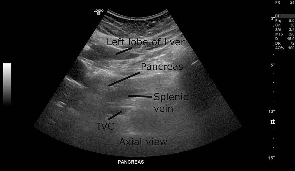
The pancreas on a convex scan, read through the left lobe of the liver as a window. The splenic vein runs along its back, the dark line that leads the probe to the gland; the IVC lies deeper still. The labels are the original’s. Image: Ptrump16, CC BY-SA 4.0.
The pancreas is the hardest of the four. It lies deep across the midline, behind the stomach, draped over the great vessels. The scan leans on a landmark and a window to find it.
The splenic vein is the landmark. It runs along the back of the pancreas, a dark line the probe follows across the belly. The gland sits right in front of that vein. Finding the vein finds the pancreas. The left lobe of the liver is the window to the body. The probe presses below the breastbone and uses the liver to carry the beam back to the gland. A drink of water in the stomach opens a further window, turning the gas-filled bag into a clear pane.
The three parts are read across. The head sits low on the right, by the loop of the duodenum and the bile duct. The body crosses the midline in front of the vein. The tail runs up to the left toward the spleen. The head and body come readily. The tail is the part the gas hides, sought with water and a turn onto the right side.
The gland is read for its make. A healthy pancreas is even, the shade of the liver or a touch brighter, with a fine duct running through it. The pancreas rewards a patient hand, coming clear head to tail once the window opens. The duct shows as a fine pair of bright lines through the body, held under about two millimetres. The gland turns coarser and brighter with age and fat, a change read against the liver. A head swollen against the bile duct, or a duct grown wide, sends the scan looking for a cause.
The spleen in the left flank
The spleen sits high and far back on the left. It hides behind the lower ribs, above the kidney on that side. The probe reaches it through the rib spaces from the flank.
The probe lies in the left intercostal spaces, low and back near the bed. A deep breath drops the spleen into view. The beam reads it as a smooth crescent, even in texture, a little brighter than the kidney below.
The full length is measured. The spleen is laid out along its long axis, from its top under the diaphragm to its tip. A normal spleen runs under about twelve centimetres. The length is taken on the longest view caught.
The tip and the top are both reached. The tip points down toward the left kidney, the spot an enlarged spleen grows past. The top tucks under the diaphragm, sought on a held breath. A small spare spleen can sit by the hilum, a round echo the size of a grape. Top to tip, measured down its length, the spleen closes the set. The vessels enter at the hilum on the inner face, the splenic artery and vein woven together. A spleen grown past its length pushes its tip toward the midline and rounds its edges. The same long view that measures the spleen catches the left kidney and the space above the diaphragm.
The vessels that thread the upper belly
The great vessels run through the upper abdomen as a map. The probe reads them both as landmarks and as organs in their own right. They lead the eye from one stop to the next.
The aorta runs down the midline, in front of the spine. It pulses, round in cross section, giving off its branches as it falls. The probe measures its width, a check for a widened wall. The coeliac trunk and the artery to the gut spring from its front. The inferior vena cava runs beside it on the right, flatter and quieter, swelling and shrinking with the breath. It carries the hepatic veins into the liver above. The two great vessels, the firm pulsing aorta and the soft breathing cava, are told apart at a glance.
The portal vein and the splenic vein lead to the organs. The portal vein runs into the liver with bright walls, the road the gallbladder and the bile duct are read against. The splenic vein runs across the back of the pancreas, the line that finds the gland. Lose the vein and the organ in front of it is lost too. The veins are the threads the whole sequence is strung on.
The spaces around the organs
The scan reads the gaps between the organs as well as the organs. Free fluid gathers in these gaps, and the upper-belly views catch the first of it. A clear look at the spaces rounds off the sequence.
The space under the right diaphragm sits above the liver. A rim of dark fluid here, between the liver and the diaphragm, is free fluid in the belly. The view that reads the liver dome reads this space at the same time.
The pocket between the liver and the right kidney is a low point of the belly. Fluid runs to it and pools as a dark stripe. The probe checks this pocket on the right, and the matching one by the spleen on the left.
The bases of the lungs sit above the diaphragm. The same high views catch a layer of fluid above the diaphragm, in the chest. A dark wedge over the liver or the spleen, above the bright diaphragm line, is a pleural effusion. The upper-belly scan reads a little of the chest for free. These spaces take only a moment, caught in the views already taken.
Tying the four together
The four organs run as one smooth sequence. The order saves time and misses nothing. The liver comes first, large and central. The gallbladder hangs from it, read next without moving far. The pancreas lies deep to the liver’s left lobe, read across the midline. The spleen waits last, out on the left flank. One breath does much of the work. A single deep breath, held, drops the liver and the spleen and steadies the field. The probe catches the dome, the gallbladder, and the spleen tip each on a held breath. The patient learns the rhythm and breathes to it. The sequence bends to the find. A lump caught in the liver is worked fully before the scan moves on. The order holds the thread, so the scan returns to where it left off. The four organs, read in turn, are covered before the probe leaves the upper belly. The whole sweep, start to last organ, settles into a rhythm the hand keeps without thinking.
The measurements that count
A few sizes are taken and saved on every scan. They turn a look into a number a later reader can trust. Each organ has its own. The liver span is measured down the right side, a guide to a liver grown large. The spleen length is taken on its long axis, the firmest sign of a swollen spleen. The bile duct is measured where it crosses the portal vein, a thin tube that widens when the flow is blocked below.
The gallbladder wall is read for its thickness, a thin line that swells when the sac is inflamed. The pancreatic duct is measured through the body, a fine channel that widens behind a block in the gland. The aorta is measured across, a check against a widened wall. These numbers, saved with the pictures, are the spine of the report. A number on the page outlasts the memory of the scan.
When an organ hides
An organ can duck the probe. The scan carries a fix for each hiding place.
Gas can bury the pancreas. A loop of bowel over the gland scatters the sound into a gray blur. A drink of water fills the stomach and clears a window. Sitting the patient up, or turning onto the side, lets the gas float off the gland.
A fed gallbladder shrinks away. A meal squeezes the sac small, its wall thick and folded, hard to read. The fix is plain: the patient comes back fasted, and the sac fills again.
A high spleen stays under the ribs. A spleen tucked deep under the diaphragm slips behind rib shadows. A deeper breath, and a probe worked into the lowest rib space, brings it down. A turn onto the right side rolls it forward into reach.
The liver dome can fall in shadow. The top of the right lobe, up under the ribs and the lung, is easy to leave unread. A breath held in, and a firm lean up under the costal margin, opens the dome to the beam. Each hiding place has its move, learned by heart, so a hidden organ is chased down in the end.
Proving the upper belly was seen
A thorough scan leaves a record that proves its reach. Each organ leaves its set of images behind. The record is read later as the proof of a complete exam. The liver leaves both lobes, the dome, and the veins. The gallbladder leaves its two planes and its wall. The pancreas leaves its head, its body, and as much of its tail as the gas allowed. The spleen leaves its length on its long axis. A measurement is saved where size carries weight, the spleen length, the bile duct, the liver span.
The gaps are owned. A tail of pancreas lost to gas is noted as not seen, so the next reader knows to chase it. An honest scan marks what it reached and what it could not, leaving no false comfort behind. The report tells the next reader what was seen and where the gaps lie.
The same set, every time
The upper-abdomen scan runs on a habit. The same four organs, in the same order, each with its full set of views. The liver swept lobe to dome, the gallbladder turned and rolled, the pancreas traced along its vein, the spleen measured down the flank. That habit builds a clear scan. What starts as a checklist becomes a reflex with the years.
The order is the safeguard. A scan run to a fixed route forgets nothing, on a slow day or a busy one. The route holds the reader to every organ and every view, where a free hand would drift and skip. The discipline of the set counts for more than any single clever angle.
A probe in a careful hand runs the four in a few minutes. The liver, the gallbladder, the pancreas, the spleen, each caught in its full set, leave the upper belly read end to end. A scan that runs the same set every time can be trusted to have seen what was there.
Common questions about the upper abdomen scanning sequence
Why fast before an abdominal ultrasound?
A fast of six hours or more clears the picture. It fills the gallbladder, so its wall and any stones stand out. It empties the stomach and settles the bowel, so less gas blocks the deeper organs. The pancreas and the aorta, buried behind the stomach, read far better on an empty belly.
What order are the upper abdomen organs scanned in?
The liver first, then the gallbladder, the pancreas, and the spleen. The liver is large and central, an easy start and a window to what lies behind. The gallbladder hangs from it, read next. The pancreas lies deep across the midline. The spleen waits last, out on the left flank. The same order each time keeps any organ from being skipped.
Why is the pancreas hard to see on ultrasound?
It lies deep behind the stomach, draped over the great vessels. Gas in the stomach and the bowel scatters the sound before it reaches the gland. The tail, tucked up toward the spleen, hides the worst. A drink of water fills the stomach and opens a window, and the splenic vein behind the gland guides the eye to it.
How does breathing help an abdominal scan?
A deep breath drops the liver and the spleen below the ribs, into the probe’s reach. Held at the right moment, it steadies the organ for a clean view. The dome of the liver, the tip of the spleen, and the gallbladder are each caught on a held breath. The patient breathes to the sonographer’s word through the scan.
What does a normal liver look like on ultrasound?
A smooth, even tissue, a shade brighter than the kidney beside it. The veins run through it as dark channels, the portal branches lined with bright walls. The edge is sharp and the surface is smooth. The probe holds the liver and the right kidney in one view to judge that brightness.
How long does a full upper abdomen scan take?
A few minutes in a practised hand. The four organs run in one smooth sequence, each with its short set of views. A clear case, on a fasted patient, goes quickly. A buried pancreas or a gassy belly adds time, with water and a change of position to win the view. The set is run in full whatever it takes, since a skipped view is a blind spot.
About the Author
Julien Mercier
Senior R&D Engineer · Medical Ultrasound Transducer Development
Senior R&D Engineer with an M.S. in Applied Physics and over 15 years of experience in medical ultrasound transducer development, specializing in the design verification and performance testing of high-frequency imaging transducers. Currently leading the development and verification of the company’s next-generation high-frequency linear-array transducer, responsible for imaging performance evaluation and reliability analysis in preclinical testing. Brings extensive hands-on experience in piezoelectric element tuning, beamforming parameter optimization, and system-level performance testing.