





































Probe shapes
Linear versus Convex versus Micro Convex Probe Image Quality Differences
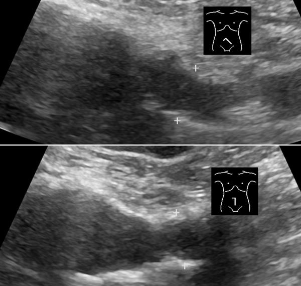
The shape of a probe’s face decides the shape of its image, the depth it reaches, and the detail it holds. A flat linear face, a broad curved convex face, and a small tightly curved micro-convex face each draw a different kind of picture, suited to a different part of the body. Picking among them starts with what the scan needs to see.
How the face shape lays down the image
A flat face fires its elements straight ahead in parallel lines, so the image is a rectangle the same width at the top and the bottom. The scan lines stay close together all the way down, which holds the side-to-side detail across the field. The flat face presses evenly on a flat surface, the skin of a neck or a limb, where it sits in full contact.
A curved face fans its lines outward, so the image is a wedge, narrow at the probe and wide at depth. The fan reaches a broad field deep in the body from a face small enough to fit a belly. The lines spread apart as they go down, so the side-to-side detail thins with depth, the cost of the wide reach. The curve rocks onto a rounded surface like an abdomen, sitting in contact along the bulge.
A small tightly curved face fans its lines from a footprint barely a few centimetres across. The image is the same wedge a larger convex face draws, opened from a far smaller window. The little face fits between two ribs, under a jaw, into the soft spot of an infant’s flank, the tight spots a broad face cannot land on flat.
Contact decides whether any picture forms at all. A face pressed flat against the skin couples its sound cleanly into the body. A face that lifts at one edge loses the sound there and leaves a dark wedge in the image, so the shape that matches the surface draws the cleaner picture before frequency or lines are weighed.
What sets the detail and the depth
The face shape travels with a frequency band, and the band sets the rest of the picture. A linear probe runs high, often 7 to 15 megahertz, since its work sits shallow and rewards fine detail. A convex probe runs low, around 2 to 5 megahertz, since its work sits deep and needs penetration. A micro-convex probe runs across a middle and low span, tuned to reach a moderate depth through a small window. The frequency each shape carries is the larger half of why the pictures differ, the face setting the field and the frequency setting the grain. Resolution follows from the pairing. The high frequency of the linear probe draws detail toward a tenth of a millimetre. The low frequency of the convex probe draws a grain several times coarser, set by the longer wavelength it runs at. Penetration follows the same frequency. The low-frequency convex beam reaches twenty centimetres and more, absorbed slowly by tissue. The high-frequency linear beam fades within four or five, absorbed fast. The micro-convex sits between, reaching a useful mid-depth. A second effect the face shape alone drives is the line density. A flat face lays parallel lines that stay evenly spaced down the field, holding the detail across the width and the depth alike. A curved face fans its lines, so they spread apart as they descend, and the picture loses its side-to-side detail toward the bottom of the wedge. The flat face holds an even sharpness across its rectangle. The curved wedge reads sharpest near the top and softens toward its edges and its depth as it widens, the breadth bought at that price. Together the frequency and the line geometry account for the whole of how the three pictures differ, before a single setting on the machine is touched.
The flat face and its picture
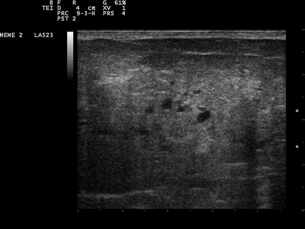
The linear probe draws the sharpest picture of the three in the first few centimetres. Its rectangular field shows a vessel wall, a nerve, a tendon at full detail across the whole width, with no thinning at the edges. A vascular study, a nerve block, a musculoskeletal look each reads on this picture. The flat face needs a flat surface to sit on, so it works the neck, the limbs, the front of the body where the skin lies even under it.
The picture stops shallow. The high frequency that draws the fine detail fades within four or five centimetres, so the linear probe shows nothing of the deep abdomen. Its field is also only as wide as its face, since the parallel lines do not spread, so a structure wider than the footprint runs off the edges of the image. For the shallow work it is built for, those limits cost nothing.
The even detail across the rectangle suits work that spans a width. A whole segment of a vessel reads in one frame, sharp from edge to edge, the flat field holding the same detail at the sides as in the middle. A long structure stays in view at full sharpness along its run, held end to end in the one rectangular frame.
The flat field shows its own artefacts plainly. A bright interface throws a comet-tail trail of repeating lines, a foreign body or a calcification marked by the tail it casts. A fluid pocket brightens the tissue under it, the posterior enhancement that names a cyst. The reader takes each as a sign of what the tissue is, the clean rectangle giving these clues room to read.
The broad curve and its picture
The convex probe draws the deep wide picture an abdomen needs. Its wedge opens from a hand-sized face to a field that holds a liver, a pregnant uterus, an aorta deep in the body. The low frequency reaches twenty centimetres and more, where the structures of the belly sit. A focused abdominal scan, an obstetric study, a deep vascular look each reads on this picture.
The detail softens with depth. The lines that fan out from the curved face spread apart as they descend, so two structures close together blur into one at depth. Near the top of the wedge they still read apart. The grain softens through the field, the depth the low frequency was chosen to reach. For the deep broad work the probe is built for, the softer grain is the detail the body allows at that depth.
The wide wedge carries artefacts of its own at depth. A strong reflector like the diaphragm mirrors a structure above it into a false copy below, the mirror image a reader learns to discount. The edge of a round structure casts a thin shadow down its side, the edge shadow that traces a cyst’s wall. Read with those in mind, the deep wedge gives a faithful map of the belly.
The small curve and its picture
The micro-convex probe draws a deep wedge from a tiny window. Its small face slips between two ribs to read the heart, under the ribs to reach the liver from below, onto the small flank of an infant where a hand-sized face would not fit. The fan opens to a useful depth from a footprint a few centimetres across, the picture a compromise the small window demands.
The image carries the marks of that small face. The field is narrow at the skin, since the fan starts from a short base, opening only as it descends. The detail runs moderate, the frequency held in the middle to reach the depth. A paediatric scan, a cardiac look between the ribs, a difficult abdomen on a small or guarded patient each reads on this picture, the small face reaching into spots a larger one cannot land on.
The small face does its main work in cardiology and at the bedside. A focused cardiac look reads the heart’s chambers and a pericardial effusion through the rib window. In a child the same small face covers a whole abdomen a hand-sized probe would overshoot, the footprint matched to a small body. The shape that fits the window is the shape that gets the picture at all.
The phased array, a fourth face
A fourth face belongs in the family, the phased array. Its footprint is the smallest of all, barely a thumbprint, and it steers a wide wedge into the body from that tiny window by firing its elements in a timed sequence. The wedge opens broad at depth from a face that fits between two ribs, the picture a cardiologist reads the beating heart on. Its near-field detail is modest, the top of the wedge narrow and the surface grain coarse, the cost of steering a wide picture from a thumbprint of a face through a rib gap to reach the beating heart below.
The newest faces add a second row of elements, or a full grid of them, a matrix array that steers the beam in two planes. The grid builds a volume, a stack of slices the machine assembles into a block of tissue it can cut and turn after the scan, and it reads two crossed planes live to guide a needle or a valve into place. The element count climbs into the thousands on these faces, the picture a matrix draws reaching past what a single row of elements could ever build on its own.
Three shapes, three pictures
Each face draws its own picture: the flat one fine and shallow, the broad curve deep and wide, the small curve deep through a tight window.
How a clinician reads the choice
The pick follows the body and the depth. A shallow structure on a flat surface, a vessel or a nerve, calls for the linear probe and its fine even detail. A deep broad target, an abdomen or a womb, needs the convex probe’s reach. A heart between the ribs or an infant’s belly, deep behind a small window, takes the micro-convex face for the access its small footprint gives.
The surface the probe sits on shifts the call along with the depth. A rounded belly takes the curved face that rocks onto it, a tight rib window the small face that fits inside, a flat neck or limb the flat face that lies even across it. The shape that holds full contact on the surface in front of it draws the cleanest picture there.
A broadband or multi-shape handheld now carries more than one of these in a single device, a face that images as a linear probe and reconfigures for a curved field, or a kit of clip-on heads. The reader picks the shape to the body and the depth, the image quality following the face that matches the work, whether that face is a separate probe or a mode on one device.
One patient shows the shapes in turn. A trauma scan reads the belly for free fluid on the convex wedge, then the neck vessels on the linear rectangle, then the heart between the ribs on the micro-convex face. Three windows on one patient, each drawn by the shape that fits it.
A service meeting a narrow range of work carries the one shape its cases need. A service meeting everything carries all three, or a device that reconfigures between them. The shapes are a set a clinician draws from by the body in front of them.
Common questions about probe shape and image quality
What is the difference between linear, convex, and micro-convex probes?
The linear probe has a flat wide face and draws a rectangular shallow picture with fine detail. The convex probe has a broad curved face and draws a deep wide wedge. The micro-convex probe has a small tightly curved face and reaches depth through a tiny window.
Why does the linear probe give the sharpest image?
Its flat face fires parallel scan lines that stay close together across the field, holding the side-to-side detail, and it runs at a high frequency, 7 to 15 megahertz, for the finest grain. The picture stays shallow as a result, fading within four or five centimetres.
Why does the convex probe reach deeper?
Its curved face fans the lines into a wide wedge, and it runs at a low frequency, 2 to 5 megahertz, that penetrates twenty centimetres and more. The detail softens with depth as the fanned lines spread apart, the price of the reach.
What is a micro-convex probe for?
A deep target behind a small window: a heart between the ribs, a liver from under the ribs, an infant’s belly. Its small tightly curved face fits where a hand-sized face cannot land flat, drawing a deep wedge from a tiny footprint.
Which probe shape is the most detailed?
The linear probe, in the shallow field it covers, since its parallel lines and high frequency hold fine even detail across the width. The convex and micro-convex draw a coarser picture, the frequency traded for the depth their work needs.
Can one handheld cover more than one shape?
Yes. A broadband or multi-shape device images as a linear probe and reconfigures for a curved field, or pairs with clip-on heads. The reader picks the shape to the body and the depth, the image quality following the matching face.

About the Author
Julien Mercier
Senior R&D Engineer · Medical Ultrasound Transducer Development
Senior R&D Engineer with an M.S. in Applied Physics and over 15 years of experience in medical ultrasound transducer development, specializing in the design verification and performance testing of high-frequency imaging transducers. Currently leading the development and verification of the company’s next-generation high-frequency linear-array transducer, responsible for imaging performance evaluation and reliability analysis in preclinical testing. Brings extensive hands-on experience in piezoelectric element tuning, beamforming parameter optimization, and system-level performance testing.