Knee Effusion Ultrasound Assessment Handheld Linear Probe
A swollen knee can be fluid in the joint, a thickened lining, or both at once. Which it is decides whether a needle helps. Ultrasound is what tells them apart. It picks out the free fluid a needle can drain from the solid tissue around it. It shows where that fluid sits before the needle goes in. That is the point of scanning a swollen knee.
Where the fluid collects
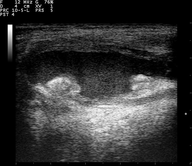
A knee effusion seen across the front of the joint. The wide dark band is fluid filling the space above the bone; the bright rounded structures below are the femoral condyles. Clear fluid lets the sound through and reads as this clean anechoic black. The text at the top left is the scanner’s settings. Image: Nevit Dilmen, CC BY-SA 3.0.
Fluid in the knee gathers in one place above all others: a pouch that runs up behind the kneecap, under the quadriceps tendon. The joint is wrapped in a synovial lining that folds into recesses around it. This suprapatellar recess is the largest fold of all. It sits high in the joint, so fluid rising inside reaches it first. When the knee fills, this is where it shows. A scan for effusion starts at this pouch.
The recess connects with the rest of the joint, so fluid moves freely in and out of it. Press on the front of the knee and the fluid in the pouch shifts away. Let go and it flows back. This free movement is the first test the scan applies.
Two smaller recesses sit at the sides of the joint. The scan checks them when the suprapatellar pouch is full, or when the view there is unclear. A small effusion can settle in these side spaces, easy to miss when only the main pouch is read.
Not every swelling at the knee is a joint effusion. A soft lump right over the front of the kneecap sits in the prepatellar bursa, a sac under the skin. It lies in front of the bone, ahead of the suprapatellar pouch. The scan places it there. A tap meant for the joint stays aimed at the joint.
Scanning the recess
The patient lies with the knee straight at first, then bent to about thirty degrees over a rolled towel. The slight bend opens the suprapatellar pouch and lets any fluid drain into it. The probe, a high-frequency linear one, sits lengthwise just above the kneecap, in line with the thigh.
In this long view the recess shows as a thin space under the quadriceps tendon, between two bright fat pads. An effusion fills this space as a dark band, far wider than the thin sliver of a normal knee. It reads plainly against the gray tissue around it.
Two moves bring out a small effusion. Pressing on the sides of the knee, or just above the pouch, milks fluid down into the recess under the probe. A gentle squeeze of the thigh muscle does the same, driving fluid from the joint up into the view.
The knee is scanned in both planes. The long view, along the thigh, shows the depth of the fluid. Turning the probe across the leg shows its width and catches fluid that sits off to one side. A pocket missed in one plane often shows plainly in the other.
Pressing with the probe is itself part of the scan. Fluid thins under the pressure and shifts aside. It returns when the pressure lifts. Tissue that stays put under the probe is not fluid at all. This compression test runs through the whole scan, at every spot where fluid might sit.
The fat pads around the recess can fool a quick look. They are soft and give a little under the probe, so a darker fat pad can pass for a thin effusion. Steady pressure tells the two apart. A fat pad dents and keeps its place and its speckled texture. Fluid flattens, flows aside, and comes back.
What an effusion looks like
A simple effusion is black on the screen. Sound passes straight through clear fluid without bouncing back, so the recess reads as an anechoic space, a clean dark band with sharp edges. The darker and cleaner the space, the more likely it is plain fluid.
The depth of that dark band is measured, straight down from the quadriceps tendon to the bone. A thin sliver, under about four millimeters, can be normal. An anechoic depth past that is counted as an effusion. The number lets the next scan be compared against this one. The probe finds far less fluid than the hand can: an effusion of around four milliliters shows on the screen, a volume too small to feel through the tissue around the joint.
Size is graded as much by eye as by the ruler, from a trace that barely lifts the tendon off the bone to a large effusion that balloons the pouch, spreads into the side recesses, and lifts the kneecap clear of the bone underneath.
An effusion shows the knee is irritated and making fluid. The cause behind it is a separate question. Trauma, wear, gout, and inflammatory disease all fill a knee. The scan measures the fluid and reads its character. The cause is pieced together from the history, the joint surfaces, and what a tap of the fluid shows.
The shape of the fluid matters too. Free fluid spreads to fill whatever space it is in, its upper edge flat or curved and shifting when the leg moves. Fluid that is walled off in a pocket of its own points to an older or loculated collection, the kind a single tap may not fully drain.
Fluid or thickened lining
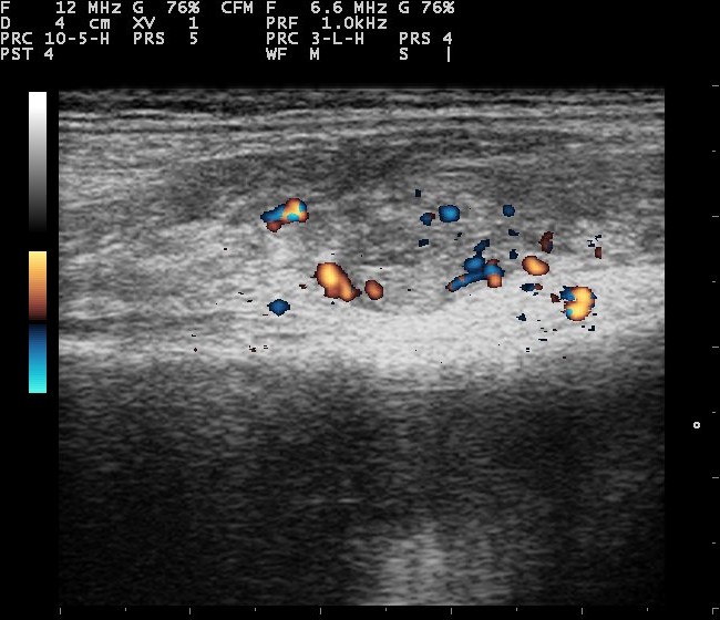
Color Doppler over an inflamed knee. The orange and blue specks are blood-flow signal, not anatomy: a joint lining this vascular is the mark of an active synovitis. Simple fluid carries no flow and stays dark. The color scale and text at the edges belong to the scanner. Image: Nevit Dilmen, CC BY-SA 3.0.
The hardest call a swollen knee asks for is whether the swelling is fluid or thickened lining. Ultrasound is built to answer it. A knee can fill with fluid. Its synovial lining can grow thick and inflamed. Often both happen at once. On the surface they feel the same, a full and tense knee. Under the probe they behave differently. Fluid is anechoic and it moves: it thins under pressure, shifts aside, and flows back when the pressure lifts. Thickened synovium is hypoechoic, a shade of gray on the screen. It stays put. Press on it and it does not flatten or flow. That one test, pressing and watching, settles the question in nearly every knee. A dark space that empties under the probe is fluid a needle can draw off. A gray mass that holds its shape is tissue a needle will not help. A needle put into thickened synovium draws nothing, or a few bloody drops from the puncture itself. The dry tap then gets read wrongly as a knee with no effusion. Knowing the swelling is solid tissue before the needle goes in spares the patient a fruitless tap. Color Doppler adds the second half of the answer. Fluid carries no blood, so it stays dark under Doppler. Inflamed synovium is alive with vessels. It lights up with color where the disease is active. A knee whose thickened lining glows under Doppler has an active synovitis, the kind seen in rheumatoid and other inflammatory arthritis. The amount of color tracks how active the disease is, scored from a quiet lining to one packed with flow. That grade is what Doppler follows when a knee is treated and watched over months, a way to read whether the drugs are working. Many knees hold some of each: a thin layer of fluid over a bed of thickened lining. The scan reads them as two findings in one joint. What it finds steers what comes next, whether that is a needle to draw the fluid off, the drugs that calm an inflamed lining, or a course of both. A tense effusion can feel as hard as solid tissue to the examining hand. Under the probe it yields and stays dark, the plain marks of fluid. The hand alone reaches none of this. It feels a single swelling and guesses at the cause. The probe names fluid and tissue for what they are. That decides whether a needle has anything to draw.
Simple or complex
Not all effusions are clear. A simple one is anechoic, black and clean, the look of a transudate or plain joint fluid. A complex one carries echoes inside it, fine dots, strands, or a layer that settles to the bottom. Those echoes are cells, blood, or debris suspended in the fluid, and what they are depends on why the knee filled.
Blood in the joint, from an injury or a clotting problem, often shows as fluid with a swirl of echoes, or a level where cells have settled to the bottom. Pus in a septic joint can look much the same, thick and full of echoes. A complex effusion is a warning the scan can raise. Naming what fills it, blood or pus or inflammation, takes a needle and a lab.
Even a clean, anechoic effusion can be infected. The look of the fluid never rules sepsis out.
This is why a complex effusion changes what happens next. A clean anechoic effusion in a well knee can be watched, or tapped at leisure. A knee that is hot and painful, its effusion full of echoes, calls for a tap soon, before a hidden infection harms the joint.
Echoes in the fluid are read with care. Turning the gain up can paint specks into clear fluid where none exist. The fluid is judged at a sensible gain. Gentle pressure helps tell real from false. True debris swirls and resettles under the probe. A speck painted in by too much gain holds still.
Behind the knee
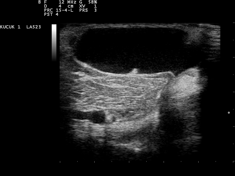
A Baker’s cyst behind the knee, the large dark pocket of joint fluid that has tracked into the bursa. A simple cyst is anechoic like this. The label at the top left is the scanner’s preset, not part of the image. Image: Nevit Dilmen, CC BY-SA 3.0.
The back of the knee is scanned as well, since fluid travels there too. A Baker’s cyst is a pocket of joint fluid that has tracked into a bursa behind the knee, between two muscle tendons on the inner side of the hollow. It fills from the joint through a narrow neck that acts as a one-way valve. Fluid passes out into the cyst and struggles to return, so the cyst can swell large on a joint that feels only mildly full.
On the screen the cyst shows as a fluid pocket with a neck pointing into the joint, the shape that tells it from other lumps behind the knee. Baker’s cysts go with the conditions that fill a knee, osteoarthritis and inflammatory arthritis among them, so a cyst behind the knee is a reason to examine the joint in front with care. A simple one is anechoic. An old one can hold debris or carry a thickened wall.
A Baker’s cyst that bursts spills fluid down into the calf. The pain and swelling that follow can mimic a clot in the leg. The scan tells them apart: a ruptured cyst leaves a tail of fluid tracking down the calf, the leg veins open and normal.
Guiding the needle
When a knee is tapped, ultrasound can guide the needle. The suprapatellar recess is the target, the same pouch the scan has already found and measured. Watching the needle enter the fluid in real time places it in the deepest pocket and keeps it clear of the kneecap, the tendon, and the bone. The fluid is seen to shrink as it drains, so the operator knows when the pocket is empty. A large, painful effusion is drawn off as much for the relief as for the sample it gives.
A small effusion is where guidance counts, since a blind tap can miss it and come back dry. Guidance helps on the way out too, confirming the pocket has drained. Under the probe, the knee is tapped in the right spot. The fluid is watched all the way out.
Knee ultrasound on a handheld unit
A knee suits a handheld scan. The joint lies close to the surface, well within the reach of a linear probe. The patient needs only to sit or lie with the leg out, with no special couch or room.
This brings the scan to where the swollen knees are: the clinic, the sports field, the bedside on a ward. A knee can be checked the moment it swells. The other knee is a moment away for comparison, a quick way to be sure a borderline space holds true fluid.
The compression test, the milking moves, the Color Doppler, all of it works the same on a small machine as on a large one. The judgment the scan asks for, fluid against tissue and simple against complex, does not depend on the size of the cart.
What the small machine asks for is the habit of pressing, milking, and looking in both planes, and of reading color before calling a knee inflamed. Followed that way, a handheld scan settles a swollen knee at the bedside as surely as any machine in the department.
Common questions about knee effusion ultrasound
What does an ultrasound show in a swollen knee?
It shows whether the swelling is fluid, a thickened joint lining, or both. Fluid reads as a dark, compressible space, usually in the pouch above the kneecap. A thickened lining reads as gray tissue that holds its shape and may light up with Color Doppler. The scan also measures how much fluid is there.
Where does fluid collect in the knee?
Mostly in the suprapatellar recess, a pouch that runs up behind the kneecap under the quadriceps tendon. Bending the knee about thirty degrees and pressing on the sides of the joint milk fluid into this pouch, where even a small effusion can be seen and measured.
Can ultrasound tell a simple effusion from blood or pus?
It can tell a simple effusion, anechoic and clean, from a complex one full of echoes that suggests blood, pus, or inflammation. Naming what the complex fluid holds, blood or pus or other, takes a needle and a lab. A hot, painful knee with a complex effusion needs that tap soon to rule out infection.
How does ultrasound tell fluid from synovitis?
Fluid is anechoic, thins and shifts when pressed, and carries no blood flow. Thickened synovium is gray, holds its shape under pressure, and lights up with Color Doppler when inflamed. A needle can draw off the fluid. The synovium stays, and it points to an inflammatory arthritis instead.
What is a Baker’s cyst?
It is a pocket of joint fluid that has tracked into a bursa behind the knee, on the inner side of the hollow. It fills from the joint and swells with it. On ultrasound it shows a fluid pocket with a neck pointing into the joint. A burst cyst spills fluid into the calf and can mimic a clot. The scan tells the two apart.
Can a handheld ultrasound assess a knee effusion?
Yes. The knee joint is shallow and within reach of a linear probe. The patient sits or lies with the leg out, and the scan runs the same pressing, milking, and Doppler as a larger machine. It works at the bedside, in the clinic, or on the field, with the other knee close by for comparison.
About the Author
Julien Mercier
Senior R&D Engineer · Medical Ultrasound Transducer Development
Senior R&D Engineer with an M.S. in Applied Physics and over 15 years of experience in medical ultrasound transducer development, specializing in the design verification and performance testing of high-frequency imaging transducers. Currently leading the development and verification of the company’s next-generation high-frequency linear-array transducer, responsible for imaging performance evaluation and reliability analysis in preclinical testing. Brings extensive hands-on experience in piezoelectric element tuning, beamforming parameter optimization, and system-level performance testing.