In Plane Needle Technique Handheld Ultrasound Guided Vascular Access
The in plane needle technique turns the probe along a vessel and slides the needle in down the length of the beam. The whole needle shows on screen as one bright line, shaft and tip together, from the skin to the vessel wall. Nothing of it is hidden. An operator who holds the needle inside the thin plane of the beam watches the tip every millimetre of the way in.
The picture it gives
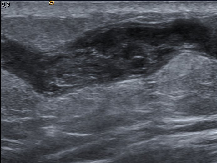
A vein seen in long axis, the lengthwise corridor the in plane approach reads, where a needle would advance as one bright line. This great saphenous vein is partly filled by a clot, dulling the clear black a healthy lumen shows. Image: Nevit Dilmen, CC BY-SA 3.0.
Turn the probe so its long edge runs along an arm and the vessel beneath stretches out lengthwise on the screen. A vein draws a long dark corridor between two bright walls, the lumen running the width of the picture. The view is the vessel in profile, a tube laid open along its length, where the cross-section would draw a circle. The needle, brought in from the end of the probe, runs down that same long axis, its bright line advancing toward the vein in full view. The marker on the probe ties the screen to the run of the vein, so the operator slides the probe up or down the arm to follow the vessel before the needle is brought in. The needle enters at the end of the probe nearest the marker, aligned to the long line of the beam from the first millimetre. The vessel chosen is one that runs straight enough to lie along the beam. A vein that curves or dives away from the plane shows only the stretch that happens to lie flat under the probe, the rest curving out of view. A straight run, a centimetre or two of vein holding one depth, is the friendliest target the long axis has.
The leading end of that line is the tip, and the eye never has to guess where it sits. A shaft entering at the side, a tip reaching toward the wall, the whole length between them drawn in one stroke of bright echo. As the needle advances, the line grows longer and its tip creeps closer to the vein, every step of the approach there to watch. This is the promise the technique is built on, a needle seen whole. Depth tells the whole story here too. The screen shows how far the tip has come and how far it has left to go, the front wall of the vein a fixed line the tip walks toward. A reader counts that gap down in plain sight, a count the single dot of the cross-section can never give. Watching the line lengthen is the rhythm of the stick. Each push adds a millimetre of bright echo at the leading end, the tip creeping toward the wall a step at a time. The operator advances on what the line shows, stopping the push the instant the tip touches the front wall, never running ahead of the picture.
One thing the long axis trades away is the company. The cross-section shows a vein with its artery and nerve all at once; the long axis shows the one vessel it runs along, with nothing to the sides of it in the picture. The operator gives up the wide map of the neighbourhood to gain the full length of the needle, a fair trade on a vessel already known to sit clear of trouble. Reading one structure at a time has a clarity of its own. With only the vein and the needle in the picture, nothing competes for the eye, the whole frame given to the one line closing on the wall of the vessel. Neither view is wrong. The cross-section and the long axis are two ways of seeing one vessel, and a fluent operator turns from one to the other as the target asks.
Keeping the needle in the plane
Here is the whole difficulty of the method.
The beam is a sheet no thicker than paper. The needle shows whole only as long as it lies flat inside that sheet for its entire length. Let the needle tilt a degree or two out of the plane and it slips half out of the beam. What was a clean bright line breaks into a faint, broken streak, or a dot, or nothing at all, the tip suddenly lost, the steel having gone nowhere, only the beam slipped off it. The picture the technique promises holds only as long as the needle and the beam are kept in perfect register, two paper-thin planes pressed exactly together. This is the skill that takes the time. The operator must drive the needle in along a line that stays inside the beam from the first millimetre to the last, and hold it there while the hand pushes and the probe answers, neither drifting off the shared plane. A needle a hair off shows as a ghost, a dim partial line that looks enough like the real thing to fool a beginner, the true tip nowhere near where the faint end seems to lie. The fix is a tiny, ceaseless tending: a small tilt of the probe to sweep its plane back onto the needle, or a small nudge of the needle into the beam, the bright full line caught again and held. A practised hand keeps the two locked together with no visible effort. A new one watches the line flicker in and out and learns, slowly, to chase it back each time. The whole needle is a gift the technique gives only to the hand that earns the alignment. None of this is guesswork once the plane is held. The needle’s whole path lies on the screen, the angle plain, the distance to the wall plain, the tip plain. The price of that clarity is the unbroken attention the alignment demands, a focus that holds through every push of the needle.
A few aids steady the line. A guide clipped to the probe holds the needle in a fixed track down the beam, trading some freedom for a needle that cannot wander out of plane. An echo-bright needle, scored to catch the beam, glows where a plain one fades at a steep angle. Neither replaces the hand that holds the alignment, the line still lost the moment the probe and the needle part company. The skill comes only with the doing. A first in plane line wobbles, the needle flickering out of the beam on every push, the early sticks slow and full of false starts. Hundreds of needles later the alignment is second nature, the line held without a thought, the tip watched in peace, the plane no longer a thing to fight for.
The longer the vessel sits below the skin, the harder the alignment grows. A shallow vein gives a short path that forgives a small wobble. A deep vein demands a long needle held true across three or four centimetres, the smallest tilt at the skin throwing the tip far off the plane down where it matters. The steeper the needle must dive to reach a deep target, the further it strains against the flat plane of the beam, and the more the picture fights to stay whole.
A steep needle is the plane’s worst enemy. A beam reflects best off steel it strikes square. A needle driven steeply meets the beam at a glancing angle, its echo cast off to the side, away from the probe. The line fades just as the angle steepens, the fade landing exactly when a deep target needs the line brightest. Some machines answer by steering their beam sideways to strike a steep needle square again, lifting the faint line back to a clean one, a help the operator on a deep line leans on hard.
Why the whole needle matters
The point of all that effort is a tip never lost. When the needle stays in the plane, its leading end is on the screen at every instant, approaching the vein in plain sight. The tip is watched as it reaches the front wall, presses it into a dimple, breaks through, and comes to rest in the middle of the lumen. At no moment does the operator advance a tip they cannot see. The front wall gives its own signal as the tip arrives. A tip pressing the near wall tents it inward, a small bowing the eye catches at once, then a give the moment the wall is pierced and the tip drops into the dark of the lumen. The operator reads that sequence like a sentence, each step seen and known. The blind alternative shows the value by contrast. A needle pushed on feel alone reaches the vein by luck and overshoots by luck, the tip’s place learned only when blood comes or fails to. The long axis trades that luck for sight, the tip known at every depth.
This is the safety the long axis is prized for. The far wall of the vessel is never approached blind, because the tip that might pierce it is in view the whole way. A needle stopped the instant its tip enters the lumen never travels on into the artery or the structure behind, the danger that the cross-section, with its hidden tip, courts on every stick. For a target where a back-wall slip is a true disaster, the whole needle in view repays the harder hand it asks. The studies bear the safety out. The in plane approach carries a lower rate of back-wall puncture than the cross-section, the tip in view the whole way the plain reason for it. On the vessels where that puncture means an artery or a lung, the gap between the two approaches is the gap between a line placed and a harm caused. The blood confirms what the line shows. A gentle pull on the syringe the moment the tip reaches the lumen brings dark blood back, the picture and the flash agreeing that the tip is home. The tip the operator sees and the tip the blood reports are one and the same, the sight and the feel of the stick in full agreement.
Depth and angle are read straight off the line. The operator sees exactly how steep the needle runs, how far the tip has to go, where the front and back walls of the vein sit along the path. A correction, a flatter angle, a slower push, is made on what the eye plainly shows. The eye stays ahead of the needle the whole way, the next millimetre planned before it is taken. The whole geometry of the stick is laid out in one bright line. An angle read wrong is corrected before it does harm. A needle pitched too steep, its tip about to overshoot the vein, is laid flatter as the operator watches, the whole path re-aimed on the screen, the eye doing the work a blind hand once guessed at.
The wire that follows the needle is watched too. After the flash of blood, the guidewire fed in through the needle shows as a bright line sliding down the lumen, seen to run along the vein, with no kink and no stray through a wall. On a central line, that glimpse of the wire safely inside the vein, before any dilator follows, is a check the long axis gives cleanly. The dilator that opens the track follows the same rule, fed over the wire under the eye, the wire seen to hold its place in the vein as the dilator passes. Each step of the line, needle and wire and dilator and catheter, is taken in view, the long axis turning a blind sequence into a watched one. A wire that strays shows at once. A guidewire that turns the wrong way at a vein junction, or buckles, or slips behind a wall, is caught on the screen before it is forced, the wire’s whole path shown the way the needle’s was. None of it is left to chance unseen.
Where it earns its place
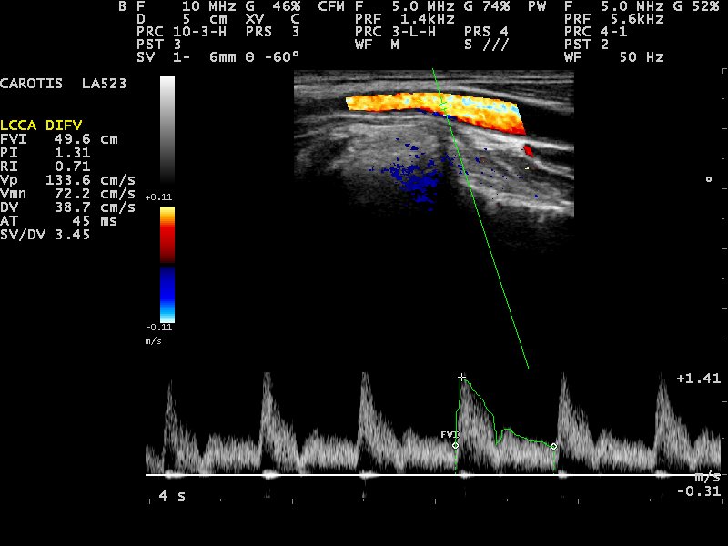
An artery on the same lengthwise view, the colour marking the blood that runs through it and the trace below its Doppler waveform. A needle guided in plane tracks down a corridor like this with its tip in sight the whole way. Image: Nevit Dilmen, CC BY-SA 3.0.
The long axis comes into its own on the targets that punish a lost tip. A central line into a great vein, the internal jugular or the femoral, runs a needle deep beside an artery and, in the chest, near the lung; seeing the whole needle to its resting place is the safeguard against the slip that would find them. An artery cannulated at the wrist or the groin asks the same care, the tip watched into the vessel and no further. The internal jugular line shows the gain plainly. Read in long axis, the vein runs as a corridor with the carotid often just beneath it, and a needle held in the plane is watched into the vein and stopped, the artery below never touched. Guidelines built around this view have driven the harm of the old blind stick down toward nothing. The femoral line in the groin runs the same way, the vein read in long axis with the artery alongside, the needle walked into the vein under the eye. Away from the chest there is no lung to fear, the artery the one neighbour to respect, and the whole needle in view keeps the tip on the right side of it. Even the subclavian line, the hardest of the three to read, can be guided in long axis under the collarbone, the needle watched short of the lung beneath, a stick once feared now done in plain view of the eye.
Beyond the vessels, the in plane view is the standard of regional anaesthesia, where a block needle is walked toward a nerve and must stop short of it, the drug laid in a ring around the nerve, the nerve itself left untouched. There the whole needle in view is the rule, no luxury, the tip watched up to the nerve and held clear. The same discipline that places a deep line safely places a nerve block, the long axis the shared language of both. A block lives or dies on seeing the tip: a millimetre too far puts the tip inside the nerve, a slip that leaves the patient with lasting damage and the operator with a complication that need never have happened. The list of targets runs long. A PICC threaded up a deep arm vein, a dialysis catheter into a great vein, a line into a vessel scarred by old sticks, each goes in more safely with the whole needle watched. Anywhere a tip pushed a hair too far would meet an artery, a lung, a nerve, or the far wall, the long axis is the approach that guards against it.
What it gives up
The whole needle is bought at a price. Centring side to side is the hard part of the long axis, since the tube on screen looks the same whether the beam runs down the middle of the vessel or along its edge, and a needle aimed true in the picture can still slide past a vein the beam was only grazing. The operator finds the vessel in cross-section first, centres on it, then turns the probe into the long axis to stick, trusting that the turn kept the beam down the vessel’s spine. The single structure in view, the longer path through the skin, and the alignment that takes weeks to master are the rest of the toll, paid gladly on the sticks that demand a tip never out of sight. The fix for the centring is a habit. Scan across first to find and centre the vein, fix it in the mind, then rotate the probe a careful ninety degrees in place, the beam kept down the vessel’s middle through the turn. A turn that drifts off-centre loses the vein on the way into the long axis, and the operator starts the find again. The longer skin path is a real cost on a shallow vein. To lie the needle along the beam it must enter further from the vessel at a flatter angle, a longer tunnel through tissue than the short steep jab the cross-section allows. On a deep vein the two paths converge, the long axis no longer the longer road. The hand that masters both views holds the real prize, free to pick the right view for each vessel as the target asks, the two a matched pair of tools for the one job.
On a handheld
The in plane stick rewards a steady, light probe. The hand that holds it has to keep two paper-thin planes locked together, the other hand driving the needle, a balance a marker-sized wireless probe makes easier than a heavy cabled head dragging on its lead. A pocket scanner run from a phone propped in the operator’s line of sight frees both hands for the fine work of holding the line in the beam. The light head is no small thing on this stick. The hand on the probe is doing delicate work, sweeping its plane by fractions to hold the needle. A heavy probe dragging on a cable fights that touch. A probe that weighs almost nothing answers the smallest move of the wrist. Some operators steady the probe hand against the patient, a finger or the heel of the hand resting on the skin, so the plane holds even as the needle pushes. A light probe makes that brace easy, the whole weight of the scan no more than the hand already bears.
The wider gain of the handheld is the same as ever, the whole apparatus shrunk to a probe and a screen that travel to the bedside, the ambulance, the clinic with no imaging room. A deep line or an arterial line that once meant a trip to a procedure suite goes in where the patient lies, the long-axis view on the phone recording the tip in the vein and the wire in the lumen as proof the line sits true. A central line placed at a remote bedside, the whole needle and wire watched in on a pocket screen, reaches a standard of safety once tied to the cart and the procedure room alone. A clip of a clean in plane stick teaches the alignment better than any words. The learner sees the line held whole, the tip walked to the wall, the wire fed home, the whole controlled sequence there to replay. A library of such clips on a phone is a training tool no cart could carry to the bedside. The same probe, wiped down, moves to the next bedside, a line or a block placed wherever a patient waits, no room booked and no machine wheeled in, the whole skill carried in a pocket.
Common questions about the in plane needle technique
What is the in plane needle technique?
It is a way of guiding a needle with ultrasound. The probe runs along the length of the vessel, and the needle is brought in from the end so the whole needle, shaft and tip, shows on screen as one bright line. It is the long-axis approach, used where seeing the entire needle matters.
How is it different from the out of plane technique?
It is the view that shows the whole needle, a line in place of a single point. Out of plane, the probe sits across the vessel and the needle crosses as one bright dot, which may not be the tip at all. In plane, the probe runs along the vessel and the whole needle shows as a line, the tip always in sight.
Why is the in plane technique harder?
Because the needle must stay inside a beam no thicker than a sheet of paper for its whole length. A small tilt out of that plane drops the needle half out of view, the clean line breaking into a faint streak. Keeping the needle and the beam locked together as the needle advances is the skill that takes practice.
When is the in plane approach used?
For targets where the tip must never be lost: a central line into a deep vein beside an artery, an arterial line, and nerve blocks where the needle stops short of a nerve. Seeing the whole needle the entire way in is the safeguard against pushing the tip through a vessel wall or into a nerve.
Can a handheld ultrasound do the in plane technique?
Yes, and a light wireless probe helps. The hand on the probe must hold the beam and the needle in the same thin plane, the other hand advancing the needle. A small probe run from a phone is easier to hold steady through that than a heavy cabled head, and it records the whole needle in the vein as proof.
About the Author
Julien Mercier
Senior R&D Engineer · Medical Ultrasound Transducer Development
Senior R&D Engineer with an M.S. in Applied Physics and over 15 years of experience in medical ultrasound transducer development, specializing in the design verification and performance testing of high-frequency imaging transducers. Currently leading the development and verification of the company’s next-generation high-frequency linear-array transducer, responsible for imaging performance evaluation and reliability analysis in preclinical testing. Brings extensive hands-on experience in piezoelectric element tuning, beamforming parameter optimization, and system-level performance testing.