The endometrium is the lining of the womb. A scan measures how thick it is. The lining shows as a bright stripe down the middle of the uterus. The thickness tells a clinician what the lining is doing. It also tells whether the lining is healthy. A scan reads the measure in seconds against the woman’s age and her cycle.
The lining and the stripe
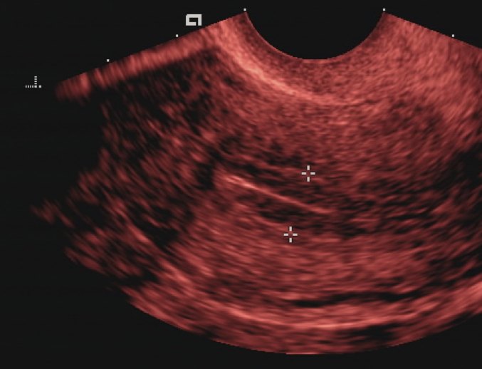
The endometrium measured on a transvaginal scan, the two caliper marks set across the lining at its thickest point. The bright stripe runs down the middle of the womb.
The womb has a lining that renews itself each month. It thickens to ready a place for a pregnancy. When no pregnancy comes, the lining sheds and bleeds away. The cycle then begins again. The lining gains thickness week by week through each cycle. The lining grows on a base that stays from month to month. The scan reads the whole of it as one bright stripe. The womb is a hollow muscle. The lining coats its inner wall. The cavity sits empty between the two walls of lining. A scan reads the lining where the two walls meet. The lining is where a pregnancy implants, a soft bed it builds fresh each month for an egg. The womb sits low in the pelvis behind the bladder. The lining runs through its centre. A reader finds the womb and then the stripe inside it. The lining sits inside the muscle wall of the womb. The muscle reads darker on the scan. The bright lining stands clear inside it. The lining stops at the neck of the womb. A reader follows the stripe down to the neck and no further. The lining is measured at its full double thickness. The two walls are added into one number. A reader never measures one wall alone. The lining is the same tissue that bleeds as a period. A scan reads it between the bleeds. The measure tracks what the lining is doing month to month.
A scan shows the lining as a bright stripe. The stripe runs down the middle of the womb. A clinician measures across the stripe at its thickest point. The number covers both layers of the lining together. The scan reads it in millimetres. The lining reflects sound brightly. It stands out against the darker muscle of the womb. A reader finds the stripe at a glance. The measure is taken along the length of the womb. The probe finds the womb’s long axis first. The stripe runs clearest in that view. A reader takes the thickness at the womb’s middle. The stripe is fullest there. A measure off to the side reads thinner than the truth. The two layers of the lining meet at a fine middle line. A reader can often see that line. It marks the centre of the measure. A reader takes more than one measure to be sure. The thickest clean reading is the one that counts.
The stripe is measured from one edge to the other. A reader takes the line where the womb sits straightest. Fluid in the cavity is left out of the measure. The two walls of the lining are caught together in one number. A clear midline view gives the truest reading. The measure is among the useful numbers a pelvic scan gives of the womb. A clinician records it with the date and the day of the cycle. The next scan reads the new number against it. The lining is read on every pelvic scan as a matter of course. A reader notes its thickness whatever the scan was for. An even stripe is the look of a healthy lining. A healthy lining reads as an even bright band. It has no lump in it and no break. A reader scans its whole length for any flaw. The lining is read against the woman before any number is judged. Her age sets the frame. Her cycle or her menopause sets the rest. The day of the cycle tells a reader what thickness to expect on the scan. The lining is read for change as much as for its number. A lining that grows on schedule is healthy. A coil in the womb sits in the cavity and marks the lining. A reader reads the lining around the bright coil. The coil thins the lining in many women. A scan early in the cycle gives the cleanest measure of a premenopausal lining. The lining is thin then and easy to read. A scan reads a premenopausal lining with the cycle day in hand. The number is judged against where the woman is in her month.
It changes through the cycle
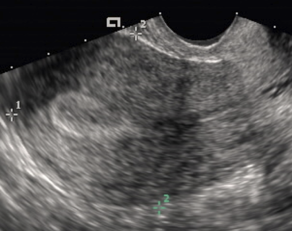
A thick secretory endometrium late in the cycle. The lining reads as a bright band against the darker muscle of the womb.
The lining’s thickness follows the menstrual cycle. A reader reads it against the day of that cycle. The table sets out the phases and the millimetres that go with each.
Endometrial thickness by setting, and what each measure means
Setting
Thickness
Meaning
Just after a period
2–4 mm
Normal, at its thinnest
Late in the cycle
12–14 mm
Normal, before a period
Postmenopausal, with bleeding
4 mm or less
Reassuring; cancer a remote chance
Postmenopausal, with bleeding
over 4 mm
Sample the lining
On tamoxifen
often raised
Read with the drug in mind
Both a thin line and a full lining are normal at the right point in the cycle. The number alone says little. The day of the cycle gives it its meaning. A reader who knows the cycle reads the lining right. Estrogen drives the lining up in the first half of the cycle. The lining grows from a few millimetres toward its fullest. A scan in this phase shows a thickening stripe. A premenopausal lining swings across a wide range with the cycle, from a few millimetres to the low teens. The cycle day settles what counts as normal. Heavy or odd bleeding before the menopause sends a woman for a scan too. A thick lining can lie behind it. A polyp or a fibroid can lie behind it. An early pregnancy thickens the lining and holds it. The lining feeds the new pregnancy in its first weeks. A scan reads a thick lining around a young pregnancy as normal. The pill thins the lining over time. A woman on it bleeds lightly. A scan reads a thin lining on the pill as the drug’s own work. A scan times itself to the cycle for a premenopausal woman. A reading early in the cycle is the cleanest. The thin lining then is easy to measure.
Mid-cycle the lining takes on a layered look. Three bright lines show with darker bands between them. A reader knows this triple line as the lining ready for a pregnancy. It marks the days around the release of an egg. After an egg is released the lining turns secretory. It softens and readies for a pregnancy. A scan shows it at its thickest in these days. The triple line is a sign of a fertile lining. It shows in the days before an egg is released. It vanishes once the egg is gone. A lining read mid-cycle is at its busiest. The triple line marks the fertile window. A reader reads a thick mid-cycle lining as the cycle’s own work. The bleeding phase shows a thin lining as it sheds. A reader times a clean measure away from the bleeding days. A measure during a period reads low and ragged.
A period strips the lining back to a thin line. The scan after a period shows the lowest number of the month. The lining then starts to build again. A reader who scans across a cycle watches the number climb and drop. The period clears the grown lining away. The scan straight after shows a thin bright line. A reader times a measure to the cycle to read it right. The lining grows under estrogen, then holds steady under a second hormone, then clears with a period. The cycle starts over each month.
The thin stripe after the menopause
After the menopause a thin lining is the reassuring one.
Bleeding after the menopause
Bleeding after the menopause is the symptom that brings the measure to the fore. The womb should be quiet by then. Any bleeding asks for a reason. A scan of the lining is the first test a clinician reaches for. The lining grows thin after the menopause. The hormones that drove it have faded. A thin lining is the expected finding in a woman past the change. Bleeding after the menopause is never ignored. It can come from a thin worn lining. It can come from a polyp. It can come from a cancer. A clinician follows a set path for bleeding after the menopause, the scan first and a sample where the lining reads thick. The four-millimetre line is drawn from large studies of women who bled. The studies followed which linings hid a cancer. The thin ones almost never did. A worn thin lining is the commonest cause of bleeding after the menopause. A polyp is the next. A cancer is behind a small share of cases. A thin worn lining can bleed of its own. The lining grows fragile after the menopause. A small vessel in it can break and bleed. A clinician asks about the bleeding first. The amount and the pattern matter. The scan reads the lining beside the woman’s account. A single bleed after years of none is the one that brings a woman in. A heavy bleed and a light spot are read the same way. Any bleeding after the menopause earns the scan. The lining is read as postmenopausal a year past the last period. The same number means more after that line. A thin lining after the menopause is the quiet a clinician hopes to see. It tells of a womb at rest. A clinician sees many women with bleeding after the menopause, and the scan sorts them quickly by the thickness of the lining.
The lining is read for its thickness in a woman who bleeds after the menopause. A thin lining points away from cancer. The American College of Obstetricians and Gynecologists holds that a lining of four millimetres or less carries a chance of missing a cancer below one in a hundred. A scan that shows a thin line lets a clinician reassure the woman without a biopsy. A measure above four millimetres sends her on for a sample of the lining. The sample reads the tissue itself. The scan and the sample together name the cause of the bleeding. The measure is taken with care in a woman past the menopause. The lining lies thin against the womb by then. A reader finds the stripe and measures it at its thickest. A lining too hard to see is sampled to be sure. A reader who cannot measure the lining says so and moves to a sample. The thin line on a confident scan is the result that reassures. A clinician reads the number, the woman’s story, and any treatment she takes, all together. The four-millimetre mark is the one number a clinician carries into every case of bleeding after the menopause. A little fluid can sit in the womb after the menopause. A reader measures the lining on each side of the fluid and leaves the fluid out. A lining can be hard to measure past the menopause. The stripe can read faint. The womb can lie at an angle. A sample of the lining is taken in the clinic with a thin tube. The tube draws a little of the lining for the lab. A wider look uses a camera passed inside the womb. A drop of salt water in the womb lifts the walls apart. The lining then shows clearly on each side. A polyp stands out against the fluid. This saline scan reads a thick lining that a plain scan left unclear. A lining over four millimetres is the sign that a sample is owed. The sample finds a benign cause in the great run of cases. The scan flags the lining. The sample settles the cause.
A thick lining found by chance is read differently. A woman with no bleeding carries a far lower risk. No single thickness flags her the way four millimetres flags a woman who bleeds. A reader notes a thick lining without bleeding and weighs it against the rest. A scan in a woman with no symptom rarely changes anything on the lining alone. A thin lining in a woman who bleeds makes a cancer a remote chance, and a clinician watches her for any return of the bleeding. Bleeding that comes back is looked at again whatever the first scan showed. A new bleed earns a fresh look. A clinician reads a thin lining as a strong reassurance. It makes a cancer a remote chance. The scan is the gatekeeper for the sample. A thin lining keeps a woman from a procedure she does not need. The sample is kept for the linings that earn it. A woman frightened by bleeding wants an answer fast, and the scan gives a strong one in minutes. The scan saves many women a needless sample. The four-millimetre line is measured the same way the world over, and a woman gets the same answer wherever she is scanned.
Treatment can thicken the lining for a known reason. Hormone therapy after the menopause builds the lining the way the body’s own hormones once did. A drug for breast cancer, tamoxifen, thickens the lining and muddies the scan. A reader reads the lining of a woman on these drugs against what the drug is known to do. Hormone therapy builds the lining unless a second hormone holds it in check. A reader reads the lining of a woman on therapy against the kind she takes. Cancer of the lining is commoner in older women past the menopause. A reader reads the same number harder in a woman of seventy. Estrogen made anywhere in the body can thicken the lining. Fat tissue makes its own estrogen. A heavier woman can carry a thicker lining for that reason. A thick lining in a young woman is read with her hormones in mind. A cycle out of rhythm can build the lining up. A thick lining at the wrong point in the cycle asks for a closer read.
The thin line is the reassuring result after the menopause. It turns a frightening symptom into a settled one. A woman who bleeds and measures thin is spared a biopsy. The scan does in minutes what once meant a wait and a procedure.
What thickens the lining
A thick lining has a handful of causes. Estrogen builds it up. A polyp grows from it as a soft lump on a stalk. Hyperplasia is an overgrowth that can lead toward cancer. Cancer itself thickens and disorders the lining. A scan reads the thickness first and looks for the cause behind it. A polyp shows as a bright lump in the stripe. A drop of fluid put into the womb can outline it. Hyperplasia is the lining grown too much. It can sit quiet or carry the cells that lead toward cancer. The sample reads which it is. A thick lining is read for its evenness. A patchy, broken-up thickening leans toward something more than a simple cause. The sample names the cause behind a thick lining. The scan measures and points the way to it. Hyperplasia comes in kinds, some of little risk and some carrying odd cells that lean toward cancer. The sample reads which kind a thick lining holds. A bright spot in the lining can be a polyp or a clot, and a reader reads it again to tell which by whether it stays put. A fibroid is a lump in the muscle of the womb. It can push on the lining and distort the stripe. A reader tells it from a thickening of the lining by where it sits. Old surgery or infection can scar the womb. The lining then reads patchy, its stripe broken in places. A reader reads it for the scar behind it. A polyp can bleed whatever its size. A small polyp can cause a big bleed. The scan finds it and the camera removes it.
A scan can point toward a cause. A polyp shows as a focal lump in the stripe. A bright feeding vessel can run into it. Hyperplasia and cancer can look alike on a scan. Only the sample of the lining tells them apart. Endometrial cancer is the worry behind the measure. It thickens the lining and breaks up its even look. A bleed after the menopause is its commonest sign. The scan and the sample catch it early. A polyp is a common cause of bleeding at any age. It is a soft growth, almost always benign. A reader finds it on the scan and sends it for removal. The cause of bleeding is found in the lining itself in the great run of cases. The scan reads the lining first. The sample reads it deeper when the scan flags it.
Measuring it on a handheld
The endocavity probe reads the lining best. It sits close to the womb with the bladder empty and draws the stripe in fine detail. A reader finds the midline and freezes the clearest view. The measure is taken on the still image in about a minute. The lining is a bold target that asks for no fine machine. The number reads the same on any good probe. The lining is one part of a full pelvic scan. The womb, the lining, and the ovaries are read in one sitting. A woman scanned for one thing has her lining read along the way, the thickness noted as a matter of course.
A handheld carries the same close probe a hospital uses. A clinic with one measures the lining at the bedside. A woman who bleeds after the menopause has her lining read in the same visit. The scan that reassures her happens there and then.
The lining measure is one of the quickest answers a pelvic scan gives. A thin line sends a worried woman home reassured. A handheld puts that answer within reach of a clinic far from a hospital. A woman who would wait weeks for a department has her lining read in one visit. The stripe is frozen and the measure stored with the image. The number travels with the woman to her next visit. A lining watched over time is read again on the same probe, the trend built where the woman is seen. Measuring a lining takes practice, and a clinician learns to hold the clean line over many scans. The lining reads in the same sweep that checks the womb and the ovaries. The measure costs only the moment it takes. A thin lining is one of the settling things a scan can show a frightened woman. The few minutes lift weeks of fear. A handheld measures the lining without sending the woman elsewhere. The scan happens in the room she is already in. The answer is hers before she leaves. The measure no longer waits on a hospital department.
Common questions about endometrial thickness on ultrasound
What is a normal endometrial thickness?
It depends on the cycle. Just after a period the lining measures about two to four millimetres. It thickens through the cycle and can reach twelve to fourteen millimetres before the next period. After the menopause the lining is thin, and a measure of four millimetres or less is reassuring.
What endometrial thickness is worrying after the menopause?
In a woman with bleeding after the menopause, a lining over four millimetres is sent on for a sample. A lining of four millimetres or less carries a chance of cancer below one in a hundred, so a clinician can reassure her without a biopsy. A thick lining found by chance with no bleeding carries a far lower risk.
How is endometrial thickness measured?
A clinician measures the bright stripe of the lining on a transvaginal scan. The line runs down the middle of the womb, and the measure covers both layers at the thickest point. Fluid in the cavity is left out. A clear midline view gives the truest number.
Why is a transvaginal probe used to measure the endometrium?
An endocavity probe sits close to the womb and draws the lining in fine detail. The thin stripe shows sharply from there. A handheld carries the close probe, so a clinic can measure the lining at the bedside.
Can tamoxifen affect endometrial thickness?
Yes. Tamoxifen, a drug for breast cancer, thickens the lining and can muddy the scan. A reader reads the lining of a woman on tamoxifen against what the drug is known to do. Bleeding on tamoxifen is looked at carefully whatever the thickness.
About the Author
Julien Mercier
Senior R&D Engineer · Medical Ultrasound Transducer Development
Senior R&D Engineer with an M.S. in Applied Physics and over 15 years of experience in medical ultrasound transducer development, specializing in the design verification and performance testing of high-frequency imaging transducers. Currently leading the development and verification of the company’s next-generation high-frequency linear-array transducer, responsible for imaging performance evaluation and reliability analysis in preclinical testing. Brings extensive hands-on experience in piezoelectric element tuning, beamforming parameter optimization, and system-level performance testing.