Deep Vein Thrombosis DVT Compression Ultrasound Handheld Diagnosis
A deep vein thrombosis is a clot sitting in one of the deep veins of the leg. Compression ultrasound is the test that finds it. The probe presses down on the vein and watches what happens. A clot is the one thing that stops a vein flattening shut under that press. That single behaviour, caught in a few seconds at the bedside, is the whole diagnosis.
The one test it rests on
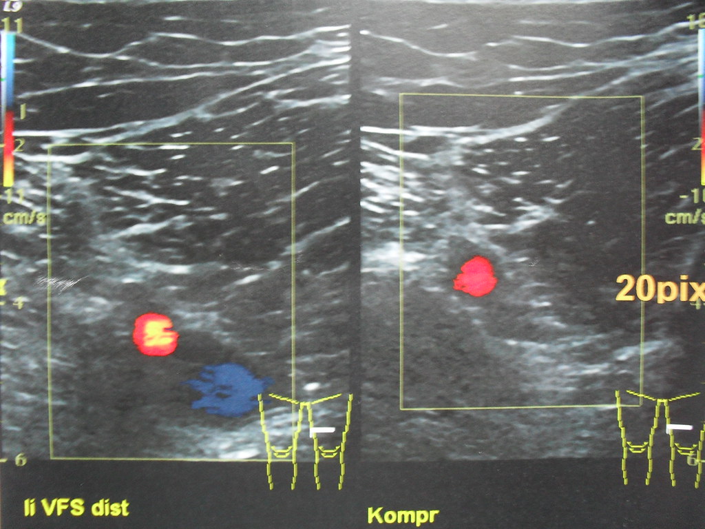
The compression test in one image. On the left, the superficial femoral vein (blue) lies beside its artery (red), both open. On the right, marked Kompr for compression, the probe presses and the vein has vanished, the artery alone left behind. A vein that disappears like this holds no clot. Image: Mr Thinktank, CC BY 2.0.
The test asks the vein a single question.
A healthy deep vein is a soft-walled tube carrying blood at low pressure. Press on it from the skin and its two walls meet, the black channel on the screen winking shut. Lift the probe and the vein springs open again. A clear vein does this over and over, the whole length the probe travels down the leg. A clot changes the picture. Fresh thrombus fills the channel with soft material close to the shade of the blood around it, hard to make out on its own. The proof is in the press, in seconds, not minutes. A clotted segment will not flatten. The two walls come down toward each other and stop, held apart by the plug inside. The vein holds a round shape under a pressure that would have shut a clear vein flat. An operator does not need to see the clot to call it. The failure to compress is the finding. A clot that fills the vein wall to wall is the easy call, the whole channel held round under a press that should have closed it. The pressure has to be real. A light touch that only dimples the skin proves nothing. The probe presses until the artery beside the vein begins to deform. That much pressure shuts any clear vein. A vein that stays open under it is a vein with a clot. A timid hand is the commonest reason the test goes wrong. A clear vein gets called blocked when the press stays too soft. The press is done in short axis, the vein a circle on the screen. A circle is easy to watch collapse. In long axis a vein can slide out from under the probe and read open by mistake. This is why the test is taught as a mechanical act first and a reading of echoes second. A beginner who can press a vein and watch it close, or fail to close, can answer the question the test exists to ask. The vein is open, or the vein is blocked.
One quick check comes first. The vein sits beside an artery. The artery has a thick muscular wall and a pulse. It resists the press. The vein is the soft one that gives way. This is the same press an operator uses to pick a vein out for a line, turned now to a different question. The press that finds a vein for a needle is the press that clears it of clot. A lump beside the vessels can fool the eye. A swollen lymph node in the groin can pass for a vein in cross-section, a round dark shape. It carries no flow through it. The surest check is to trace the structure along the leg. A vein runs as one continuous channel the whole way down. The probe follows it from the groin into the thigh, the dark tube never giving out.
The answer comes fast at the common sites. A clot holds the vein open through the firmest press a patient can bear. There is little middle ground in the groin or behind the knee. The vein shuts, or it does not. The femoral vein runs double in many legs. Two channels often lie side by side at the groin, where a textbook draws one. A clot can sit in just one of the pair. An operator who presses its mate alone meets a clear vein and a false all-clear. A check for two channels at the groin belongs in a careful press. Not every clot fills the whole vein. A clot stuck to one wall leaves a slit of channel beside it. The vein squashes part way and stops on the clot. A partial compression counts the same as a vein that will not move at all. The walls meet where the vein is clear. They stay propped apart where the clot sits. The certainty of the press is its gift. A blocked vein is rarely a matter of opinion. The walls meet, or they stand apart. A photograph shows which. A test with so little grey area travels well to hands that scan only now and then. A vein that will not yield is a vein to act on, even with a clot no one can see.
Everything rides on that answer. A clot in a deep leg vein can break loose and travel to the lung. A test that finds the clot sends the patient to treatment. A test that clears the leg sends the patient home. The press of a probe stands between those two roads. The press works inside a chain of decisions. A score from the history and the exam sorts patients into higher and lower risk. A blood test for clot fragments clears many low-risk legs with no scan at all. The compression press settles the patients left in doubt. The test earns its place inside that chain. A found clot starts a clock. Blood thinners begin within the hour in many places, before the clot can grow or throw a piece. A press that takes two minutes can move a patient from a waiting room to a treatment that protects a lung. Few bedside tests carry that much weight in so little time. A clot found at the bedside is a clot moved to treatment within the hour.
Where the probe presses
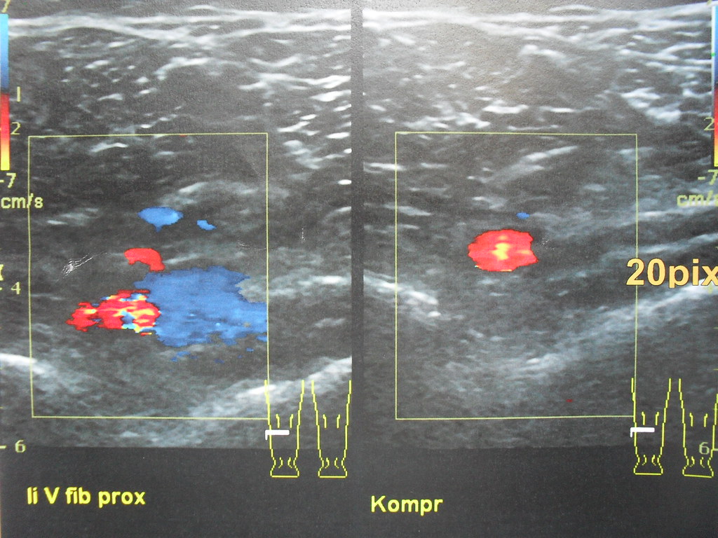
The same press at a deep calf vein. The left panel shows the vein in colour beside its artery. The right panel, again under compression, shows the vein squashed away to nothing. A clot is what would have held the vein open against this press. Image: Mr Thinktank, CC BY 2.0.
The exam works two points on the leg. The first is the groin. The common femoral vein runs there, just below the crease, beside its artery. The probe sits across the vein in short axis and presses, then walks down a few centimetres, pressing every centimetre of the way. A clear vein closes flat at each step. Each press takes about a minute in a trained hand. The groin press starts high. The great saphenous vein joins the deep system here, at the saphenofemoral junction. A clot can sit right at this join. The press begins above it and walks down through the common femoral vein into the thigh. The leg is set up to fill the veins first. The patient lies with the head raised a little, the leg turned out and bent at the knee. Blood pools in the deep veins and opens them wide. A full vein presses cleaner than a flat one. The setup is half the test.
The second point is the back of the knee. The popliteal vein runs through the hollow behind the joint. The patient bends the knee a little to open the space. The probe presses the vein there the same way it pressed the groin. The popliteal vein splits below the knee into the calf veins. The press follows it down to that split. A clot in the popliteal vein, or right at the split, falls inside the two-point reach. Below the split the calf veins begin. The focused exam lets them go. The knee is read from behind. The patient rolls toward the far side, or sits with the leg hanging, the popliteal hollow turned up to the probe. The vein lies above the artery in this view, nearer the skin than the artery is. The press reaches it first.
Two points cover the ground that counts. A clot in the common femoral vein or the popliteal vein is the kind that threatens the lung. Studies put this two-point press close to a full scan of the whole leg for patients with symptoms. The trade is a faster test against a small blind spot lower down the calf. The choice of two points is not arbitrary. A clot above the knee carries the real danger to the lung. The common femoral vein and the popliteal vein catch nearly all of those. A clot found at either point sends the patient straight to treatment. The exam aims its effort where a missed clot costs a life. One vein branches off and leaves the press behind. The deep femoral vein dives into the thigh muscle just below the groin. The two-point exam reaches its mouth and goes no further. A clot deep in this branch is rare. It rarely threatens the lung. The names trip people up here. The femoral vein is a deep vein. Its old name, superficial femoral, still trips people up. A clot in it is a true deep vein thrombosis. The calf question divides clinicians. Some scan the calf veins in full, holding that a calf clot can climb. Others press only the two points and bring the doubtful patient back in a week. Both paths have a place in the guidelines. The two-point press with a planned repeat is the one a handheld serves best.
Reading the clot
A fresh clot has a look of its own. It is soft. Its shade sits close to flowing blood, easy to miss by eye alone. It swells the vein wider than the vein on the other leg. A vein standing fatter than its own artery is a quiet warning before the press even confirms it. It shifts a little in the flowing blood, not yet stuck fast to the wall. The press is what gives a fresh clot away. A clot has a top and a bottom the probe can map. The highest point it reaches is the mark that counts for the lung. A clot creeping up toward the groin is the one that turns urgent. Some fresh clots wave a loose tail. The head of the clot sticks to the wall. The tail floats loose in the blood, its end swinging. A tail like this worries a clinician, since a loose piece can break away. The sight of it sharpens the urgency of the treatment. The fat, full look belongs to a fresh clot. The swelling settles when the clot shrinks down. A wide, soft, uncompressible vein is the picture of an acute clot. A fresh clot is the dangerous one, loosely held and ready to move. It is the finding the test must never let slip.
An old clot looks nothing like a fresh one. Months on, it shrinks. It grows tough. It reads bright on the screen, its surface rough. It draws in a thick, scarred wall. A vein damaged this way may never fully close again, the old scarring left behind. A reader weighs that history before calling a stiff vein a new clot. An old clot leaves the vein changed for good. Channels can bore back through the old clot over months, the blood finding a way through a scarred tube. The valves inside the vein take damage along the way. A leg with an old clot often swells for years, the vein never the smooth tube it was. Thin strands cross the channel where a clot once healed, the webs of a recanalized vein. A thickened wall, bright on the screen, tells of an old fight. None of these is the smooth, soft, easily shut vein of a clear leg. Telling this from a fresh clot keeps a patient off treatment they do not need. The other leg is the best ruler. A quick look at the matching vein in the well leg shows what normal looks like for this patient. A vein twice the width of its mate is plain at a glance. The comparison turns a vague picture into a plain call.
Colour flow adds a second look. A clear vein fills with colour where the blood runs through it. A blocked segment shows a gap in the colour. Sometimes colour squeezes in a thin line around the edge of a clot, a clue to a vein not yet fully blocked. These are aids. The compression is the test. Colour has its traps. A slow trickle of blood can fill with colour. The colour then masks a partial clot. A colour setting turned up high paints flow where there is barely any. A reader leans on colour for a quick look, then trusts the press for the answer. The grey-scale press outranks the colour every time the two disagree. A vein with no colour is not always blocked. A slow flow, a cold limb, or a low setting can empty a clear vein of colour. The calf squeeze is what tells the two apart, a clear vein lighting up on the push.
Breathing leaves its mark too. The flow in a clear deep vein keeps shifting from one breath to the next. A flat, unchanging signal can mean a blockage higher up, above the point the probe sits on. This sign backs up the press. A squeeze of the calf is the way to test the colour. A firm push at the calf, or the foot, drives a slug of blood up the deep vein. In a clear vein the colour floods bright at once, then settles. A weak answer, or none, points to a blockage between the squeeze and the probe. A deep breath, or a bear-down, swells the leg veins, the pressure in the belly holding the blood back. A vein that stays flat through all of this can be blocked somewhere above the probe.
What the test can miss
The two-point press has one real blind spot. A clot that sits only in the calf veins, below the knee, can hide in the gap between the two pressed points. A calf clot can climb into the thigh over the days that follow. That risk is why a negative scan in a patient who still hurts is often pressed again a week later, to catch the clot that has climbed into reach. The blind spot has a second part higher up. A short stretch of vein runs deep through the thigh inside a tunnel of muscle, the adductor canal. The probe presses it poorly there, the vein buried too deep for a clean squash. A clot in that stretch can slip a two-point exam. A whole-leg scan run by a trained sonographer presses every centimetre. No gap is left for a clot to hide in. The two-point press trades that completeness for speed and reach. The arms are not immune either. A clot can form in the deep veins of the arm, around a line left in too long, or after hard effort. The same press finds it, a clot in an arm vein holding it open against the probe. A clot can hide above the groin as well. The iliac veins run up into the pelvis, out of reach of a press through the belly. A clot sitting only there leaves both leg points clear. A swollen leg with clear femoral and popliteal veins points the search higher, into the pelvis, where other scans take over. A swollen leg is not always a clot at all. A burst cyst behind the knee, a torn muscle bleeding into the calf, a spreading skin infection, each swells a leg and mimics a DVT. The same probe that presses the veins can spot a cyst or a pocket of blood in a moment.
On a handheld
The compression test suits a handheld better than almost any other scan. The answer is a yes or a no. The vein closes, or it stays open. A pocket probe pressed in the groin shows that as plainly as a cart in a vascular lab. A clinician at a bedside, in an ambulance, or on a remote ward can press the two points and reach an answer in minutes. The skill lies in the pressing itself. Speed is what counts in the night. A patient with a swollen leg at three in the morning can have the dangerous question answered at the bedside, with no wait for the imaging department to open. The formal scan can wait for daylight once the lung is out of danger. A handheld has honest limits here. The two-point press is its strength. A full survey of every calf vein asks more time and a cleaner picture than a pocket probe gives in a hurried bay. A clinician uses the handheld to answer the urgent question and sends the doubtful leg on for a full study. The tool fits the first look. A short protocol keeps the handheld honest. Press the common femoral vein from the groin crease down. Press the popliteal vein from the knee hollow to the calf split. Squeeze the calf and watch the colour at each point. A clinician who runs the same steps every time misses fewer clots than one who improvises.
The reach of the small probe changes who gets the test and when. A swollen, painful leg once meant a wait for a slot in the imaging department. A probe and a phone bring the test to the patient within the hour. The clip of the vein closing, or failing to close, saves to the phone as proof for the chart and for the doctor who decides what comes next. The press is a skill a clinician picks up fast. The hand finds the vein, presses, and reads the answer in one motion. A doctor or a nurse trained on the two points can run the test between other jobs. A leg cleared at the bedside spares the patient a trip and a long wait. A colleague checks the saved clip later, the closing vein plain on the small screen. The record carries weight on a busy ward. A vein pressed and filmed at two in the morning can be reviewed by the day team with no second scan. The clip shows the pressure applied, the vein closing or holding, the colour answering or not. A picture settles a question a written note leaves open. A clear answer at the bedside can change the course of a patient’s night. A saved loop of the press carries the finding to whoever decides the treatment. The test crosses roles as easily as places. An emergency doctor, a ward nurse, a rural clinician, each can learn the two-point press in a short course. The binary answer leaves little room for a split reading. A clear close is a clear close on any trained pair of hands. The cost of the tool puts this test in places a vascular lab never reached.
Common questions about DVT compression ultrasound
How does compression ultrasound diagnose a DVT?
The probe presses on a deep vein. A clot is what keeps the vein from flattening shut under the pressure. That failure to compress is the sign of a clot, even when the clot itself is hard to see.
What is two-point compression?
It is a focused exam that presses the vein at two places: the common femoral vein in the groin and the popliteal vein behind the knee. These are the sites where a clot does the gravest harm, the ones that feed the lung. For patients with symptoms, this two-point press performs close to a scan of the whole leg.
Can compression ultrasound miss a clot?
Yes. A clot sitting only in the calf veins, below the knee, can fall in the gap between the two pressed points. Some calf clots later climb into the thigh. For that reason a negative scan in a patient who still has symptoms is often repeated about a week later.
How is an old clot told from a new one?
A fresh clot is soft, swells the vein, and sits close to the shade of flowing blood. Age changes all of that: the clot shrinks, reads bright on the screen, and pulls in a thick, scarred wall. A vein scarred by an old clot may never fully compress again, which a reader weighs before calling a new event.
Can a handheld ultrasound check for a DVT?
Yes. The compression test gives a yes or no answer that a pocket probe shows as clearly as a larger machine. A clinician presses the femoral vein in the groin and the popliteal vein behind the knee, then saves the clip as proof. The handheld brings the test to the bedside within the hour.
About the Author
Julien Mercier
Senior R&D Engineer · Medical Ultrasound Transducer Development
Senior R&D Engineer with an M.S. in Applied Physics and over 15 years of experience in medical ultrasound transducer development, specializing in the design verification and performance testing of high-frequency imaging transducers. Currently leading the development and verification of the company’s next-generation high-frequency linear-array transducer, responsible for imaging performance evaluation and reliability analysis in preclinical testing. Brings extensive hands-on experience in piezoelectric element tuning, beamforming parameter optimization, and system-level performance testing.