





































Doppler modes
Color Doppler versus Power Doppler in Handheld Ultrasound
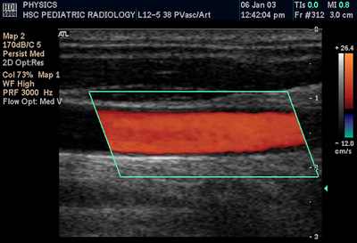
Color Doppler and power Doppler are two ways the same machine paints blood flow onto a greyscale image. Both read the Doppler shift, the change in frequency a moving target gives the echo it returns, and both lay a colour over the moving blood. They differ in what they take from the shift. Color Doppler reads the speed and the direction of the flow. Power Doppler reads the strength of the signal alone. Each shows a side of the flow the other leaves out, and a clinician picks the mode by what the question turns on.
The Doppler shift both modes read
Blood that moves toward or away from the probe changes the frequency of the sound it reflects, the way a passing siren rises and falls in pitch. The size of that shift follows the speed of the flow, the angle between the beam and the vessel, and the frequency the probe runs at. A beam aligned with the flow reads the full shift. A beam square across the flow reads almost none, since the movement carries neither toward nor away.
The machine samples the shift at each point in a chosen box and turns it into colour. What it does with the sampled shift is where the two modes part. One mode keeps the speed and the sign of the shift. The other keeps only how much signal came back. The same returning echoes feed both, read two different ways.
How color Doppler reads the flow
Color Doppler turns the shift into a speed and a direction at every point in the box. Flow toward the probe paints in one colour, conventionally red, and flow away paints in another, conventionally blue, with a brighter shade for faster flow. A vessel lights up along its course, an artery pulsing with the heartbeat, a vein running steady. A narrowed segment shows a bright jet of fast flow. A leaking valve shows a spray of colour running the wrong way. The mode reads the flow by sampling it many times a second, at the pulse repetition frequency, and that sampling sets a ceiling. A flow faster than half the sampling rate, the Nyquist limit, wraps around and reads as the wrong direction, the aliasing that paints a fast jet in a mottle of red and blue mixed together. The operator lifts the scale, the pulse repetition frequency, to push that ceiling up for a fast jet, then drops it to fill in a slow vein, the scale matched each time to the flow expected. The reading also leans on the angle between the beam and the flow, since the shift carries the speed only along the beam. A beam square across a vessel, at ninety degrees, reads almost no shift and can miss the flow altogether, so the operator angles the probe or steers the colour box to bring the beam well off ninety. The shade shown is the speed along the beam, which the machine corrects toward the true speed once the operator marks the vessel’s line on the screen. Speed and direction are the whole point of the mode. A study that turns on how fast blood moves, the jet through a tight valve, or on which way it runs, the reflux down a vein, reads on color Doppler, since those are the numbers it draws and the greyscale image alone cannot.
How power Doppler reads the flow
Power Doppler turns the shift into a single number, the strength of the Doppler signal, and ignores its speed and its sign. It maps that strength in one colour, a wash that marks where flow exists rather than how fast it moves. The display shows the presence and the amount of flow, the vascularity of a tissue, painted as a single warm colour over the grey.
Power Doppler drops the speed and the sign, and that loss is what makes it sensitive. It gathers the weak signal from slow flow and tiny vessels that a speed reading smooths away, reaching perfusion several times fainter. It does not alias, since it never measures speed against a sampling rate. It holds up at almost any beam angle, since signal strength stays even where the beam runs square to the flow. The cost shows as a flutter artefact, a flash of colour when the probe or the patient moves, with no speed or direction given.
The sensitivity to faint flow is the point of the mode. A question about whether a tissue is perfused at all, or about the fine vascularity of a small structure, reads on power Doppler because it catches the flow a speed-reading mode would miss.
The trade is set the moment the mode is chosen. Speed and direction, or faint-flow sensitivity, is the line between them.
What each mode is reached for
Color Doppler is reached for the questions of speed and direction. A heart study reads a valve’s leak and a chamber’s flow, the direction telling forward flow from regurgitation. A vascular study reads the speed through a carotid narrowing and the direction of venous flow for reflux. A vessel mapped before a line shows its pulsing, the artery and the vein each read by the way it flows. Wherever the answer is a velocity or a direction, color Doppler draws it.
Obstetric and venous work leans on the direction too. The cord and the placental vessels read for their flow, the fetal heart’s chambers shown filling and emptying. A deep vein is checked for the reflux that marks a failed valve, the way the blood runs the whole finding.
Power Doppler is reached for the questions of presence and amount. A testis is checked for flow to rule torsion in or out, the perfusion present or absent the whole answer. An inflamed joint lining, a tendon, a thyroid nodule shows its fine vascularity, the amount of flow marking active disease. A transplanted kidney is read for perfusion to its edges. Wherever the answer is whether and how much flow exists, power Doppler draws it.
The amount of flow grades disease as well as finding it. A synovitis is scored by how much colour fills an inflamed joint lining, the quantity tracking how active the disease is. A tumour shows its vascular pattern, a fine rim or a dense core, on the mode that catches the faint vessels feeding it.
Many studies run both in turn. A scrotal scan opens on power Doppler for the faint flow of a torsion question, then turns to color Doppler to read the direction and speed where a vessel is found. A thyroid nodule is mapped on power Doppler for its vascularity, then on color Doppler to read a feeding vessel’s flow. The two modes answer different halves of one study, and a reader switches between them as the question shifts.
Reading numbers off the flow
Colour and power lay flow over the picture, and the numbers come from a third reading, the spectral trace. A sample gate set on a vessel plots the speed against time, a waveform that rises with each heartbeat and falls between. The shape of that waveform carries the finding, and its peak, the peak systolic velocity, is the figure a carotid narrowing is graded by, a number in centimetres per second that climbs higher the tighter the narrowing grows.
From the same trace the machine reads a set of ratios. The resistive index weighs the peak of flow against its trough, a number that rises when the vessel beyond the gate is stiff or blocked, read on a transplanted kidney or a torsed testis. The pulsatility index and the systolic-to-diastolic ratio carry the same reading into obstetrics, an umbilical artery’s waveform marking a thriving placenta or a failing one. The colour finds the vessel, and the trace puts a number on its flow.
The artefacts each mode shows
Each mode shows artefacts a reader learns to set aside. Colour blooms past the vessel wall when the gain runs too high, a wash spilling into the tissue around the flow, settled by dropping the gain until the colour sits inside the vessel. A flash artefact throws colour across the whole box when the probe or the patient moves, a burst the wall filter is raised to quiet. Power Doppler shows that flash readily, one face of the sensitivity that makes it so good at catching faint flow.
Some artefacts are read as signs rather than cleared. A twinkling artefact, a flicker of mixed colour behind a hard surface, marks a kidney stone or a calcification the greyscale alone can pass over, a clue a reader hunts for on purpose. Aliasing itself, the wrap of colour at a fast jet, points to the narrowed spot where the flow speeds up, the artefact aimed straight at the lesion. A reader takes each mark for what it is, a fault to clear or a sign to read.
How the modes run on a handheld
A handheld unit carries both modes as a tap on the screen. The same probe and the same returning echoes feed each, so a clinician switches from one to the other without changing anything in the hand, the machine reading the stored signal the other way. The colour box is dragged over the area of interest and sized to it, a smaller box holding a faster frame rate.
The settings each mode needs are few and shared. The pulse repetition frequency is set high for the fast flow of an artery and low for the slow flow of a vein or a perfusion question, the scale matched to the flow expected. The colour gain is raised until flow fills the vessel and dropped just below the point where noise speckles the box. A wall filter cuts the slow thump of moving tissue, set low when the hunt is for the faint flow power Doppler is run for.
The colour box is shaped to the work. It is steered to bring the beam off ninety degrees to a vessel running across the screen, and an angle-correction cursor is laid along the vessel so the machine reports a true speed, corrected up from the lower figure a square beam alone would return. A narrow box set just over the vessel of interest keeps the frame rate high, since the machine has fewer lines to sweep, the picture refreshing fast enough to follow a pulsing flow.
The handheld brings both modes to the bedside in one hand. A torsion question answered with power Doppler in an emergency room, a valve leak read with color Doppler at a clinic bed, the flow painted on the screen where a cart-bound machine could not easily go. The two modes that once meant a console now ride in a pocket probe, each a tap away.
Other ways to read the flow
Newer modes read flow in ways that sit beside the two. B-flow draws the blood itself from its echoes without colour at all, shown as moving grey specks that trace the channel and its walls cleanly. Contrast-enhanced ultrasound goes further still, where a microbubble agent given into a vein lights the smallest vessels of a tumour or an organ deep in the tissue, its perfusion graphed as the bubbles wash in and out over time. A third route, microvascular imaging, strips the background clutter to map the finest flow as a still picture. Each reads the same blood the two colour modes do, reached by a different path.
One signal, two readings
Color Doppler reads the speed and the direction. Power Doppler reads how much flow is there.
Common questions about color and power Doppler
What is the difference between color Doppler and power Doppler?
Color Doppler reads the speed and the direction of blood flow, painting flow toward the probe in one colour and away in another. Power Doppler reads only the strength of the flow signal in a single colour, marking where flow exists rather than how fast it moves.
Why is power Doppler more sensitive to slow flow?
It drops the speed and direction and keeps only signal strength, so it gathers the weak signal from slow flow and tiny vessels that color Doppler smooths away, picking up perfusion several times fainter. It pays with no speed or direction and a flutter artefact on motion.
When is color Doppler the right choice?
When the answer is a velocity or a direction: a jet through a narrowed valve or vessel, reflux down a vein, or telling an artery from a vein by how each flows. Color Doppler draws the speed and the sign those questions turn on.
When is power Doppler the right choice?
When the answer is whether and how much flow exists: perfusion of a testis in a torsion question, the fine vascularity of an inflamed joint or a thyroid nodule, or flow to a transplanted kidney’s edges. Power Doppler catches the faint flow color Doppler would miss.
Does power Doppler show direction?
No. It drops the sign of the Doppler shift to gain sensitivity, so it shows presence and amount of flow in one colour with no direction and no speed. A study needing direction switches to color Doppler.
Can a handheld probe do both?
Yes. Both modes read the same returning echoes, so a handheld carries each as a tap on the screen, the same probe switching between them. Many studies run both in turn, power Doppler for faint flow and color Doppler for speed and direction.

About the Author
Julien Mercier
Senior R&D Engineer · Medical Ultrasound Transducer Development
Senior R&D Engineer with an M.S. in Applied Physics and over 15 years of experience in medical ultrasound transducer development, specializing in the design verification and performance testing of high-frequency imaging transducers. Currently leading the development and verification of the company’s next-generation high-frequency linear-array transducer, responsible for imaging performance evaluation and reliability analysis in preclinical testing. Brings extensive hands-on experience in piezoelectric element tuning, beamforming parameter optimization, and system-level performance testing.