Cervical Lymph Node Ultrasound Scanning Handheld Linear Probe
A lymph node in the neck is a small filter on the body’s drainage. It swells to fight an infection in the throat or the skin. It can swell too when a cancer seeds it from nearby. A probe sorts the one from the other by the node’s shape, its bright fatty center, and the way its blood flows.
The hilum is the heart of the read
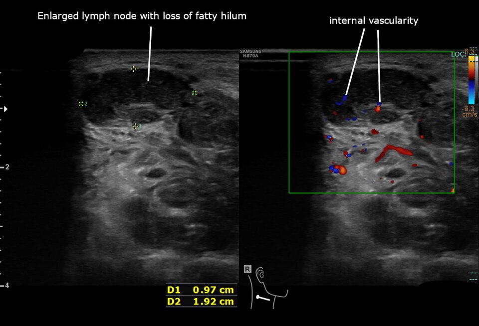
An enlarged neck lymph node on ultrasound, its fatty hilum lost. The left panel shows the node, a dark round mass with no bright central streak; the calipers and the figures, 0.97 by 1.92 cm, are the original’s measurements. The right panel lays color Doppler over it and marks the scattered internal vascularity, the flow of a turned node. Image: Cerevisae, CC BY-SA 4.0.
A lymph node is read by a handful of features. The shape comes first. A node that is long and thin, oval like a bean, reads as a calm node. The length runs more than twice the width. The probe turns the node in two planes to judge that shape. The hilum comes next. A healthy node carries a bright streak at its center, a wedge of fat where the vessels enter. That bright hilum is the surest mark of a friendly node. The eye looks for it on every node. The hilum can sit as a full bright wedge, or as a thin bright line, by the size of the node. A node that keeps its bright hilum is read with calm. The border comes after. A calm node has a smooth thin edge, sharp against the tissue around it. The flow comes last. Color Doppler laid on a calm node shows a few vessels running out from the hilum, branching like a small tree from the center. That tree of flow rises from the middle and fans outward. It is the flow of a node doing its work. These four marks, the oval shape, the bright hilum, the clean edge, the central flow, paint the picture of a reactive node, the kind that swells with a sore throat and settles in its own time. A reactive node can sit in a chain of its like, several small calm nodes in a row, all bean-shaped and bright-cored. Such a node needs no more than a note and a date to look again. The worry begins where these marks fade. A node that rounds up, its length falling toward its width, draws a second look. A node that loses its bright hilum, darkening through the center, draws a harder one. A node whose edge turns bulging, or whose flow springs from the rim in scattered streaks, draws hardest. Dead tissue inside a node, a dark pool that the sound passes through, draws hardest of all. The hilum sits at the heart of the read. A node that holds its bright hilum is friendly until something else speaks up. The needle is aimed at the node that has lost it. A node is judged on all its marks together, never on one alone. The whole skill of the neck scan is the reading of that bright central streak, and the company it keeps.
The sweep of the neck
The scan of the neck follows a route. The patient lies back with a pillow under the shoulders, the chin lifted and turned. The neck is stretched open. A high-frequency linear probe runs over it, around ten to fifteen megahertz. Plenty of gel and a light hand keep the picture clean.
The probe works through the neck by levels. It starts under the chin and runs back along the jaw. It follows the great vessels down the side of the neck. It sweeps the hollow above the collarbone. It crosses the midline over the thyroid. Each level is covered in turn, so no node is missed.
Every node of note is read in two planes. The probe lays the node out along its length, then turns across it. The shape, the hilum, the edge, and the flow are read on each. The node is measured across its short axis, the width that grows when a node turns round.
The far side of the neck is read to match. A node on one side is held against the nodes of the other. A known cancer steers the search. A scan for a throat cancer reads hardest at the levels that throat drains to.
Telling a node from its neighbors
A neck holds more than nodes. The scan first makes sure a lump is a node at all. A few neighbors can pass for one at a glance.
A vessel can look like a node in one plane. The probe turns across it. The round shape draws out into a long tube. Color Doppler fills it with flow. A pulse or a steady stream marks a vein or an artery, and the search for a node moves on.
A salivary gland sits where nodes sit. The submandibular gland fills the space under the jaw. The tail of the parotid hangs below the ear. Each reads as a soft even gland with its own duct. A gland is far larger than a node, and paler.
A cyst can sit among the nodes. A smooth black space low in the neck can be a branchial cyst. One at the midline that moves with a swallow can be a thyroglossal cyst. The clear fluid and the thin wall set a cyst apart from a solid node.
A thyroid nodule can bulge out and pass for a node. The probe traces it back to the gland. A lump that springs from the thyroid is read as a thyroid nodule, on its own track.
The reassuring node
A reactive node is the common find in any neck. It is the node that swells to fight an infection of the throat, the teeth, the ear, or the scalp. The scan comes to know its calm look well.
It keeps the shape of a bean. The length runs well past twice the width. It lies along the neck, with no urge to round up.
It carries a bright hilum. A streak of fat sits at its center, bright against the darker rind around it. The vessels of the node enter through that hilum. The bright streak is the badge of a node at ease.
Its flow rises from the hilum. Color Doppler shows a few vessels branching out from the center, like a tree from its trunk. The flow keeps to the middle and fans outward.
A reactive node can grow large. A throat infection swells it past a centimeter, with the bean shape, the bright hilum, and the central flow all held. Such a node is noted and left, with a date to look again if it stays up.
The node that worries
Some nodes wear a different look. The scan reads these for the signs of a cancer or a deep infection. Each sign is a step away from the calm reactive node.
A worrying node rounds up. Its length falls toward its width, so it sits as a ball, its bean shape gone. The rounding is one of the earliest signs the scan can catch.
It loses its bright hilum. The streak of fat at the center fades. The node turns dark through and through. A node gone wholly dark, with no bright center, is a node that calls for a needle.
Its edge can turn. A node whose edge bulges, or breaks out into the fat around it, points to a cancer pushing past its shell.
Dead tissue can show inside it. A pool that the sound passes through, dark and structureless, marks necrosis. Necrosis sits in a node taken by a throat cancer, or by tuberculosis. It is a sign that always calls for a closer look.
Bright specks can dot it. Tiny points of calcium scattered through a node point toward a cancer of the thyroid, carried there from the gland. The scan that finds them turns at once to the thyroid.
Size is read last
Size is the weakest guide of all. A node’s size follows the trouble that woke it. A sore throat can swell a friendly node large. The danger in a node shows in its shape, its hilum, and its flow, at any size.
The scan still measures every node of note. It records the short axis, the width across the node. A short axis past a centimeter is noted, more so high in the neck where nodes run a touch larger. The number feeds the record, so a node can be tracked over time.
Change over time carries weight. A node that grows from one scan to the next calls for a closer read, even when each single reading looks bland. A node that shrinks with a course of antibiotics shows its hand as a reactive node. The scan that follows a node reads its trend as much as its look.
The patient behind the node
A node is read with the patient in mind. The age, the time, and the feel of a lump steer the scan before the probe is laid on.
A tender node that came up fast, over days, points to a reactive node from an infection. A hard painless node that has grown over weeks points toward a cancer. A run of rubbery nodes in a young adult, with night sweats or weight loss, points toward a lymphoma. The story sets the scan looking for the look that fits, or the look that breaks the pattern.
The metastatic node
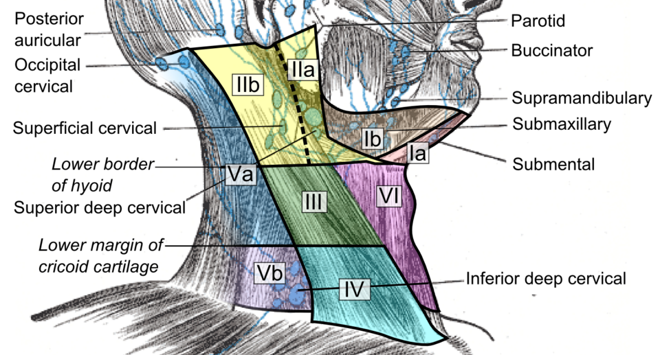
A schematic of the lymph node levels of the neck, colored from under the chin down to above the collarbone. The level of a node points back toward the patch it drains: the upper neck to the throat and tonsil, the low neck to the chest and the belly. This is a drawing, not an ultrasound scan. Diagram: public domain (CC0).
A cancer can travel to a neck node from a nearby home. The scan reads a metastatic node against the primary that fed it.
A node from a throat or mouth cancer runs dark. It rounds up. It can hollow out into necrosis, a dark pool of dead tissue at its core. It sits at the levels that drain the throat, along the great vessels of the neck. A node from a tonsil cancer of the kind tied to the wart virus can fill with fluid, a smooth cystic node that can pass for a simple cyst. A cystic node high in the neck of an adult is read with care, and sampled.
A node from a thyroid cancer carries its own mark. It can hold bright specks of calcium, or fill with fluid into a bright-walled cyst. A cystic or speck-flecked node low in the neck turns the scan to the thyroid gland.
A node low above the collarbone carries a longer reach. The lowest nodes of the neck drain the chest and the belly. A hard node there, on the left side above all, can carry a cancer from far below. It sends the search down into the body.
The node in lymphoma
Lymphoma fills the nodes in its own way. The scan learns its look apart from the reactive node and the metastatic one.
A lymphoma node runs near-black. It can read so empty of echoes that it mimics a pocket of fluid. The sound still meets tissue inside it. A careful eye reads the faint texture there.
It often holds its hilum early. A lymphoma node can keep a thread of its bright center as it swells and rounds. A growing, near-black node is sampled even when that thread remains.
Its flow runs rich. Color Doppler lights a lymphoma node up, with vessels at the center and around the rim both. The pairing of a near-black node and a brisk flow is a flag for lymphoma.
Lymphoma nodes gather in numbers. They stud a level, or several levels, as a chain of dark rounded nodes of a like size. A run of such nodes, in a young patient above all, sends the scan toward a core of tissue for the answer. A fine-needle sample can flag a lymphoma. The type of lymphoma needs the build of the whole node. A core of tissue, or a node taken out whole, gives the pathologist that build.
The matted nodes
Some nodes clump together as a mass. The scan reads the way nodes sit against their neighbors.
Tuberculosis mats the nodes of the neck. A run of nodes fuses into a clump, their edges lost against each other. Dark pools of necrosis sit within. The skin and fat around them swell. The planes turn hazy. A matted, necrotic clump, in the right patient, points to tuberculosis. A matted clump can soften to a cold abscess, a pool of fluid with no heat to it. It can track to the skin and open a sinus. These mark a long granulomatous disease.
A cancer can mat the nodes as well. A node that breaks through its shell joins to its neighbors and to the tissue around it. The matting of cancer marks a node that has spread past its capsule, a harder case for the surgeon.
The plane around a node carries the read. A node that drags a vessel as the probe pushes has grown beyond its capsule. The probe tests each suspect node for that grip on the tissue around it.
The vessels of a node
Color Doppler reads the vessels of a node. The pattern of the flow sorts the calm node from the turned one as well as any single sign.
A friendly node draws its flow from the hilum. The vessels enter at the center and branch outward, a neat tree from a single trunk. The flow that rises from the hilum is the flow of a working node.
A turned node draws its flow from the rim. New vessels crawl in through the capsule, scattered, with no neat center to them. Flow that springs from the edge of a node, or runs in a chaotic tangle, is the flow of a node gone wrong.
Lymphoma runs its own brisk flow, rich at the center and the rim both. The reading of the flow joins the shape and the hilum, never standing alone. A node is judged by the whole of its look, the vessels read as one part of it.
The levels of the neck
The neck is mapped in levels, and the level of a node carries a clue. Each level drains its own patch of the head and the body.
The nodes under the chin and jaw drain the mouth, the lips, and the front of the tongue. The nodes along the upper neck drain the throat, the tonsil, and the back of the tongue. The nodes low in the neck and above the collarbone drain the deepest, reaching the chest and the belly.
A node sends the search to the patch it guards. A hard upper-neck node turns the scan and the scope to the throat and tonsil. A speck-flecked lower node turns it to the thyroid. A firm node above the left collarbone turns it to the belly and the chest below. A hard node low on the left, at the root of the neck, carries an old name, the signal node of a cancer deep in the belly. A node there sends the search down the gut and into the chest.
The level also sets the size that counts. A node high in the neck runs a touch larger in health than one low down. The scan reads each level against its own normal.
Sampling the node
The scan finds the node that needs an answer, and guides the needle that gives it. A node with a turned look is sampled under the probe.
The needle is watched the whole way. The probe holds the node on the screen. The needle comes in from the side, its tip in view. The tip is set in the solid part of the node, clear of the vessels. A sample is drawn from the living rind, away from any dead center.
The kind of sample fits the question. A fine-needle sample names a cancer carried from the thyroid or the throat. A core of tissue, a thicker thread, serves a node that may hold a lymphoma, where the build of the node must be read. The scan places either with the tip in sight. A first sample can come back too thin to call. The scan brings the needle back to a fuller part of the node, or to a second node, for another pass. A sample read at the bedside, with the needle still in the room, shows whether enough has come up.
The node guides more than the needle. A node marked as the roundest, the darkest, its hilum gone, is the one the needle is aimed at. The scan picks the node, and the part of it, that gives the truest answer.
Past the probe’s reach
The neck scan has a clear reach. It reads the nodes within that reach. The rest it points toward another test.
The nodes it can reach lie near the skin. The deep nodes, behind the throat or down in the chest, lie out of the probe’s view. A search that needs those nodes turns to a CT or an MRI of the neck and the chest.
The look of a node guides the read. The final word comes from elsewhere. A turned look raises the odds of a cancer. The kind of cancer, and its certainty, come from the needle and the microscope.
The primary can hide from the scan. A node can carry a cancer whose home the neck scan never finds. A search for that hidden primary runs into the throat with a scope, and through the body with a scan. The neck node is the clue that opens the hunt.
The scan keeps its place after a cancer is treated. A neck once cleared of a cancer is swept again at each visit, for a node that has crept back. A new round node, or a node regrown at an old site, brings the needle back. The scan watches the neck at set times for years, for as long as the watching is needed.
The streak at the center
A neck node scan comes back to one bright streak. The hilum, that wedge of fat at a node’s center, is the sign the whole read leans on. A node that wears its bright hilum sits at ease. The read pauses on the node that has lost it. Find the hilum, and a neck full of nodes sorts itself.
The reading runs in a set order: the shape, then the hilum, then the edge, then the flow. Each feature is weighed on its own. Together they place a node on a line that runs from plainly reactive to plainly turned. The levels and the size fill in the rest.
Nearly all the nodes a scan meets in a neck are friendly. A bean shape, a bright hilum, a flow from the center: these mark a filter at work, owed no more than a date to look again. The few that have rounded and gone dark, the streak lost, are the ones drawn out for a needle. A finger feels a lump in the neck. The probe reads it for what it is, a node at work or a node that has turned.
Common questions about cervical lymph node ultrasound
What makes a neck node look reactive?
A bean shape, a bright fatty hilum at the center, and a flow that branches from it mark a reactive node. The node is swollen to fight an infection. The scan reads each node against that calm look and marks the ones that depart from it. It measures the node and notes its level in the neck. It guides a needle into any node that needs an answer.
How can ultrasound tell a reactive node from a cancer?
By the shape, the hilum, and the flow. A node that keeps a bright fatty hilum, in an oval shape, with a flow that branches from the center, reads as reactive. When a node turns, the bright streak goes. The node rounds up. It darkens through the center. Its flow scatters to the rim. The signs shift the odds. A needle gives the certain answer.
Does the size of a node matter?
Size matters least. A sore throat can swell a friendly node well past a centimeter. The shape, the hilum, and the flow carry the read at any size. A node that grows from one scan to the next calls for the closest look.
What does a hard lump above the collarbone mean?
A node low above the collarbone drains the deepest. The lowest neck nodes reach the chest and the belly. A firm node there, on the left side above all, can carry a cancer from an organ far below. A new hard node above the collarbone is read with that long reach in mind, and sent for tissue.
Why does a node turn round when it is cancerous?
A lymph node in health is twice as long as it is wide, a thin filter set along the neck. A cancer seeded inside it grows in every direction at once. The node swells from within and fills toward a ball. Its length closes on its width. The rounding is one of the earliest marks the scan reads.
Does a swollen neck node always mean cancer?
No. The common swollen node is a reactive one, fighting an infection of the throat, the teeth, or the ear. It keeps its bean shape, its bright hilum, and a flow from the center. It settles once the infection passes. The scan judges a node by its shape, its hilum, and its flow. A friendly look clears it, swelling and all.
About the Author
Julien Mercier
Senior R&D Engineer · Medical Ultrasound Transducer Development
Senior R&D Engineer with an M.S. in Applied Physics and over 15 years of experience in medical ultrasound transducer development, specializing in the design verification and performance testing of high-frequency imaging transducers. Currently leading the development and verification of the company’s next-generation high-frequency linear-array transducer, responsible for imaging performance evaluation and reliability analysis in preclinical testing. Brings extensive hands-on experience in piezoelectric element tuning, beamforming parameter optimization, and system-level performance testing.