Central Venous Catheter CVC Ultrasound Guided Insertion Handheld
Putting a central line into the internal jugular vein used to be a blind procedure, the needle aimed by feel and surface landmarks toward a vein sitting right next to the carotid artery. Ultrasound ended the guessing. The operator now watches the vein, the artery next to it and the needle tip on a screen as the line goes in, which is why the scan has all but replaced the old landmark method at the neck.
What a central line is and why blind placement is hard
A central line ends in a large central vein, usually the superior vena cava right above the heart. It carries treatments too strong or too long-running for a small arm vein. The internal jugular vein in the neck is the common route, a wide vein that runs down beside the carotid artery.
A central line is needed when a treatment would burn a small vein, when a patient cannot eat and must be fed into a vein, or when dialysis has to run through a large vessel. The internal jugular is one of three main sites, along with the subclavian vein under the collarbone and the femoral vein in the groin. Each has its place. The neck vein is the one ultrasound reads best.
The old way of placing the line was blind. The operator felt for landmarks, the pulse of the artery and the angle of the neck muscles, then pushed the needle toward where the vein should be. The method worked often enough, since the vein usually sits in a known spot. The trouble is that the vein does not always sit where it should.
The vein does not lie in the same place in everyone. Studies of neck anatomy find the internal jugular straight in front of and to the side of the carotid in only about a third of people. In the rest the vein is shifted out to the side, sitting straight in front, or overlapping the artery. A blind needle aimed at the textbook spot can miss the vein, or pass through it into the artery behind.
Why ultrasound replaced the blind method
Ultrasound changed central line placement by showing the vein directly. The operator sees the vein on the screen and watches the needle enter it, with no guessing from surface landmarks. The numbers behind the shift are large. The American College of Surgeons reports that more than five million central lines are placed each year in the United States, that the older methods carried a complication rate over 15 percent, and that up to a third of blind attempts failed to reach the vein on the first try. Mechanical harms, the punctured artery and the collapsed lung, accounted for up to a fifth of the complications. Trials that compared the two methods head to head show the difference. Studies of internal jugular lines report ultrasound reaching the vein in around 94 percent of patients, against about 79 percent for the landmark method. Reported complication rates fall the same way, from around 17 percent by the landmark method to under 5 percent with ultrasound. A review pooling many trials found that ultrasound cut serious complications by more than half and cut failed attempts by a wide margin. This depends on doing it live. Marking the vein and then setting the probe down to push the needle blind, the static method, helps far less than watching the needle go in under the beam. Real-time guidance is what the evidence points to. The scan also shortens the time to reach the vein and cuts the number of needle passes. Each pass avoided is one less chance to strike something it should not. The gain is largest in the patients hardest to stick, those with short necks, scarring from old lines, or veins collapsed by low blood volume. Where the landmarks give nothing to feel, the scan still finds the vein. The picture holds across the studies. Guidance is strongest at the internal jugular vein, where the evidence is clearest and the guidelines now name ultrasound the standard. The benefit is smaller and less certain at the other sites, the subclavian and the femoral veins, where the anatomy is harder to image. None of this makes the scan a guarantee. A vein can still be missed and an artery still hit, since the operator has to read the screen right and keep the needle in view. What ultrasound removes is the guesswork of aiming at a spot that may not hold the vein. The needle goes in under sight, no longer by feel.
Setting up before the stick
A central line goes in under full sterile cover, since the catheter stays in a deep vein for days. The operator gowns and gloves. The probe is wrapped in a sterile sleeve with gel so it can sit in the field without contaminating it. This setup is the same with or without ultrasound, the probe cover being the one addition.
Position helps the vein. Tipping the patient head-down, the Trendelenburg position, fills the internal jugular and widens it, which makes a larger target and lowers the chance of drawing air into the vein. Turning the head slightly away opens the neck. Too much turn can roll the vein over the artery.
Telling the vein from the artery
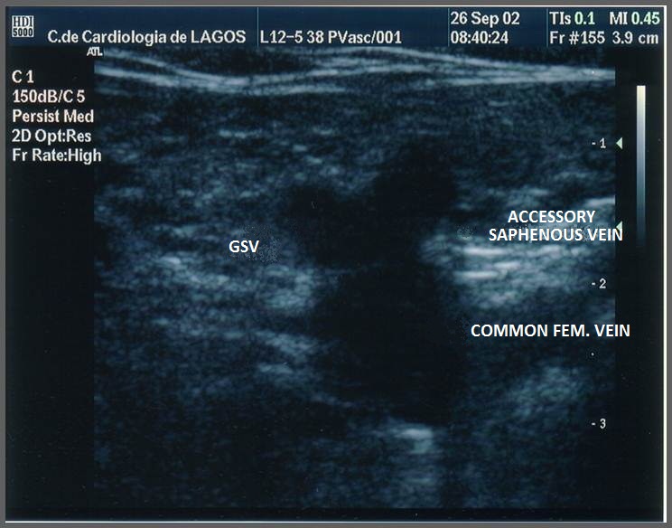
A transverse vascular scan, here at the groin, with a vein and an artery side by side. The neck looks much the same. The vein is the one that squashes flat under the probe.
The first job under the probe is to tell the vein from the artery, since they lie side by side and the line must go into the vein. Four signs separate them. The vein is squashed flat by gentle pressure from the probe. The artery, thick-walled and under high pressure, holds its round shape. The artery pulses with the heartbeat. The vein lies still. Color Doppler shows the brisk flow of the artery and the slower flow of the vein. A breath-hold and bear-down, the Valsalva, swells the vein and leaves the artery unchanged.
Pressure is the quickest test. It is the one used first. A vein gives way under the probe and disappears, then springs back when the pressure lifts. An artery barely changes. The operator presses lightly through the scan, since heavy pressure flattens the vein out of sight and hides the target.
The feel under the probe settles any doubt. A vein widens as the patient breathes out and narrows breathing in, a sway an artery does not show.
Getting this wrong is the error to fear. A catheter that ends up in the carotid artery can cause a stroke or a heavy bleed. The compression test takes a second and settles the question, which is why it is done before the needle goes near the skin.
Finding the internal jugular vein
The scan starts with the probe across the neck, low down near the collarbone. The vein and the artery show as two dark circles in cross section, the vein usually the larger and the one that flattens under pressure. The operator slides the probe up and down the neck to find the level where the vein is widest and sits clear of the artery.
The level that works best is where the vein sits beside the artery, clear of the spot directly above it. A vein lying straight over the artery is a hazard, since a needle through the back wall of the vein lands in the artery beneath. Sliding and angling the probe often finds a point where the two move apart.
Depth matters as well. The internal jugular runs shallow, a centimeter or two under the skin, well within reach of a high-frequency probe. The operator notes the depth and the angle before the needle goes in, so the path is set in advance.
The vein can be traced down the neck to where it joins the subclavian vein behind the collarbone. That low point sits close to the lung, so the stick is usually made higher up, where the vein is clear of the lung and easy to compress.
Guiding the needle into the vein
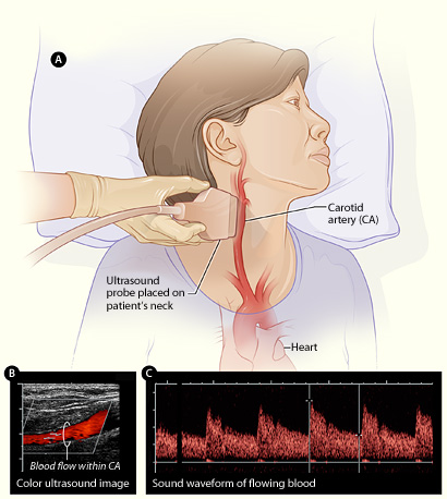
An illustration of scanning the neck. The probe sits over the carotid artery and the internal jugular vein beside it, the view used to guide the needle into the vein.
With the vein found, the needle advances under live view. The operator keeps the vein centered on the display and moves the needle toward it, watching for the tip to reach and enter the vein. The exact way the needle is lined up with the probe, along the beam or across it, is a technique of its own, with its own pages. What matters here is that the needle is watched all the way, never pushed blind.
The sign that the needle is in the vein is a flash of dark blood drawn back into the syringe, with the tip seen inside the vein at the same moment. The two together, the blood and the picture, confirm the vein before anything more is done. A flash of bright pulsing blood means the artery. The needle comes straight back out.
Placing the catheter
Once the needle is in the vein, the catheter goes in over a guidewire, a method called the Seldinger technique. A thin wire is threaded through the needle into the vein. The needle comes off, leaving the wire in place. The catheter, sometimes after a dilator widens the track, slides down the wire into the vein. The wire then comes out, leaving the catheter behind.
Ultrasound has a part to play past the first stick. The probe can check that the guidewire sits inside the vein before the catheter goes over it, catching a wire that has strayed into the artery while it can still be pulled. It can scan the vein along its length to confirm the wire runs the right way, up the neck toward the heart.
The wire goes in only so far.
A wire pushed deep into the heart can set off a run of irregular beats, so the length fed in is watched and held short of that. The wire is drawn back if the rhythm stirs.
The steps after the wire are done by feel and by the marks on the catheter, since the depth to the vena cava is known from the patient’s size. The scan has done its main work by the time the wire is in. It got the needle into the right vessel, which is the step where central line trouble usually starts.
The complications ultrasound helps avoid
The harms ultrasound guards against are the ones that come from a misplaced needle. The worst at the internal jugular is a punctured lung, a pneumothorax, when the needle goes too deep and too low and catches the lung below the neck. Seeing the depth on the screen keeps the needle short of it.
Hitting the carotid artery is the other main harm. A pass through the vein into the artery, or a needle aimed wrong from the start, can tear the artery and bleed. Guidance lowers this by showing the artery to keep clear of and the vein to enter. It also catches a wire that has gone into the artery before the large catheter follows.
Infection and clot come later, past the placement. Ultrasound does less for these. A clean first stick with fewer passes still lowers the chance of both. The main value of the scan is a safe entry. That first step is where the blind method did its harm.
Air drawn into the vein is one more harm to guard against. A breath at the wrong moment can pull air through the needle or the open catheter into the vein, an air embolism. The head-down position and care with the open hub lower the risk. The scan helps here only by way of the faster, cleaner placement it allows.
Confirming the line on a handheld unit
After the line is in, ultrasound can help confirm it. A quick scan of the chest looks for the sliding sign that rules out a pneumothorax, faster than waiting for an X-ray. On a handheld unit, the same probe that guided the stick does this check at the bedside, the placement and its confirmation done without leaving the room.
Common questions about ultrasound-guided central lines
What is an ultrasound-guided central venous catheter?
It is a central line placed while the operator watches the vein and the needle on an ultrasound screen. The tube ends in a large vein near the heart and carries treatments too strong for a small arm vein. The scan shows the vein directly, with no need for surface landmarks.
Why is ultrasound better than the landmark method for central lines?
It shows the vein, which does not always sit where the landmarks predict. Studies report ultrasound reaching the internal jugular vein in around 94 percent of patients against about 79 percent for the blind method, with complication rates falling from around 17 percent to under 5 percent.
How does ultrasound tell a vein from an artery?
Four signs separate them. The vein flattens under gentle probe pressure. The artery holds its shape. The artery pulses and the vein does not. Color Doppler shows the flow in each. A Valsalva breath-hold swells the vein.
Which vein is used for an ultrasound-guided central line?
The internal jugular vein in the neck is the usual choice, with the strongest evidence for ultrasound guidance. It runs shallow beside the carotid artery. The subclavian and femoral veins are also used, with the benefit of ultrasound less certain there.
What complications does ultrasound guidance help avoid?
It lowers the chance of a punctured lung by showing the depth, and the chance of a carotid artery injury by showing the artery to avoid and the vein to enter. It can also catch a guidewire that has strayed into the artery before the catheter follows.
Can a handheld ultrasound place a central line?
Yes. A handheld probe shows the vein, guides the needle in real time, and can check the chest for a pneumothorax afterward. The placement and its confirmation can be done at the bedside with one portable unit.
About the Author
Julien Mercier
Senior R&D Engineer · Medical Ultrasound Transducer Development
Senior R&D Engineer with an M.S. in Applied Physics and over 15 years of experience in medical ultrasound transducer development, specializing in the design verification and performance testing of high-frequency imaging transducers. Currently leading the development and verification of the company’s next-generation high-frequency linear-array transducer, responsible for imaging performance evaluation and reliability analysis in preclinical testing. Brings extensive hands-on experience in piezoelectric element tuning, beamforming parameter optimization, and system-level performance testing.