A pinched nerve at the wrist can be seen and measured. The median nerve swells where it is squeezed in carpal tunnel syndrome. Ultrasound shows that swollen nerve. It puts a number on the swelling. That number makes the diagnosis from a picture. The scan reaches the cause as well, the thickened lining or the lump crowding the nerve. A nerve conduction test never sees that. A handheld probe lays the nerve on the screen at the bedside, in the minute the patient holds the wrist out.
The carpal tunnel and the median nerve
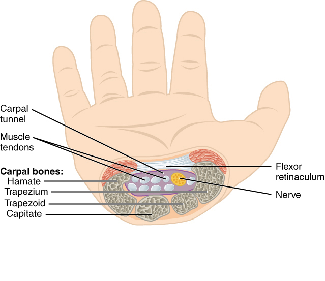
A cross-section through the wrist. The carpal tunnel is the space under the flexor retinaculum, floored and walled by the carpal bones. The median nerve, in yellow, lies at the top of the tunnel among the flexor tendons. This is the crowd the nerve is squeezed in. Diagram: OpenStax, CC BY 3.0.
The carpal tunnel is a short passage at the front of the wrist. Its floor and walls are the small bones of the wrist, curved into an arch. A tough band, the flexor retinaculum, roofs the arch and closes the tunnel over.
Through the tunnel run the tendons that bend the fingers and thumb, nine in all, packed close. The median nerve runs with them, the one soft nerve among the firm tendons. It lies just under the roof of the tunnel, close to the skin, where the probe finds it without trouble.
The median nerve carries feeling from the thumb, the index, the middle, and half the ring finger. It also works the muscles at the base of the thumb. Squeezed in the tunnel, it gives the numbness, tingling, and weakness of carpal tunnel syndrome. Just past the tunnel the nerve sends a small branch to the thumb muscles, the branch whose loss wastes the mound at the base of the thumb. The scan reads the main trunk at the wrist, where the pressure falls.
What carpal tunnel syndrome is
Pressure in the tunnel falls on the median nerve. The tendons and their linings swell, the tunnel has no room to give, and the nerve takes the squeeze. Numbness and tingling follow in the fingers the nerve serves.
The trouble wakes the patient at night, the hand numb and shaken to bring it back. It bites at fine work, at holding a phone, at gripping a wheel. The thumb, index, and middle fingers carry the worst of it. The little finger, served by another nerve, stays clear.
Left long enough, the squeeze wastes the muscle at the base of the thumb and dulls the feeling for good. Grip weakens. Small objects slip from the hand. These late signs mark a nerve pressed for months or years. By then the nerve and the muscle may not come fully back, even after the pressure is lifted. An early scan is what spares the patient that lasting loss.
The story and the examination point to the tunnel. The scan confirms it, measures how hard the nerve is pressed, and looks for what is doing the pressing. A diagnosis suspected at the bedside is settled at the bedside.
Some wrists are squeezed for a reason. Pregnancy, an underactive thyroid, diabetes, and rheumatoid disease all swell the tunnel’s contents. Heavy, repeated use of the hands plays a part. Often the tunnel is built tight from the start, the cause left unnamed.
Two bedside tests raise the suspicion. Tapping over the nerve at the wrist can shoot a tingle into the fingers. Holding the wrist bent for a minute can bring the numbness on. Neither test is sure on its own. The scan turns the suspicion into a measured finding.
Scanning the wrist
The patient sits with the forearm up, the palm open, the wrist resting flat. The probe lies across the front of the wrist, in short axis, at the crease where the hand meets the arm. This cut shows the tunnel end-on, the nerve and the tendons in cross section.
The median nerve shows as a cluster of fine dark dots in a brighter frame, a honeycomb of nerve fascicles. It sits just under the bright band of the retinaculum, above the tendons. Two bony landmarks fix the level: the pisiform on the little-finger side, the scaphoid on the thumb side. The nerve is read where these bones mark the tunnel’s mouth.
The probe is kept square to the nerve. Tilted off the right angle, the nerve darkens and can fade into the tissue around it, the same artifact that troubles any tendon scan. A square beam holds the honeycomb bright and its border sharp for the measurement to come.
The nerve is followed in the long view as well, the probe turned along its length. In this plane the nerve runs as a striped band. The step where it swells at the tunnel mouth shows as a bulge along its course. The short view takes the measurement. The long view maps the swelling up and down the nerve.
Measuring the nerve
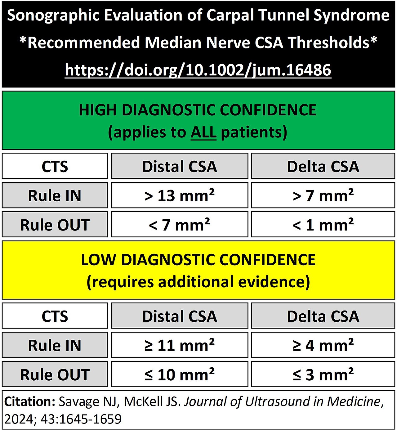
Published cutoffs for the median nerve’s area at the wrist, from a 2024 review. These are a table of numbers, not an ultrasound image. They show how the threshold shifts: a high-confidence rule-in sits well above the lower figures, around which a normal nerve and a pinched one can overlap. The delta is the wrist-minus-forearm difference. Table: DrNathanJSavage, CC0, after Savage and McKell, J Ultrasound Med 2024.
The size of the median nerve is the number the scan is built to find. A healthy nerve is slim. A nerve squeezed in the tunnel swells above the squeeze, where the press dams it like water behind a wall. That swelling is what the scan measures. The measure is the cross-sectional area, the area of the nerve seen end-on. The operator traces the border of the nerve on the screen, just inside its bright rim, and the machine returns the area enclosed. The bright rim the trace follows is the nerve’s own sheath. The area inside it is what swells under pressure. The rim is followed close. A trace that runs wide of it or short of it shifts the number. The median nerve is small enough that a millimeter of slack on the border counts. The trace is made at the mouth of the tunnel, at the level of the pisiform bone, where the swelling is greatest. A nerve area above about ten square millimeters marks a nerve under pressure, the figure that carries the diagnosis. The cutoff is read as a guide. A normal nerve and a pressed one can sit close to it, and the threshold itself moves between studies, scanners, and patients, so the number is weighed alongside the symptoms and the rest of the scan. The trace is repeated to be sure, the area read two or three times and averaged, since a loose or tilted border throws the number off. A bright, square image and a careful trace are what make the measurement reliable. The swelling has a plain cause. Pressure stops the nerve draining, fluid backs up inside it, and the fascicles thicken, so the nerve reads both larger and darker than a healthy one. The swelling sits at the inlet and eases higher up the forearm, a step in size the scan can follow along the nerve. The scan catches the change as a number a clinic can track. A nerve measured today can be measured again after a splint, an injection, or an operation, the area watched as it settles toward normal over the months that follow. A nerve conduction test reports that the nerve conducts slowly. The scan reports how swollen it is, in plain millimeters a patient can be shown on the screen. That number, traced at the mouth of the tunnel, is the heart of an ultrasound for carpal tunnel syndrome.
The wrist and the forearm
One number on its own can mislead. Some people carry a fat nerve, some a thin one, and a single area can read high or low on the person’s own build alone. A second measure steadies the first.
The nerve is measured a second time higher up, in the forearm, away from the tunnel. There the nerve sits at its normal size, untouched by the squeeze. The area at the wrist is set against the area in the forearm. The wrist figure divided by the forearm figure gives a ratio.
A ratio above about one and a half points to a nerve swollen at the wrist beyond its own normal. The ratio measures the nerve against itself, so it holds steady where a single area can mislead. A swollen wrist nerve on a person with naturally large nerves still shows in the ratio.
Grading the squeeze
The size of the swelling grades the squeeze. A nerve a little over the line, with mild symptoms, marks an early, lighter case. A nerve well above it, swollen and flattened, with night waking and a weak grip, marks a heavier one. The number puts the case on a scale a clinic can read.
The grade steers the treatment. A light case is held with a splint worn at night and a change in how the hand is used. A heavy case, with a fat nerve and wasting muscle, points sooner to an injection or to releasing the tunnel. The scan puts a measure on the grade.
The grade rests on more than the one number. A nerve heavy with Doppler flow, flattened hard in the tunnel, and swollen well past the cutoff all lean the same way. The signs gathered together place the squeeze more surely than any one of them alone.
The nerve and the tendons
The median nerve sits among the flexor tendons. A beginner can take one for the other. The nerve and the tendons look much alike at a glance, both oval and pale. The scan tells them apart by pattern and by movement.
By pattern, the nerve is a honeycomb, a mesh of fine dark fascicles in a bright frame. A tendon is a tight weave of bright parallel lines, more uniform and more sharply bright. The honeycomb of the nerve, once seen, is hard to miss.
By movement, the test is to ask the fingers to bend. The tendons slide up and down under the probe as the fingers curl and straighten. The nerve barely shifts. This glide marks the tendons and leaves the nerve standing out as the one structure that holds still.
Anisotropy helps in the telling. The tightly woven tendon dims sharply when the beam tips off square. The nerve dims far less. Rocking the probe makes a tendon flash dark and bright, the nerve holding steadier through the tilt. The difference is one more way to name the nerve among its neighbors.
How the nerve moves
A healthy median nerve does not sit still in the tunnel. It glides up and down a little as the wrist and fingers bend. It shifts to the side to make room as the tendons crowd past it. The nerve slips and slides through its tunnel as the hand goes about its work.
A pressed nerve loses that freedom. Swollen and bound in the crowded tunnel, it moves less than it should, tethered where it once slid. The scan follows the nerve as the patient opens and closes the hand and watches how far it travels. A nerve that hardly stirs adds to the case for a tight tunnel.
The movement is read in the same short-axis view, the nerve watched as the fingers curl and straighten. A nerve that slides freely and flattens with ease points away from a squeeze. A nerve held fast and stubbornly round points toward one. The glide is one more thread in the picture the scan builds, read together with the size, the shape, and the flow.
What the scan shows that nerve tests cannot
A nerve conduction test measures how well the nerve carries its signal. It reports a nerve slowed by pressure. It says nothing about what is doing the pressing. The scan answers that second question, the one a test of conduction leaves open.
Often no single cause stands out. The nerve is crowded by a tunnel that runs tight, and nothing more. Some wrists hold a reason the scan can find. The lining around the tendons can thicken and swell, a tenosynovitis that fills the tunnel and leaves the nerve no room.
A lump can crowd the nerve, a ganglion cyst from a wrist joint or a tendon sheath, round and fluid-filled on the screen. A swollen tendon, a build-up of deposit, an extra muscle slipping into the tunnel, each can take the space the nerve needs. The scan names the intruder.
The cause can also lie in the body at large. The tunnel lining swells in rheumatoid disease and in long-standing diabetes. In a patient on dialysis for many years a protein builds up in the soft tissue and presses the nerve. A wrist held in a healing fracture can narrow the tunnel for good. The scan reads the crowded tunnel the same way each time. The finding points back to the illness behind it.
Finding the cause changes the treatment. A tight tunnel with a swollen nerve is treated as the common syndrome, with a splint, an injection, or a release. A cyst or a mass calls for its own removal. The scan sorts the wrist that needs surgery on the nerve from the wrist that needs something else taken out. Naming a cyst or a mass spares the patient a tunnel release that would never have touched the real trouble.
Variants and traps
Two variants catch the unwary. The median nerve sometimes splits into two bundles, a bifid nerve. Each bundle measured alone reads small. The two areas are added to give the nerve’s true size. A bifid nerve passed off as a single small one hides a real swelling. An anomalous muscle is a third surprise, a belly of muscle reaching into the tunnel where the scan expects only tendon and nerve, taking room the nerve can ill spare.
An artery sometimes runs in the tunnel with the nerve, left over from the growing hand, a persistent median artery. On the gray picture it can pass for a third bundle of nerve. Color Doppler settles it at once, the artery lit with the flow of blood the nerve has none of. A touch of Doppler keeps the count of nerve and vessel straight.
Flattening and blood flow
The swelling at the mouth of the tunnel is the main sign. Two more back it up.
Deeper in the tunnel the squeezed nerve is pressed flat, wider than it is tall. A nerve that swells at the inlet and flattens within fits the pattern of the syndrome. The bulge of the nerve against the underside of the retinaculum adds to it, the roof of the tunnel pushed up over the swelling.
The third sign is in the blood. Color Doppler lights a pressed nerve with vessels a healthy nerve does not show, the inflamed nerve running with new flow. A heavier flow leans toward a tighter squeeze. The link is a loose one. The flow adds to the other signs.
The signs read together. A nerve swollen at the inlet, flattened in the tunnel, and lit with Doppler is a nerve under real pressure. One sign alone can be borderline. The three together settle it.
Ultrasound and the nerve test
The nerve conduction test has long been the reference for carpal tunnel syndrome. It passes a small current and times the nerve’s reply, a direct measure of how well the nerve still works. It reads the function of the nerve.
Ultrasound reads the nerve’s shape. It measures the swelling, sees the flattening, and finds the cause, none of which the conduction test reaches. The two tests answer different questions, one how well the nerve works, the other what has happened to it and why.
The scan brings real strengths to the bedside. It is quick and painless. The conduction test takes longer and gives a sting. The scan shows the patient the swollen nerve on the screen. It finds the cyst or the swollen lining a current test would miss. It asks for no special room and costs little to run. A patient who dreads the needle test, or who cannot sit through it, is met by a scan that hurts not at all and answers in a minute.
The two work best in step. A scan can screen a wrist, confirm a clear case, and point to a cause, with the conduction test held for the doubtful or the severe. Many clinics now reach for the probe first and the needle test second. The nerve is both seen and measured before it is wired.
After the release
Surgery for carpal tunnel syndrome cuts the band that roofs the tunnel, the flexor retinaculum, and lets the walls spread and the nerve breathe. Ultrasound checks the work. It looks for the cut ends of the band, parted where the blade passed, and for a nerve that has eased back toward its normal size.
The scan also reads a release that has fallen short. A band left part-cut still roofs the tunnel and still presses the nerve. New scar can bind the nerve in its old bed. A nerve that stays swollen, or symptoms that come back after a quiet spell, send the wrist back under the probe to find the reason.
The measured area gives the before and the after a single scale. A nerve fat at the inlet before the operation, read slimmer in the weeks after it, shows a tunnel truly opened. The same number that made the diagnosis follows the cure. A wrist that fails to improve declares itself in a figure that will not fall.
Carpal tunnel ultrasound on a handheld unit
The wrist suits a handheld scan. The median nerve sits a few millimeters under the skin, in easy reach of a linear probe. The patient rests the forearm on a table, palm up, and the tunnel comes straight onto the screen.
This brings the test to the clinic room, with no trip to a separate department. The hand that complains is scanned the moment it is held out. The other wrist is a turn of the arm away, its nerve measured for a healthy comparison on the same patient. Both hands are often scanned at one sitting, since the squeeze rarely keeps to a single wrist. A follow-up scan costs only the minutes it takes, with no booking and no separate visit.
The handheld probe also guides the needle. An injection of steroid into the tunnel works better placed under sight, the drug laid beside the nerve and clear of it. The same scan that finds the swollen nerve steers the treatment into the right space.
What the small unit asks for is a steady hand and a square beam: the nerve found at the tunnel mouth, traced clean for its area, and checked against the forearm. A handheld probe used this way measures a pressed nerve at the bedside, the number a department machine would give read on the spot.
Common questions about carpal tunnel ultrasound
How does ultrasound diagnose carpal tunnel syndrome?
It measures the cross-sectional area of the median nerve at the mouth of the carpal tunnel, where a pressed nerve swells. An area above about ten square millimeters points to carpal tunnel syndrome. The scan also looks at the wrist-to-forearm ratio and at any cause crowding the nerve.
What is the normal size of the median nerve?
A healthy median nerve at the wrist measures below about ten square millimeters in cross section. A nerve swollen above that, especially with a wrist-to-forearm ratio over about one and a half, points to a nerve under pressure. The cutoff is a guide, read alongside the symptoms.
How is the median nerve told from the tendons?
The nerve has a honeycomb pattern of fine fascicles. The tendons are a tighter, brighter weave. On asking the fingers to bend, the tendons glide up and down under the probe, the nerve barely moving with them. The still, honeycomb structure is the nerve.
What can ultrasound show that a nerve conduction test cannot?
It shows the cause. A nerve conduction test reports that the nerve is slowed. It does not report why. Ultrasound finds a swollen tendon lining, a ganglion cyst, a mass, or an anomalous muscle crowding the tunnel, and it separates these from the common tight-tunnel syndrome.
Does ultrasound replace the nerve conduction test?
No. The two answer different questions. The conduction test measures how well the nerve works; ultrasound measures its shape and finds the cause. A scan can screen and confirm and point to a cause, with the conduction test kept for doubtful or severe cases.
Can a handheld ultrasound scan for carpal tunnel syndrome?
Yes. The median nerve is shallow and within easy reach of a linear probe. The patient rests the forearm on a table, palm up, and the wrist is scanned in a minute. The handheld probe also guides a steroid injection into the tunnel under sight.
About the Author
Julien Mercier
Senior R&D Engineer · Medical Ultrasound Transducer Development
Senior R&D Engineer with an M.S. in Applied Physics and over 15 years of experience in medical ultrasound transducer development, specializing in the design verification and performance testing of high-frequency imaging transducers. Currently leading the development and verification of the company’s next-generation high-frequency linear-array transducer, responsible for imaging performance evaluation and reliability analysis in preclinical testing. Brings extensive hands-on experience in piezoelectric element tuning, beamforming parameter optimization, and system-level performance testing.