Abdominal Ultrasound AIUM Practice Parameter Handheld Convex Probe
The AIUM Practice Parameter for abdominal ultrasound is a published standard that sets out what a complete scan of the abdomen must cover, how it should be recorded, and who should carry it out. Without a standard of this kind, the same request at two clinics can come back as two quite different studies. Published by the American Institute of Ultrasound in Medicine, it is the document that makes one abdominal scan mean the same as the next.
What sends a patient for the scan
A scan is ordered for a reason. The parameter begins there, with the complaints and findings that justify a look. Pain in the belly or the flank heads the list. A mass felt under the hand, an organ thought to be enlarged, a set of liver or kidney blood tests gone abnormal, a yellow tinge of jaundice, a suspicion of fluid in the belly, the follow-up of a lesion already found: each is a reason the parameter recognises. The clinical question that rides on the request is meant to shape the study that answers it. A good request states that question plainly. A request that asks only for “an ultrasound” leaves the operator to guess. A guessed-at question is the start of an incomplete answer.
The list the word “complete” stands for
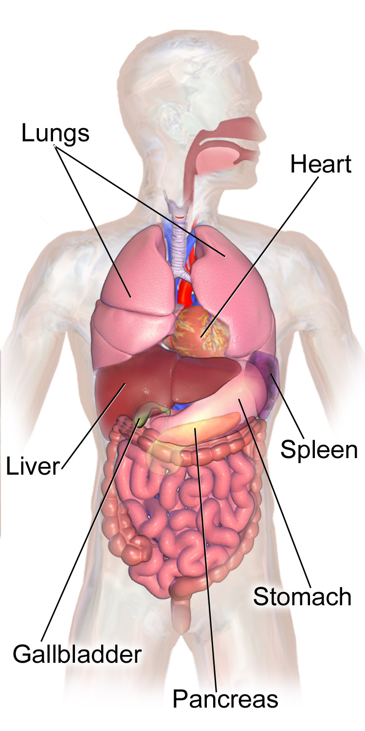
The organs of the trunk, among them the abdominal ones the standard names: the liver and the gallbladder, the spleen, the stomach, and the pancreas. The kidneys and the great vessels lie deeper, against the back wall, and join the same list. The lungs and the heart, above the diaphragm, fall outside the abdominal exam. Illustration: BruceBlaus, CC BY-SA 4.0.
At the centre of the parameter sits a list of organs. A study earns the word “complete”, as the parameter defines it, only when it has looked at the liver, the gallbladder with its bile ducts, the pancreas, the spleen, both kidneys, and the great vessels along the back wall, the aorta and the inferior vena cava. The bladder and the bowel are brought in where the clinical question reaches them.
The point of fixing the list is plainness. A report that says “abdominal ultrasound” carries the promise of that whole list behind it. Leave the spleen unmentioned, or the pancreas unsought, and the study has fallen short of what its own name claims. The list draws the line between a complete study and a partial one wearing a complete study’s name.
Reading the patient before the probe
A good study starts before any gel is laid down. The parameter notes the preparation that gives the cleanest pictures, because the cleanest pictures are where the standard is easiest to meet.
For the upper abdomen, an empty stomach is the first preparation. A fast of several hours fills the gallbladder, so its wall and its stones stand clear, and it settles the bowel gas that would otherwise blur the pancreas and the deep vessels. Where the bladder is part of the question, a full bladder gives the window the lower abdomen needs.
The patient is positioned and asked to breathe to order. A turn onto one side brings an organ out from behind the ribs; a held breath drops the liver and the spleen into reach. None of this is incidental to the standard. A study that fights poor preparation is more likely to leave an organ half seen. The parameter’s whole aim is the organ seen whole.
The standard meets the patient who cannot prepare. An emergency leaves no time to fast; a frail patient cannot turn or hold a breath. The parameter bends to these, asking the operator to win what views the conditions allow and to record what they did not. Good preparation makes the standard easy to meet. Without it, the same standard is harder won.
Two planes and a measurement
The parameter does not stop at naming the organs. It says, for each, how thoroughly it must be seen. The rule that runs through it is plain. An organ is read in two planes. The sizes that carry meaning are measured and recorded. A single view can deceive: a structure cut one way can hide what a cut across it reveals, and only the pair tells the truth.
The liver is swept in its long and its short axis, both lobes brought in, its texture judged and its span noted where enlargement is in question. The spleen is laid out along its length and measured, the figure being the firmest sign of a swollen spleen. Each kidney is seen in two planes, its length taken, its cortex compared with the liver or the spleen beside it.
The gallbladder is read in two planes as well. The parameter asks for it in more than one position of the patient, so a stone is rolled and caught. Its wall is assessed. The common bile duct is measured where it crosses the portal vein.
The pancreas is followed from head to tail as far as the overlying gas allows, its duct noted. The aorta is seen in length and in cross section, its diameter measured against the threshold for an aneurysm. The vena cava is checked alongside it. The bowel, where the question reaches it, is surveyed for a thickened loop or a mass, with gas keeping much of it from the probe.
None of this is left to the eye alone. A measurement saved with the image turns a soft impression into a number a later reader can hold the next study against. The insistence on two planes and a figure is what lets two scans, months apart, be compared.
The back wall and its organs
The parameter’s title pairs the abdomen with the retroperitoneum, the compartment along the back wall, and the pairing is no accident. The organs that lie there round out the complete study.
The kidneys are the chief of them. Each is measured for length and read for the thickness and brightness of its cortex, the rind that grows thin in an ageing or scarred kidney. The collecting system is checked for the dark dilatation that means a blockage below. A stone, a cyst, a mass, a swelling: each leaves its mark on the renal picture.
The aorta runs down the midline in front of the spine. The parameter asks for it in two planes, measured across, because a widening past the threshold is an aneurysm that changes a patient’s care at once. The vena cava lies beside it, soft and flat, its calibre a rough gauge of the pressure in the right heart.
The adrenal areas, above the kidneys, are looked at where the question calls for them, even if a normal adrenal is often too small to see. The point of the retroperitoneal sweep is the same as the rest: the compartment is covered, so nothing in it is missed for never having been sought. The retroperitoneum is easy to slight, lying deep and behind the gut, and the parameter names it for that reason. What is easy to skip is what a standard must insist on.
Complete, or limited
Not every scan needs the whole list. The parameter says so plainly. It marks out a second kind of study, the limited or focused examination, aimed at a single question. A right-upper-quadrant scan for gallstones, a check of the kidneys for obstruction, a look at a known liver lesion to see whether it has grown: each is a limited study by design, complete for its purpose, with no need to sweep the whole abdomen.
The parameter asks that a limited study declare itself as such. A report should say plainly that the scan was focused, so no reader mistakes a narrow look for a full one. The danger the parameter guards against is the silent gap, the organ left unscanned and unmentioned, read later as if it had been cleared.
The choice between complete and limited follows the clinical question. A first presentation of vague belly pain calls for the whole list. A follow-up of one known cyst calls for that cyst and little more. The parameter leaves room for both, on the condition that the study names which one it is.
The machine, and the care it asks
The parameter is plain about the tools. An abdominal study calls for a real-time scanner and a curved or sector probe, run at the highest frequency that still reaches the depth. For adults that usually falls between two and five megahertz, the range that reaches the back wall and still holds detail on the way.
The frequency is set as high as the patient’s depth allows, lower in a large patient, higher in a slim one. The skilled operator changes the setting to the body in front of the probe, never holding to a single one. The right picture comes from the hand that adjusts, not the dial left where it sat.
Safety rides alongside. Ultrasound carries no ionising radiation, and its risks are small. The parameter still holds to the principle of keeping exposure as low as reasonably achievable. The power is kept no higher than the study needs, and the probe dwells no longer than the picture asks. A standard that cares about quality cares about this too.
Cleaning belongs to the care as well. A probe that touches one patient is wiped down before it touches the next, by the same standard whatever its size. A pocket scanner is cleaned like any other. The small machine carries the same duty of a clean probe and a steady screen as the cart it might replace.
What the images have to prove
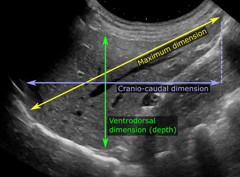
A liver measured on ultrasound. The colored arrows and labels were added to the image to mark the dimensions a complete study records, the long axis and the depth. A figure like this, saved with the picture it was taken from, is what lets a later scan be compared with this one. Image: Mikael Haggstrom, CC0.
A scan that is not recorded did not, for the record, happen. The parameter ties its organ list to a documentation standard: each organ named on the list leaves a representative image behind, stored and labelled. The pictures are the proof that the sweep was made.
The labelling matters as much as the picture. An image marked with the organ and the side, the plane and the orientation, can be read by someone who was not in the room. An unlabelled image proves little, since no later reader can be sure what it shows.
Measurements are saved alongside the pictures. The liver span, the spleen length, the bile duct, the kidney length, the aortic diameter: where the parameter calls for a figure, the figure is stored with the image it was taken from. A number without its picture, or a picture without its number, leaves the next reader half-equipped.
The aim of all this is a study another clinician can trust without repeating. A complete, labelled, measured record lets a scan done once be read, compared, and acted on by people who never met the patient. The documentation is what turns a private look into a shared medical fact. A study with gaps in its record is read with caution, since what was not captured cannot be checked.
The kept record answers to more than the next scan. It is the account a later question is settled by, when a finding is disputed or a change is sought. A study held only in the operator’s memory cannot do this. The parameter asks for a record that stands on its own, read the same by anyone who opens it.
Who may hold the probe
The parameter speaks to people as well as pictures. A scan of this kind is meant to be performed by a trained sonographer or physician, and interpreted by a physician qualified to read it. The skill of the hand that drives the probe, and of the eye that reads the screen, belongs to the standard itself.
The point is not gatekeeping for its own sake. An abdominal scan is only as good as the person making it: a missed window, a skipped organ, a misread measurement can all pass unseen on the screen. The parameter sets a floor under who should be trusted to make and to read the study, so the picture’s quality does not rest on chance. The clause names a duty, not a privilege. The hand that cannot find the spleen, or the eye that misreads its size, fails the patient as surely as a skipped organ would.
The report that closes it
The study ends in words. The parameter expects a written report, issued in good time, that records what was examined, what was found, and what could not be seen. The report is the part of the study that travels to the clinician who ordered it.
A good report answers the question that was asked. It states the indication, walks the organs of the list, gives the measurements, and ends with an impression that a treating doctor can act on. Where the scan fell short, where gas hid the pancreas or a patient’s size dimmed the deep field, the report says so. It names the gap, where a silence would read as normal.
The report and the images are kept together as the record of the study. Months later, when the same patient returns, that record is what the new scan is measured against. A scan without a clear report has done only half its work.
The report carries the operator’s name and the date, and it stands behind the study as a signed account. A finding flagged as urgent reaches the ordering clinician fast, by a route the standard expects to exist. The written word, in the end, is what the patient’s next doctor reads, long after the probe has been set down.
Beyond the gray picture
The parameter’s core is the gray-scale sweep. It leaves the door open to more where a case asks for it. Color and spectral Doppler join the study to read the flow in a vessel or a mass: the patency of the portal vein, the flow in a kidney, the blood within a lump that the gray picture cannot grade alone.
Other tools sit one step further out. Contrast agents can light up a liver lesion that the plain scan leaves uncertain. They add to the study and leave its backbone intact. The parameter treats them as additions a complete exam may reach for when the question summons them.
The lesson is that the standard names a floor of gray-scale coverage and then makes room above it. A complete abdominal study is the organ list seen and recorded. The flow and the contrast are added where they earn their place in answering the question asked.
The floor, not the rulebook
The parameter is careful about its own authority. It describes itself as minimum criteria for a complete examination. It is not, in its own words, a legal standard of care. The distinction is deliberate. It tells the reader what a good study includes. It stops short of binding every clinic to one rigid recipe.
What this means in practice is a floor, not a cage. A scan may do more than the parameter asks, guided by the case in front of it. The parameter sets the least a study should be, below which it should not fall, and leaves the skilled operator free to go further.
The softness is honest about medicine. A patient too large, a belly too full of gas, an emergency that allows only a focused look: the parameter bends to these, asking that the study say what it could and could not reach. It is a standard built to be met in the real world, where ideal conditions are rare.
This is the quiet achievement of the document. It turns a vague order into a defined, recordable, comparable study, and still leaves room for judgement. A scan that meets it can be trusted by a clinician who never saw it made, in a way a scan with no standard behind it cannot. A guideline that calls itself a floor still carries weight: it does not command, it defines, and the defining is enough to make a scan answerable to something outside the room.
The wording rewards a closer read. By calling itself a floor, with no ceiling above, the parameter avoids two traps at once. It does not let a lazy study hide behind “I did what I could”, since the floor is named and fixed. It does not force a rigid recipe onto a case that needs more, since the room above the floor is left open. The two together make a standard that lifts the weak study and leaves the strong one free.
Where a handheld scan stands
A handheld, battery-powered scanner raises a fair question: can a study made on a pocket device meet a standard written in the era of the cart? The parameter answers through what it requires. The size of the box does not enter into it. It asks for organs covered, planes taken, measurements made, images stored. A small machine that does all of this meets the standard, whatever a larger one beside it might skip.
The handheld form has real strengths against the list. Its convex probe reaches the depth the abdomen needs, at the two-to-five-megahertz range the parameter expects. It saves and labels images and measurements like any machine. It brings the scan to the bedside, the clinic, the field, where a fixed cart cannot follow.
The honest limits belong to the operator and the conditions. The form of the machine is not where they lie. A pocket device in an untrained hand falls short of the standard, as a large one would. A gassy belly hides the pancreas on any machine. The qualifications clause applies to the handheld exactly as to the cart.
What the handheld changes is reach. The bar stays where it was. The same list, the same planes, the same record are asked of it. A clinic that could not house a large scanner can still run a complete abdominal study to the standard, on a device that fits a coat pocket.
The parameter, in the end, is indifferent to the machine. It cares whether the liver was seen, the gallbladder rolled, the aorta measured, the findings written down. A handheld scanner that answers yes to all of these has met the same standard as any other. The reach this buys is the heart of the matter for a clinic far from a hospital, where a complete study to the same standard can be run on a device that fits a coat pocket, and read by a specialist a continent away.
What the standard is for
Strip the parameter to its core, and a plain idea remains. An abdominal ultrasound is a promise: that a defined set of organs was looked at, in defined ways, and the looking was written down. The standard is what holds the promise to its word.
For the patient, this is what lets one scan speak to the next, and one clinic’s study be read at another. For the clinician, it is the difference between a report that can be acted on and a picture that has to be taken on faith. The document does its work out of sight, in the sameness it imposes on a thousand different scans.
None of it asks for a particular machine, a particular room, a particular brand. It asks for the liver seen and the aorta measured and the gaps owned, by a hand that knows what it is doing. Meet that, on whatever device is to hand, and the scan has earned the name it carries. That, and nothing more exotic, is what the parameter asks, and what a careful operator on any machine can give.
Common questions about the AIUM abdominal practice parameter
What is the AIUM practice parameter for abdominal ultrasound?
A published standard from the American Institute of Ultrasound in Medicine. It sets out the minimum a complete ultrasound of the abdomen should include: which organs are examined, how they are recorded, and who should perform and read the study. It describes itself as minimum criteria for a complete exam. It does not set a legal standard of care.
Which organs are in a complete abdominal ultrasound?
The liver, the gallbladder and the bile ducts, the pancreas, the spleen, both kidneys, and the aorta and inferior vena cava along the back wall. The bladder and the bowel are added where the clinical question reaches them. A study that skips one of these is no longer a complete abdominal exam by the standard.
What is the difference between a complete and a limited abdominal ultrasound?
A complete study covers the whole organ list. A limited or focused study answers a single question, such as gallstones in the right upper quadrant or a follow-up of one known lesion. The standard allows both, on one condition: a limited study must say plainly that it was focused, so no organ is taken as cleared when it was never scanned.
Does a handheld ultrasound meet the AIUM standard?
It can. The standard asks for organs covered, planes taken, measurements made, and images stored, with no requirement about the size of the machine. A handheld scanner with a convex probe in the two-to-five-megahertz range can do all of this. The limits that matter are the operator’s skill and the patient’s body, the same on any machine.
Why must abdominal ultrasound findings be documented?
Because a scan that is not recorded cannot be trusted or compared later. The standard ties each organ on the list to a stored, labelled image, with measurements where size matters. The pictures and the report prove what was examined and what was found, so another clinician can act on the study without repeating it.
Who should perform and read an abdominal ultrasound?
A trained sonographer or physician should perform it, and a qualified physician should interpret it. The standard counts the operator’s skill as part of the exam itself. A scan is only as reliable as the hand that makes it and the eye that reads it, so the parameter sets a floor under both.
About the Author
Julien Mercier
Senior R&D Engineer · Medical Ultrasound Transducer Development
Senior R&D Engineer with an M.S. in Applied Physics and over 15 years of experience in medical ultrasound transducer development, specializing in the design verification and performance testing of high-frequency imaging transducers. Currently leading the development and verification of the company’s next-generation high-frequency linear-array transducer, responsible for imaging performance evaluation and reliability analysis in preclinical testing. Brings extensive hands-on experience in piezoelectric element tuning, beamforming parameter optimization, and system-level performance testing.