An abdominal aortic aneurysm is a ballooning of the aorta, the body’s main artery, where it passes down through the belly. Ultrasound screening looks for it by measuring one number, the width of the vessel, and that number is the whole of the test. The danger it guards against is a quiet one: an aneurysm widens slowly across years, with no pain and no sign, until the day it bursts and kills in minutes. One quick scan, taken years ahead, is what stands in the way.
Who is offered the scan
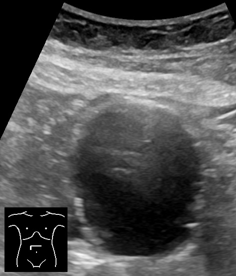
An abdominal aortic aneurysm in cross-section. The large dark circle is the widened aorta, seen end-on as the screen catches it below the kidneys. The width is read straight across this round view, from one outer wall to the other. Image: Mikael Haggstrom, CC0.
Screening picks its targets. The aneurysm is a disease of older men who have smoked. The evidence for screening is strongest there, so the offer goes out, in many programmes, to men at sixty-five, with a single scan for any man who has ever smoked. A family history of the aneurysm widens the net. The scan is offered a single time, and a normal result ends it, since an aorta clear at sixty-five rarely turns dangerous later. National programmes in several countries send the invitation by post, a single appointment of about fifteen minutes.
Women are screened by selection, where smoking or a relative’s aneurysm raises their odds. The point of aiming the scan is plain: a one-time look at the right people catches the quiet aneurysms when they are still small enough to watch, long before any of them turns dangerous. The aim is narrow on purpose. A test spread to everyone raises alarm out of proportion to the few aneurysms it turns up, so the offer holds to the group the evidence covers. The man at highest risk is often the one least likely to seek a scan, which is why the invitation goes out to him before he ever thinks to ask.
What makes the wall give way
The aorta is a pressure pipe. Every heartbeat drives a pulse of blood against its wall, sixty and more times a minute, for a lifetime. The wall holds through elastic layers that stretch and recoil with each beat. Age, smoking, and high pressure wear those layers thin. The aorta is the widest artery in the body, carrying every drop of blood the heart sends out. Its wall meets that load with three layers, and an aneurysm is a slow failure of the middle one.
Where the wall weakens, it stretches. The stretching thins it more, the thinner wall stretches further, and a slow, widening bulge sets in. This is the aneurysm: a length of aorta blown out past its proper width, growing by fractions of a millimetre a year, painless the whole way. A wider tube, under the same pressure, takes more force on its wall than a narrow one, so a bulge that has started tends to keep growing. The growth runs slowly for years, then can quicken once the aneurysm is wide. Speed of growth, as much as size, weighs on the decision, since a fast-stretching wall is a wall close to its limit.
The bulge almost always sits low, in the stretch of aorta below the kidneys’ arteries, above the split into the legs. That is where the screen looks hardest. A short segment, easy to reach with a probe pressed into the lower belly, holds nearly all the aneurysms the screen needs to find.
The risk runs along a few clear lines. Smoking is the heaviest, a habit that weakens vessel walls for decades after the last cigarette. Age and male sex add to it, and a parent or sibling with an aneurysm raises a person’s own odds. High blood pressure drives the wall harder with every beat. The screening rules are drawn straight from this list, aimed at the people the aneurysm favours. Each risk on the list multiplies with the others, so an older male smoker with a family history stands at the sharp end of the screening case.
Finding the vessel
The aorta hides in plain sight. Lay a curved probe across the upper belly, press gently to nudge the gut gas aside, and a thick-walled, pulsing circle appears in front of the spine. That circle is the aorta. Beside it, to the patient’s right, sits the vena cava, with thinner walls that flatten easily under the probe. Trace the aorta down. It runs ahead of the spine, a little left of the midline, until it splits low in the belly into the two vessels of the legs. The screen reads it along that whole run, from below the ribs to the split. A thin patient gives the aorta up easily, the vessel sitting close under the probe. A heavy or gassy belly hides it deeper, and patience with pressure, or a second try after a fast, brings it into view. The spine behind gives a steady landmark, a bright curved line the aorta always rides in front of.
The shape it takes
Many aneurysms swell the whole ring of the wall, so the aorta widens evenly into a smooth spindle. This is the common shape, the fusiform aneurysm, a vessel ballooned along its length. The screen measures it across its widest point. The widest slice of the spindle is the one that counts, and the probe slides along the bulge to find it before the calipers are set.
A few bulge from one side only, a pouch ballooning off the wall like a blister. This saccular shape is less common, and a reader notes it, since a lopsided pouch can carry its own risk. Either way, the rule holds: measure the widest outer width, wherever the bulge reaches it. The shape carries a hint of the cause. A smooth fusiform swelling speaks of ordinary wall disease. A sharp-necked pouch can follow an injury or an infection of the wall. A spindle can run several centimetres along the aorta, its swell gentle enough that the eye needs the calipers to catch the widest point.
The one measurement that matters
One width decides everything here. With the aorta held in cross-section, the probe measures it from the outer edge of one wall to the outer edge of the other, front to back. Outer wall to outer wall: the figure takes in the whole vessel, its lining and any clot stuck inside, down to the far wall. The cross-section is squared first, the vessel turned into a clean circle on the screen, since an oval cut would stretch the width and read too high.
The clot is the reason the rule is strict. A thrombus can line a wide aneurysm, leaving a narrow open channel down its centre. Measure only that channel, and a dangerous aorta gets called safe. The width that counts is the width of the whole vessel, clot and all. The lesson is burned into every operator: the outer wall holds the true width.
The number is taken twice. Once across, in the round cross-section, where the beam must sit square to the vessel to read it true. Once along, in the long view, as a check. A tortuous aorta, cut at a slant, reads wider than it is, so the probe is squared up until the smallest honest width is found. The two views guard each other, since a single plane can flatter or exaggerate a width that the other corrects. The long view also shows how far the bulge runs up and down the vessel, its length as much as its width.
The figure that comes out is the diameter, in centimetres. It carries the whole weight of the scan. That number is written down to the millimetre, the anchor every later scan is compared against.
What the number means
Below three centimetres, the aorta is normal, and the screen is done. At three centimetres the vessel takes the name aneurysm. From there the number sets the path. The three-centimetre line is a convention, drawn where the risk of trouble first becomes real enough to track. A normal aorta in an adult runs around two centimetres, so three marks a clear half-again of stretch.
A small aneurysm, three to four centimetres, is left in place and watched from a distance. A medium one, four to five, is watched more closely. Past five, the watch tightens to a few months at a time, because every extra millimetre of width adds to the risk of bursting. Each band carries its own rhythm of watching, the steps tightening toward the line of repair. A man told his aorta sits at four centimetres is not in danger that day, only on a list to be watched with care.
At five and a half centimetres, the watching ends. An aneurysm this wide is referred for repair, by open surgery or a stent run up from the groin, before it can rupture. A smaller aneurysm that is growing fast, more than a centimetre in a year, is referred on the same logic. The number, and the speed it is changing, together decide between watching and cutting. The five-and-a-half line is itself a balance, set where the danger of leaving the aneurysm outweighs the danger of the operation to fix it. In women, whose vessels start smaller, repair is sometimes considered a little sooner. The cutoff is a guide, weighed against the patient’s age, fitness, and wishes before any operation is set.
What else the scan reads
The diameter rules the screen. The probe still sees more on its way down. The arteries to the legs, a little past the aortic split, can carry aneurysms of their own, and a careful sweep follows the aorta into them. An iliac artery bulging past its normal width gets the same watch as the aorta above it.
The neck of the aneurysm matters to whoever may repair it. The length of normal aorta between the kidneys’ arteries and the top of the bulge decides whether a stent can seat there. The screen notes it, a small service to the surgeon who may follow.
Inside the vessel, the probe picks out the clot that lines so many aneurysms, a layer of old thrombus packed against the wall. It reads the flow down the open channel with color Doppler. On rare occasions it catches a flap of torn lining waving in the stream, the mark of a dissection, a different emergency that the same probe can name.
The iliac arteries deserve their own look. Past the aortic split, these two vessels run down into the pelvis, and they can balloon out as the aorta does, sometimes alone, sometimes alongside an aortic aneurysm above. A measurement past about one and a half centimetres marks an iliac aneurysm, watched on its own schedule. The sweep that follows the aorta down does not stop at the split. The split sits low, near the brim of the pelvis, and the probe drops to find it before turning to each leg vessel in turn.
Calcium in the wall throws its own shadow. Years of disease lay down hard plaques along the aortic wall, bright on the screen, each one casting a dark stripe behind it that can hide a slice of the vessel. A reader works around the shadows, angling the probe to fill the gaps, until the whole width is seen past the calcium. Heavy calcium can defeat the scan at times, the wall lost behind a band of shadow, and that is one of the moments the screen hands the question to a CT. The CT sees past bone, gas, and calcium alike, a backstop for the aorta the probe cannot fully reach.
How often to look again
The interval follows the width. A borderline aorta, a shade under three, is checked again after some years. A small aneurysm is seen every year or two. A medium one, yearly. A wide one, every few months. The bigger the aneurysm, the shorter the leash, until it crosses the line to repair. The intervals are not arbitrary. Each is set so a slow-growing aneurysm cannot cross into danger between one scan and the next. A faster grower is pulled onto a tighter schedule.
Where the measurement misleads
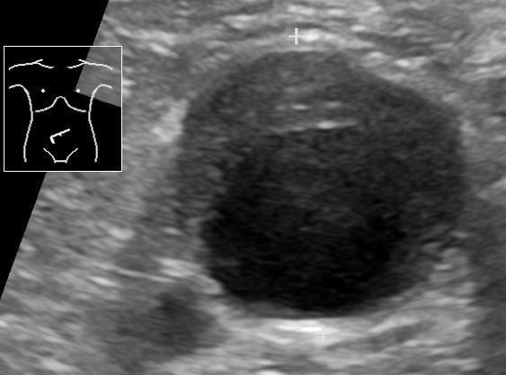
An aneurysm measured across its width. The calipers, set at the top and bottom, span the full diameter from outer wall to outer wall. A layer of old clot lines the inside of the wall here, the mural thrombus the rule warns about: measuring only the open channel would call this aorta far smaller than it is. Image: Mikael Haggstrom, CC0.
A number this important can lie, and a careful operator knows the lies. Each trap has a fix, and a reader who knows the four reads the number with a clear conscience.
Gas is the first. A belly full of bowel gas can hide the aorta or blur its walls, so the probe leans in with steady pressure to push the gas aside, or the patient comes back after a fast. Pressure applied slowly eases the gas aside and spares the patient, and rolling the patient onto a side can open a window the flat-on-the-back view denied.
Slant is the second. An aorta that bends and twists, cut at an angle, looks wider than its true width. The fix is to square the probe to the vessel and take the narrowest clean measurement.
Clot is the third. A thrombus can hush the pulsation and soften the walls until a large aneurysm reads like a quiet vessel. The cure is the outer-wall rule: measure the whole width, clot and all.
The fourth is the wrong vessel. The vena cava beside the aorta can fool a hurried eye, until a press flattens it and a glance at the pulse and the spine sets the two apart.
When it has burst
The screen exists to prevent one event: the rupture. A burst aneurysm floods the belly with blood and drops the pressure fast. Such a patient has only minutes. The whole machinery of screening exists for this one moment it hopes never to meet.
This is the moment a bedside scan turns from screening to rescue. An older patient with sudden back or belly pain and a falling pressure is scanned in seconds. A wide aorta on the screen, in that patient, is treated as a rupture until proved otherwise, and the patient goes straight to the operating room. Three things raise the alarm before the probe even touches the skin: pain in the belly or back, a pulse felt in the abdomen, and a pressure that is sliding.
The scan shows the aneurysm plainly. The rupture itself, the blood leaking back behind the belly, is harder to see, and its absence on the screen proves nothing. The rule is blunt: a big aneurysm and a crashing patient need no further proof. Speed is the whole of it here. A patient with a leaking aneurysm lives or dies by how fast the diagnosis is made and the operating room is reached. A bedside scan can shave the minutes that decide it, naming the aneurysm before the patient leaves the resuscitation bay.
Watching a repaired aorta
A repaired aneurysm is not a finished story. A stent graft, run up from the groin to line the aorta from within, can loosen or leak over the years, letting blood seep back into the old sac. This leak, the endoleak, swells the sac again, and a swelling sac is a warning the scan is built to catch.
So a stented aorta is scanned at intervals for life. The sac is measured as the native aorta once was, watched for any sign that it is filling again. Color Doppler hunts the telltale jet of blood where it should not be. The screen that found the aneurysm becomes the screen that guards the repair.
Not every leak behaves the same way. Some seep from the ends of the graft, where it meets the native aorta. Some feed back through small branch vessels that fill the sac in reverse. The scan, with color flow, tries to find where the blood enters, a clue to how urgent the leak is and how it might be fixed.
The schedule is its own discipline. A stented aorta is checked soon after the repair, again within the first year, then year by year for as long as the patient lives. A sac holding steady or shrinking is the sign of a sound repair. A sac creeping wider sends the patient back toward the vascular team. A stable sac at five years is strong reassurance the graft is holding, the kind of answer a yearly scan is built to give.
Ultrasound carries much of this watching, sparing the patient the dye and the radiation of a CT at every visit. The contrast scan and the CT keep their place for the leak that ultrasound cannot pin down. For the routine year-by-year look, the probe in the clinic does the work. The shift saves a patient years of dye loads, a real gain for someone scanned every year across a long life.
A scan that fits the pocket
Few scans suit a handheld machine as well as this one. The target is large, the measurement is one width, and the skill is quickly learned. A curved probe on a pocket device finds the aorta and reads its diameter as well as any cart. The accuracy holds up. In trained hands, a pocket scanner measures the aorta within a millimetre or two of a full machine, close enough to sort the normal from the aneurysm and the small from the large. The few aortas that sit near a threshold, or that hide behind gas, are sent on for a formal scan to settle the figure.
So aortic screening has spread out of the radiology room. A primary-care clinic, an emergency bay, a rural outpost: each can run the scan on a device that travels in a coat, and refer onward only the aortas that cross a line. For a patient far from a hospital, that reach is the difference. A quiet aneurysm caught on a pocket scanner at sixty-five is a rupture prevented at seventy. A few hours of training carry a clinician a long way, since the target is forgiving and the measurement is one simple width.
The width and the wait
Strip the scan to its core and almost nothing is left to it: find the aorta, measure across, read the number against a few thresholds, and decide whether to watch or to act. A person could be taught the steps in an afternoon. The plainness is the point. A test this simple can run almost anywhere, by almost anyone trained to it, on almost any machine. The hardest part is remembering to do it at all, to put the probe on the belly of every man the rule names.
The power is in the timing. The aneurysm that ruptures was, years before, a small dilation a thirty-second scan would have caught. Screening moves the discovery from the day of the rupture to a quiet morning long before it. A small dilation found today is a catastrophe disarmed years ahead of time.
A man turns sixty-five. A probe crosses his belly for half a minute. A number comes up under three, and he is sent home for good; or it comes up above, and a watch begins that may, one day, save his life. The whole of it stands on one width, honestly measured. One number, read with care, stands between a man and the quiet aneurysm that would otherwise find him unprepared.
Common questions about aortic aneurysm screening
Who should be screened for an abdominal aortic aneurysm?
Men aged sixty-five to seventy-five who have ever smoked gain the clearest benefit, and a one-time ultrasound is the standard offer. A family history of the aneurysm is another reason to be scanned. Women are screened by selection, where smoking or a close relative’s aneurysm raises the risk.
What size of aorta counts as an aneurysm?
Three centimetres across. Below that, the aorta is normal. At three centimetres or wider, it is an aneurysm, measured from the outer wall to the outer wall, front to back. Many aneurysms found by screening sit between three and five and a half centimetres, below the size that calls for surgery.
How is an aortic aneurysm measured on ultrasound?
Across the vessel, from outer wall to outer wall, in the front-to-back direction. The figure takes in the whole aorta, including any clot lining it, down to the outer wall. A second measurement along the vessel confirms it. A tortuous aorta is squared up to the probe, so a slanted cut does not read too wide.
When does an aortic aneurysm need surgery?
At five and a half centimetres across, an aneurysm is usually referred for repair, before the risk of rupture climbs too high. A smaller aneurysm that grows quickly, more than a centimetre in a year, is referred on the same grounds. Below that size, the aneurysm is watched at intervals set by its width.
How often is a small aneurysm rescanned?
A wider aneurysm is rescanned more often. A small one, three to four centimetres, is seen every year or two. A medium one, four to five, is seen yearly. Past five centimetres, the scan is repeated every few months, because each step up in width adds steeply to the rupture risk.
Does a normal aortic screen mean it is safe for life?
Largely, yes. An aorta under three centimetres at sixty-five rarely grows into a dangerous aneurysm later, which is why one normal screen is enough for nearly all men. A borderline aorta, a shade under three, may be rechecked after some years. The one-time scan, caught at the right age, settles the question for a lifetime in nearly all who pass it.
About the Author
Julien Mercier
Senior R&D Engineer · Medical Ultrasound Transducer Development
Senior R&D Engineer with an M.S. in Applied Physics and over 15 years of experience in medical ultrasound transducer development, specializing in the design verification and performance testing of high-frequency imaging transducers. Currently leading the development and verification of the company’s next-generation high-frequency linear-array transducer, responsible for imaging performance evaluation and reliability analysis in preclinical testing. Brings extensive hands-on experience in piezoelectric element tuning, beamforming parameter optimization, and system-level performance testing.