





































AAO American Academy Ophthalmology Ultrasound Guidelines Key Points
Ophthalmic ultrasound is held to a set of standards. The bodies that govern eye care, the American Academy of Ophthalmology among them, set out when the scan is called for, how it is done, and how its findings are written down. These standards turn a probe on the eye into a reliable test, one whose findings hold up from one clinic to the next. The key points run through the whole exam, from the reason it is ordered to the report that closes it.
Why the standards matter
Ophthalmic ultrasound carries real weight in the clinic. The picture it returns can send a patient to the operating room, open a cancer work-up, or settle a worry and send them home the same afternoon. A number taken loosely, off a poor image or a wrong setting, can point all of that the wrong way. The eye leaves little room for a second guess once the plan is set. The course that follows can be hard to walk back: an eye opened in theatre, a course of radiation begun, a cancer chased down or missed. So the scan is held to a set of standards. They exist so the same eye, scanned by different hands on different machines, returns the same answer. They put a floor under the exam that every clinician builds on, from a trainee with a pocket probe to a specialist at a console. The points are few in number. They stay the same whether the eye is calm in a clinic chair or bleeding in a trauma bay at midnight.
The American Academy of Ophthalmology is the largest body of eye physicians in the world, with tens of thousands of members across more than a hundred countries. It publishes Preferred Practice Patterns, the consensus statements that steer eye care from cataract through glaucoma to retinal disease. It runs the teaching courses and the open references that train clinicians in the scan. The ultrasound societies sit alongside it, setting practice parameters for how an eye exam is performed, recorded, and reported. The frequency for each probe, the safety ceiling for the eye, the contents of a proper report: each is spelled out in a parameter a clinician can look up by name. Together these bodies form the guidance that ophthalmic ultrasound is measured against. None of it rests on one person’s habit. It is the settled practice of a whole field, written down and revised over the years. The parameters are reviewed on a set cycle, retired or rewritten when machines and evidence move on, so the guidance a clinician follows keeps pace with the machines in the room.
The guidance lives in how the scan is taught and done, built into the exam as a set of habits. A clinician who learns the eye exam well learns the standards with it, without reading them off a page at the bedside. The habits show in small choices: the preset picked at the start, the planes swept in order, the gain held at a set level, the calipers placed the same way each time. The points that matter are few. They hold across machines from different makers, across the gain and depth a reader dials in, and across the conditions the scan is asked to sort out. A clinician who keeps them turns a grainy grey picture into a finding a surgeon can open an eye on. Letting them slip can send a whole plan of care astray, off the strength of one bad image. The same eye read twice by the same standard reads the same both times. That sameness is the whole point of a standard.
The standards also draw a line around what the scan is for. A test ordered out of habit, with no question behind it, wastes the patient’s time and the clinician’s. It can turn up an incidental mark nobody knows what to do with, and start a chase after nothing. The guidelines tie each scan to a clinical question written down before the probe touches the lid. The eye that can be seen plainly, the disease the slit lamp already shows in full, gains little from a scan over it. The eye gone dark, the structure too deep for light to reach, the length no ruler can take: these are where the scan does real work. Ordered for the right reason, it repays the minute it takes many times over. The scan stands beside the clinical exam, the dilated look and the slit lamp, filling the gap they leave when the view is blocked.
When the scan is called for
The first key point is the reason. The commonest reason is a view that has gone dark. A dense cataract, a bleed in the vitreous, blood pooled in the front of the eye after an injury: each blocks the light an examiner needs to see the back of the eye. The fundus that should show the retina shows only a grey blur or nothing at all. Ultrasound looks where light cannot, passing through the opaque eye to read the wall behind it. The guidelines name this opaque eye as the classic call for the scan, the case where no slit lamp and no ophthalmoscope can reach. A patient who has lost sight in a quiet white eye, with no view to the back, is the patient the scan was made for. A scarred cornea clouds the view as surely as a cataract does. A hyphema fills the front chamber with blood after a blow, and the same blockage follows.
When the back of the eye is hidden, the scan answers the questions that decide care. Is the retina detached, lifted off the wall it should lie against. Is there a solid mass sitting on the choroid. Is a fragment of metal or glass lodged in the vitreous. Is the space behind the lens filled with blood or with the pus of an infection. Each is a finding that changes the next step, from an urgent trip to theatre to a course of watching and waiting. The standards hold the scan to these clear, answerable questions, the ones a decision can rest on. A scan run as a vague look around, with no question in mind, is the kind the guidance steers away from. Each question takes a clean yes or no, the kind a next step can be hung on.
The scan is called for well beyond the opaque eye. It measures the length of the globe before a lens implant, a number to a tenth of a millimetre a cataract surgeon cannot do without. An error of a tenth of a millimetre in that length shifts the lens power by about a quarter of a dioptre, enough to leave a patient in glasses they had hoped to shed. It sizes a tumour and follows its height across months of treatment. It checks the optic nerve behind the globe when the pressure inside the head is in question. It maps a detached retina before surgery so the operator knows the shape and the tether to expect. It looks into a socket behind a prosthesis, and at the muscles of an eye pushed forward by thyroid disease. Each use is tied, in the guidance, to a question the scan can answer and a decision the answer will change. The breadth is wide. The rule behind every use is the same: a clear question, asked before the probe goes down.
The modalities the standards cover
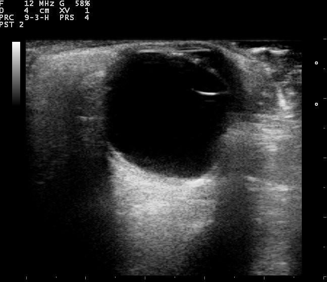
The guidelines speak of more than one scan. The B-scan is the two-dimensional picture, the slice through the eye that shows the shape and the place of a structure, run at around ten megahertz for the depth the eye needs. The standardized A-scan is a single line of spikes, taken at a fixed tissue-sensitivity gain so the height of each spike means the same from one machine to the next. That fixed setting is what the word standardized carries: a calibration that lets a reader judge a tissue by its reflectivity. The two often run together in one sitting. The picture locates a structure, and a careful look at its spikes helps tell what fills it, a solid tumour or a fold of detached retina. The standardized method rests on three habits: placing a finding in the eye, watching how it moves, and measuring the height of the echoes it returns.
Other tools sit under the same standards. High-frequency biomicroscopy runs at thirty-five to fifty megahertz, drawing the front of the eye, the drainage angle and the ciliary body, in a detail a standard probe cannot reach. A biometry A-scan measures the length of the eye for a lens implant, by gentle contact or through a small fluid-filled shell that floats off the cornea. Each tool runs at its own frequency, with its own preset and its own job the guidance spells out. The clinician picks the one that fits the question in hand, since a probe meant for the front of the eye shows the back of it poorly. The standards keep each tool to the work it does best, and name the setting it does that work at. A color overlay can show blood moving inside a mass or confirm flow in the vessels behind the eye. Where the media are clear, an optical biometer reads the length with a beam of light.
| Item | Figure | Note |
|---|---|---|
| B-scan frequency | about 10 MHz | the cross-sectional picture |
| Standardized A-scan | about 8 MHz, fixed gain | reflectivity of tissue |
| Biometry A-scan | about 10 MHz | axial length to 0.1 mm |
| High-frequency biomicroscopy | about 35–50 MHz | the angle and front segment |
| Normal axial length | about 22–25 mm | the lens-implant number |
| Acoustic output | lowest of any scan | set for the open eye |
| Globe rupture | a contraindication | no pressure on the eye |
| Every exam | images, measurement, report | kept on the record |
The eye’s safety ceiling
One point stands above the rest: the eye is scanned with more care than any other target in the body. It sits open to the beam, with no bone to shield it and little blood flow to carry off the heat that sound deposits in tissue. So the guidelines hold ophthalmic ultrasound to the lowest acoustic output of any application, a ceiling set far below the limit for a scan of the belly or the heart. An ophthalmic preset caps the energy at the source. The thermal and mechanical indices stay near the floor of the scale, where the lens and the retina take no harm. A globe that may be cut or ruptured is left untouched, with no probe pressed on an eye that might burst under it. Gentleness is the first rule the standards teach, ahead of every other.
Doing the exam to standard
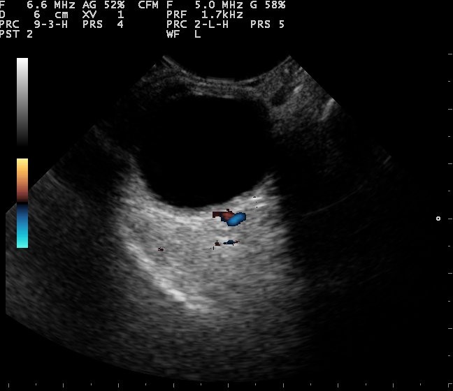
A scan done to standard starts gently. The probe rests on a closed, gel-covered lid, the eye looking ahead beneath it, under a drop of topical anaesthetic where a contact scan calls for one. A clear film and a generous layer of gel carry the beam in with no air trapped between the probe and the eye. The probe lies light on the lid, never pressing on the globe under it. The patient is settled back, the room dimmed enough to make out the faint greys on the screen. These small steps are not fuss. They keep the eye safe and the picture honest, and the guidance counts them as the opening moves of the exam. The probe is wiped clean between patients, the film fresh each time, so nothing passes from one eye to the next.
The eye is swept in set planes. The probe moves through the globe in one plane, then turns ninety degrees and moves through it again, so nothing slips through the gap between two slices. The examiner names where each slice lies against the clock face of the eye, twelve o’clock above, six below, the marker on the probe held to a known meridian. A finding is then pinned to its place, by its depth into the eye and its hour on the clock, for the surgeon who will follow the scan into theatre. The standards ask for this orderly sweep so a reading can be repeated, by the same hand an hour later or another hand the next day. A scan taken at loose, random angles cannot be matched against itself. A finding is crossed in one sweep and run along in another, the two together fixing it in space.
Movement is part of a full exam. The patient is asked to look up, then down, then from side to side, while the probe holds still and watches the structures shift. A detached retina sways on its tether each time the gaze turns. Loose blood swirls through the vitreous and drifts back down. A solid mass stays anchored where it grows. The guidance builds this dynamic look into the exam, since a single frozen frame can hide what a moment of motion makes plain. The way a structure moves, or holds still, is often the clue that names it. An examiner who scans only the resting eye throws away half of what the scan can show.
Both eyes belong in a careful exam. The fellow eye gives a healthy baseline to set the troubled one against, a same-patient yardstick no textbook picture can match. The same gain, depth, and focus are carried from one eye to the other so the two can be set side by side. A measurement is taken more than once and averaged into a single number. The standards lean hard on this repetition, since a value that lands the same way twice is a value a clinician can act on. A lone reading, taken once and never checked, is the kind that hides an error in plain sight. The few extra seconds a repeat takes are the cheapest insurance in the whole exam. In a tumour followed across visits, the new height is set against the height saved at the last one. A change of half a millimetre is taken seriously.
The settings are chosen for the eye, not left on whatever preset came up last. The gain is set so a faint membrane shows up against a vitreous that stays dark. The depth is set to take in the whole globe and the orbit a little behind it. The focus is brought to the level of the finding so its edges read sharp. An ophthalmic preset holds the machine to the gentle output the eye calls for. A clinician who sets the machine with care draws a cleaner picture and a truer number. The guidance counts that setup among the skills of the exam, equal to a steady hand on the probe. A gain left wrong paints noise that mimics disease or buries a thin membrane in the dark.
Who performs and reads the scan
A scan is only as good as the hand that takes it. The guidelines set who may perform and read an eye ultrasound. An ophthalmologist trained in the scan, or a technician working under one, holds the probe and gathers the images. Some technicians carry a formal credential in ocular ultrasound and biometry, earned by examination and by logged hours of scanning. The reading is signed by someone qualified to stand behind it. Training is written down and kept on record. The standards treat the skill of the operator as part of the test itself, since a clean answer depends on the hands that take it. A picture is only as true as the person who tuned the machine to take it. The credential goes by names like registered ophthalmic ultrasound biometrist, a standard a reader earns by examination and renews on a cycle.
The skill takes practice to build. Finding the structure, setting the machine, studying the picture and the spike trace together: each is learned over many supervised scans. A clinician new to the eye works beside one who knows it until their readings agree, eye after eye. The guidance favours this slow handover, the path that turns a probe into a tool a team can rely on. A short course alone does not make an echographer of anyone. The standards ask for hands-on hours, logged and signed by a supervisor, before a reader works an eye on their own. The number of scans behind a reader is part of what a signed report is worth. Many training programs set a count in the hundreds before a trainee works an eye without a hand over their shoulder.
The reader knows the limits of the scan. A poor image is called poor and taken again, with the gain and the angle reset. A scan that cannot answer its question is followed by one that can, a CT, an MRI, or a referral to a specialist eye unit. The standards ask the reader to say plainly what the scan shows and what it leaves open. An honest reading that admits a doubt serves the patient better than a confident one that papers a doubt over. Knowing when the scan has reached its edge is as much a part of the skill as reading the picture in front of it. An examiner who never says I am not sure is one to watch with care. A finding that cannot wait, a fresh detachment or a ruptured globe, is carried to the treating team at once, ahead of the written report that follows.
The record, the report, and the handheld
Every exam leaves a record. The images are saved, the measurements written down, the settings noted beside them. A finding is captured with the calipers in place, so the next clinician sees exactly what was measured and where it sat. The record holds the date, the eye, the reason for the scan, and the name of the reader who took it. The guidance lists what a full record carries: the facility, the indication, the technique and its limits, a comparison with any earlier scan, and the reader’s signed reading. A report then closes the loop, stating the question the scan was asked, the findings, and what they mean for care, in plain terms the treating clinician can act on without a second call. The standards count the report as part of the exam, the step that turns a grey picture into a decision. A scan with no clear report is a scan left half done.
The handheld scanner carries these standards to the bedside. A probe the size of a phone runs the same B-scan a wheeled console runs, on a screen a clinician holds in one hand. The key points hold wherever the scan happens: the clear reason, the gentle technique, the chosen settings, the saved image, the written report, the trained reader behind it all. A small machine changes where the scan can be done. The standard it is held to stays the same. A scan in a tent, a rural ward, or a clinic far from an eye unit answers to the same key points as a scan in a teaching hospital. That is what lets a finding taken on a pocket probe travel, and be trusted, all the way to the surgeon who acts on it. A device that fits in a coat pocket puts the standard within reach of a clinic that could never wheel in a console. A pocket probe and a hospital console answer to the very same page of guidance.
Common questions about ophthalmic ultrasound guidelines
When is an eye ultrasound called for?
When the question cannot be answered by looking into the eye. A dense cataract, a vitreous bleed, or blood in the front of the eye hides the back from view. The scan looks through it for a detached retina, a mass, or a foreign body. It is also used to measure the eye before a lens implant, to follow a tumour from visit to visit, and to check the optic nerve when the pressure inside the head is in doubt.
Why is the eye held to a lower safety limit than the rest of the body?
The eye is scanned at the lowest acoustic output of any ultrasound exam. It sits open to the beam, with no bone over it and little blood flow to carry off the heat that sound leaves behind. An ophthalmic preset on the machine holds the output low for the eye. The probe rests light on the lid and never presses. A globe that might be ruptured is not scanned at all.
What are the A-scan and the B-scan used for?
The B-scan is the two-dimensional picture, the slice that shows the shape and place of a structure inside the eye. The standardized A-scan is a single line of spikes, taken for the height and pattern of the echoes a tissue sends back. The picture finds a structure. The spikes help tell what it is made of. The two are often run together in the same sitting.
Who should perform and read an eye ultrasound?
Someone trained in the scan and working to a standard. An ophthalmologist who knows the exam, or a technician under one, holds the probe and takes the images. A qualified reader signs the findings. Training is logged and kept on record. A clinician new to the eye learns it over many supervised scans before reading on their own.
Does a handheld scanner meet the same guidelines?
Yes. A handheld probe runs the same scan a console does. The same points apply: a clear reason, a gentle technique, an ophthalmic preset, a saved image, a written report, a trained reader. The small size changes where the scan can be done. The standard it is held to stays the same.

About the Author
Julien Mercier
Senior R&D Engineer · Medical Ultrasound Transducer Development
Senior R&D Engineer with an M.S. in Applied Physics and over 15 years of experience in medical ultrasound transducer development, specializing in the design verification and performance testing of high-frequency imaging transducers. Currently leading the development and verification of the company’s next-generation high-frequency linear-array transducer, responsible for imaging performance evaluation and reliability analysis in preclinical testing. Brings extensive hands-on experience in piezoelectric element tuning, beamforming parameter optimization, and system-level performance testing.