





































Anterior Chamber Depth Measurement Glaucoma Handheld Ophthalmic Ultrasound
The anterior chamber is the small pool of clear fluid at the front of the eye, the space between the cornea and the iris. Its depth, measured from the back of the cornea to the front of the lens, is a number that matters out of all proportion to its size. A shallow chamber crowds the drain that keeps the eye’s pressure in check. A crowded drain is the setup for angle-closure glaucoma, an attack that can blind an eye in a day. Measuring that depth, and judging the angle it sets, is work a handheld ultrasound does in a minute at the front of the eye.
The space behind the cornea
The eye makes a clear fluid called aqueous, day and night, behind the iris. The fluid flows forward through the pupil into the anterior chamber, washes the back of the cornea, and drains out through a ring of fine mesh where the iris meets the cornea. That ring is the drainage angle. The whole system holds the eye at a steady, gentle pressure, the pressure that keeps it firm. The eye makes and drains this fluid at the same slow rate, a teaspoon’s worth turned over every few hours. The balance of the two holds the pressure in a narrow healthy band. A blocked drain breaks that balance fast.
The depth of the anterior chamber is set by how far back the iris and the lens sit. In most eyes the cornea arches well clear of the lens. The chamber is deep, around three millimetres or a little more. The drainage angle in such an eye is wide open, the mesh in plain reach of the fluid. The pressure stays where it should. The eye is at no special risk. A healthy pressure sits in a band roughly between ten and twenty-one. The wide-open angle of a deep eye lets the fluid leave as fast as it is made. Such an eye can be dilated, examined, and operated on without a second thought about the angle.
A shallow chamber is the one to watch. When the iris and the lens sit forward, the cornea-to-lens depth drops. The iris crowds toward the drainage angle. A depth under about two and a half millimetres is the figure that raises concern. In such an eye the angle is narrow, the mesh half-hidden behind the bunched-up iris. The drain is one step from being blocked. The shallowest chambers run under two millimetres. The lens of an older eye, swollen and thickened over a lifetime, is the usual reason a once-safe chamber turns shallow. A depth that was three millimetres at forty can be two and a half at seventy.
Depth and pressure are not the same thing. A shallow eye can read a normal pressure for years. The danger is the angle’s narrowness, the standing risk that the drain will shut all at once. A shallow chamber is a loaded spring. The depth is what tells the clinician how tightly it is wound. On a shallow eye a normal pressure is no all-clear. The depth carries a warning the pressure reading leaves out.
All of this turns on one measurement. The depth of the anterior chamber, read in millimetres, places an eye on the scale from safe to dangerous. It is a number any handheld ultrasound can take, on any eye, in under a minute. A shallow figure flags an eye for a closer look at the angle and a careful hand with every drop and every dilation. The figure means the same thing in any clinic, on any machine, a hard number anyone can act on. It passes from one reader to the next with no explanation needed. A depth of three is a depth of three, in any pair of hands.
Measuring the depth
The depth is measured the same way the length of the eye is measured, with a pulse of sound. A probe sends sound into the eye along its axis. The sound bounces off the back of the cornea, then off the front of the lens, and returns two echoes a moment apart. The gap between those two echoes, turned from time into distance, is the depth of the anterior chamber. The machine reads it out in millimetres on the spot. The same biometry sitting that reads the length of the eye reads this depth as well. The corneal echo and the lens echo are two of the spikes on the trace. A few readings are taken and averaged, the way any careful measurement is.
The depth is steadiest when nothing presses on the eye. A probe set straight on the cornea can dent it and read the depth a touch short, the same trap that shortens an axial length. So the careful way floats the probe in a small bath of fluid, off the cornea, the sound crossing the gap with nothing pressing the eye. The number that comes back is the true depth, the one a clinician acts on. A small cup of saline held against the eye keeps the probe a clean distance off the cornea. The measurement is taken with the patient lying back, looking straight up at a fixation light. A tenth of a millimetre changes the figure on a borderline eye, so the touch is kept light.
A shallow chamber, a crowded angle
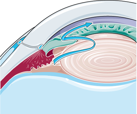
A shallow chamber and a narrow angle go together. The depth at the centre of the eye and the width of the angle at the rim rise and fall as one. The shallower the chamber reads at the centre, the narrower the angle runs at the rim. A depth read off the scan is a quick stand-in for the gonioscopy a busy clinic may not have time for. The one number at the centre speaks for the angle the clinician cannot easily see. An angle-closure attack can take an eye in hours, faster than almost any other eye disease. That speed is why finding the narrow angle early matters so much. A depth measurement is the cheapest way to find the eyes at risk before any harm is done.
Some eyes are built shallow from the start. A small, far-sighted eye packs the same parts into less room, so its chamber runs shallow and its angle narrow. The lens thickens through life and creeps forward, taking another fraction of a millimetre off the depth in each decade. Angle-closure runs higher in women, in older people, and in some families and populations more than others. A short, hyperopic eye in an older patient is the classic shallow chamber. Far-sightedness and a shallow chamber tend to come as a pair. A careful clinic learns to look harder at the small, long-sighted eye. A parent who lost an eye to an attack is a strong reason to measure the children’s depths early. Eyes from East Asian and Inuit backgrounds carry the risk more often, a pattern worth knowing in a mixed clinic. The first attack in a life usually lands between the fifties and the seventies, the years the lens has thickened most. A depth taken at the first sign of trouble dates the risk before it ripens.
The angle does not close on its own. The trigger is most often a small jam at the pupil. Aqueous heading forward has to slip between the iris and the lens to reach the pupil. In a shallow eye those two press close. Fluid backs up behind the iris and pushes it forward, bowing it like a sail. The bowed iris falls against the angle and seals the drain. Pressure has nowhere to go. It climbs. The jam at the pupil has a name: pupillary block. It is the commonest road to a closed angle. The bow of the iris it makes shows on the scan as a forward curve, the iris arching away from the lens toward the angle.
The scan reads the setup before the spring is sprung. A depth measured shallow, an angle seen narrow, an iris that bows forward on the picture: these mark the eye that could close. Finding them early lets the eye be treated before any attack, often with a tiny laser hole in the iris that lets the trapped fluid through. The measurement turns a hidden risk into a treatable one. The laser hole, a peripheral iridotomy, takes a minute and gives the trapped fluid a way around the pupil. With the jam relieved, the iris falls back. The angle opens. The same depth is taken again after, to confirm the chamber has deepened.
When not to dilate
One rule comes straight off the depth reading: think hard before dilating a shallow eye. Dilating drops widen the pupil. They also bunch the iris into the angle. On a shallow eye with a narrow angle, those drops can tip it into a full attack, an acute angle-closure brought on by the very exam meant to help. A quick depth check, or a look at the angle, before reaching for the dilating drops is what tells a clinician which eye is safe to open up. The shallow eye is dilated with care, or with a plan to watch the pressure after, or not at all until the angle is dealt with. The drops in question are the everyday dilating ones, used in clinics by the thousand. On most eyes they are entirely safe. The depth reading is what marks the few eyes where they are not, so the dilation is held or watched closely. A note in the file, shallow angle, no dilation without cover, follows the patient from visit to visit. Every clinician who opens that file is warned before reaching for a drop.
When the angle slams shut
An acute angle-closure attack is one of the few true emergencies of the eye. The pressure rockets in hours, far past anything a healthy eye sees. The eye turns red and goes rock-hard. The vision fogs, with coloured haloes around lights. The pain is fierce, often with a headache and a sick stomach to match. The pupil sits half-open, fixed, no longer answering the light. An eye in this state can lose sight within a day. The pressure in an attack can run three or four times normal, high enough to feel the eye harden under a gentle touch. Water forced into the cornea clouds it, dimming the view further. The whole picture is unmistakable to anyone who has seen it once.
The diagnosis is mostly clinical, read off the hard red eye and the high pressure. Ultrasound has its part even so. On a clouded, painful eye it confirms the shallow chamber and the closed angle behind the haze of a swollen cornea. It rules out another cause pushing the iris forward, a tumour or a mass behind it. The scan reads the front of the eye when a swollen cornea hides the angle from a direct look. The whole read takes a few seconds on an eye that pain and a clouded cornea have put beyond the usual exam. It is also the test that catches the rarer attack driven by a mass behind the iris, the one that needs a different treatment altogether.
Speed decides the eye. The pressure has to come down fast, with drops and pills that turn off the fluid and pull water out of the eye. A laser hole in the iris then breaks the jam at the pupil and lets the angle fall open. The scan that confirmed the shallow closed angle is also what checks the other eye, the fellow eye that shares the same shallow build and the same risk of an attack. Both eyes are usually treated, the second before it ever closes. Most of the time the fellow eye has not yet had an attack of its own. Treating it early, with the same laser hole, heads that attack off before it starts. One acute attack is, in effect, a warning about two eyes. Pilocarpine, an old drug that pulls the pupil small, drags the iris out of the angle in the thick of an attack. The pressure-lowering drops and pills buy the time the laser needs. The laser then makes the fix permanent.
| Item | Figure | Note |
|---|---|---|
| Normal anterior chamber depth | about 3.0–3.5 mm | cornea to lens, on the axis |
| Shallow, at-risk depth | under about 2.5 mm | the figure that raises concern |
| Narrow angle on gonioscopy | about 20° or less | the angle that can close |
| Healthy eye pressure | about 10–21 mmHg | the band the drain holds |
| Pressure in an attack | 3–4 times normal | the eye goes rock-hard |
| UBM frequency | about 35–50 MHz | the angle in fine detail |
| The fix | a laser iridotomy | a pinhole that opens the angle |
Seeing the angle itself
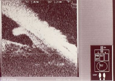
Seeing the angle itself takes a finer scan. Ultrasound biomicroscopy, or UBM, runs at a very high frequency and trades depth for detail, drawing the front of the eye in fine slices. On a UBM picture the cornea, the iris, the angle, and the ciliary body behind the iris all stand out clearly. The clinician takes the exact width of the angle, the bow of the iris, the spot where it touches the wall. It is the closest thing to looking straight into the angle. UBM works at thirty-five to fifty million cycles a second, many times the frequency of a standard eye scan. That high pitch buys fine detail at the front of the eye, at the cost of reach into the back. The probe is held just off the eye in a small water bath, the same gentle way the depth is taken.
UBM catches the traps a depth measurement alone can miss. Some angles close from a different cause: a ciliary body that sits too far forward and shoves the iris root into the angle, a setup called plateau iris. In plateau iris the crowding sits out at the rim, where a central depth reading does not reach. UBM sees the ciliary body where no other scan can. It names the cause the depth alone would hide. The finer scan is the one for the hard cases. Several fine numbers come off a UBM picture, the width of the angle and the gap between iris and wall measured in degrees and microns. These feed the decision to treat or to watch. The scan that sees the ciliary body is the one that settles an angle a depth measurement leaves open to question. What UBM settles is whether to watch an angle or treat it, a call made on a clear picture. The hardest angles are the ones it handles best.
The handheld at the front of the eye
A handheld scanner puts the depth measurement at the front-of-eye exam, at the chair, in a clinic anywhere. The probe is small, the bath of fluid quick to set up, the number on the screen in a minute. A clinic with no large biometer still gets a true anterior chamber depth on every eye it needs one for. The figure goes into the same record as the pressure and the rest of the exam. The whole rig is a probe, a small cup, and a screen, run off a battery at the chair. A depth taken where the patient sits saves the trip to a biometry suite. The number lands in the notes beside the pressure the same day. The same probe that takes the axial length and the lens power takes the chamber depth in the same minute. One sweep of the front of the eye gives all three. A small clinic runs a full biometry off a single handheld.
What the scan does best is spot risk before it bites. A shallow depth on a routine eye, in a patient with no symptoms at all, is a flag worth raising. It marks the eye to dilate with care, the eye to send for a laser iridotomy, the eye to watch in the years to come. Catching a narrow angle in a quiet clinic is what heads off the three-in-the-morning emergency. Many of the world’s angle-closure attacks fall on eyes never checked for a narrow angle. A simple depth, taken once in mid-life, would flag a good share of them in time. The handheld brings that check to clinics that have never had a way to make it. The check costs nothing but a minute and a smear of gel. A single shallow figure, flagged once, can spare an eye a lifetime of trouble.
The same depth matters before any surgery at the front of the eye. A cataract operation works in the anterior chamber. A shallow chamber leaves the surgeon less room to move. The depth taken beforehand warns of the tight eye and shapes the plan for it. The number that flags an angle-closure risk is the same number a cataract surgeon wants in hand before the first cut. Removing the thickened lens, the cataract operation itself, deepens the chamber and opens the angle, a cure for some angle-closure along with the cloudy lens. The depth taken before that surgery shows how shallow the working space will be. It guides the surgeon’s hand from the first move.
The depth measurement has its bounds. It is one number. It is not the whole angle. A borderline depth still needs a proper look at the angle to settle it. The scan does not measure the pressure or grade the optic nerve, the other halves of a glaucoma work-up. It flags the eye at risk and times the closer look. A shallow figure is a reason to examine more closely. The diagnosis itself waits on the fuller look. A depth measurement does not say how high the pressure is, nor whether the optic nerve has already taken damage. Those come from the pressure check and the look at the nerve head. The scan adds the one piece those miss: the shape of the front of the eye and the angle hidden in it.
So the small pool of fluid behind the cornea, and the millimetre or two that sets its depth, reads out as a number on a handheld screen. That number speaks for the angle a clinician cannot easily see, flags the eye that should not be dilated blind, and finds the narrow angle in time to open it with a pinhole of laser. A measurement that takes a minute at the front of the eye is what stands between a quiet shallow chamber and a blinding attack. A pinhole of laser, placed in time, is all it takes to keep that angle open for life. Finding the eye that needs it is the depth measurement’s whole gift. The small probe is where that measurement is made.
Common questions about anterior chamber depth and glaucoma
What is the anterior chamber depth, and why does it matter?
It is the depth of the fluid-filled space at the front of the eye, from the back of the cornea to the front of the lens, normally around three millimetres. A shallow depth, under about two and a half millimetres, crowds the drainage angle where fluid leaves the eye. A crowded angle can close, sending the pressure up in an attack of angle-closure glaucoma. The depth is an early read on that risk.
How is the depth measured with ultrasound?
A probe sends a pulse of sound along the axis of the eye. It returns an echo from the back of the cornea and another from the front of the lens. The gap between the two, turned from time into distance, is the depth. The reading is most accurate when the probe floats in a small bath of fluid, with nothing pressing on the cornea to shorten it.
Why not dilate an eye with a shallow chamber?
Because the drops that widen the pupil also bunch the iris into an already narrow angle. On a shallow eye that can tip the angle shut and bring on an acute attack, the very thing the exam was meant to avoid. A quick depth check before dilating shows which eye is safe to open up and which needs care or a plan to watch the pressure after.
What does ultrasound biomicroscopy add?
UBM is a very high-frequency scan that draws the front of the eye in fine detail. It shows the angle, the iris, and the ciliary body behind the iris, structures a depth reading alone cannot resolve. It catches an angle crowded by a forward ciliary body, a setup called plateau iris. A central depth reading does not reach the rim where that crowding sits. It is the closest thing to looking straight into the angle.
Does the scan diagnose glaucoma on its own?
No. The depth is one number, an early flag. It is not the whole picture. The scan does not measure the eye’s pressure or judge the optic nerve, the other parts of a glaucoma work-up. A shallow reading marks an eye to examine more closely, to dilate with care, and often to send for a preventive laser hole in the iris. The diagnosis is settled by the full eye exam.

About the Author
Julien Mercier
Senior R&D Engineer · Medical Ultrasound Transducer Development
Senior R&D Engineer with an M.S. in Applied Physics and over 15 years of experience in medical ultrasound transducer development, specializing in the design verification and performance testing of high-frequency imaging transducers. Currently leading the development and verification of the company’s next-generation high-frequency linear-array transducer, responsible for imaging performance evaluation and reliability analysis in preclinical testing. Brings extensive hands-on experience in piezoelectric element tuning, beamforming parameter optimization, and system-level performance testing.