Developmental Dysplasia Hip DDH Infant Ultrasound Handheld
Developmental dysplasia of the hip, or DDH, is a hip that has not formed properly in a baby, with the socket too shallow to hold the ball of the joint firmly. Ultrasound is the test that finds it in the early months of life. A baby’s hip is still soft cartilage then, the kind of tissue ultrasound shows clearly. From a short scan, a clinician measures how well the socket is formed, watches the joint for looseness, and catches a hip that needs treatment before it grows hard to fix.
What DDH is, and why ultrasound
The hip is a ball-and-socket joint. The ball is the head of the thigh bone. The socket is a cup in the side of the pelvis, called the acetabulum. For the joint to work, the socket has to be deep enough to hold the ball in place. In DDH the socket is too shallow, so the ball sits loosely, rides to the edge, or slips out altogether. A deep cup holds the ball through every kick and stretch of infancy. Most babies are born with a hip that does just that.
Caught early, DDH is easy to treat. In the first months a baby’s hip is still forming. A shallow socket can be guided to deepen with a simple harness that holds the ball in place. Left unfound, a dislocated hip leads to a limp, a difference in leg length, and arthritis in early adult life. The whole point of screening is to find it before the easy fix is lost. The harness works only on a hip still young enough to mould.
Ultrasound is what makes early screening possible. A newborn hip is mostly cartilage, the kind of tissue that does not show on an X-ray for the first months of life. Ultrasound reads cartilage well, so it pictures the whole joint from the start. A short scan shows the shape of the socket, the position of the ball, and the looseness of the joint, all the things screening needs. From a side-lying baby, the whole joint comes into one clear view.
DDH gives little warning on its own. It causes no pain in a baby, and often no outward sign at all. A hip can be dislocated under the skin with nothing to feel on a quick check. The scan is what brings that hidden hip into plain view, early enough for a harness to set it right. Found this early, a dislocated hip can be coaxed back into shape.
A hip made of cartilage
The infant hip suits ultrasound perfectly. It sits shallow, just under the skin on the side of the hip, within easy reach of a probe. It is made of cartilage, which sound passes through cleanly. A small probe laid on the side of the hip reads the whole joint in fine detail. No part of the joint lies out of the probe’s reach.
Cartilage is the key. The ball of a baby’s hip, and much of the socket rim, are cartilage in the early months, turning to bone only later. Ultrasound shows that cartilage plainly, the soft ball and the cartilage roof of the socket alike. The scan reads the joint as it actually is in a baby, soft parts and all. The soft cartilage ball reads clearly on the screen, in fine detail.
This is why ultrasound owns the early window. Ultrasound needs no bone to read a joint, so it pictures a baby’s hip through the months it is still mostly cartilage. For the first months, when DDH is found and fixed most easily, it is the tool that sees the joint clearly. The window closes later, once the bone grows in. By the time bone takes over, the screening has long been done. That early window is exactly when a hip is most ready to be reshaped.
The Graf method and the alpha angle
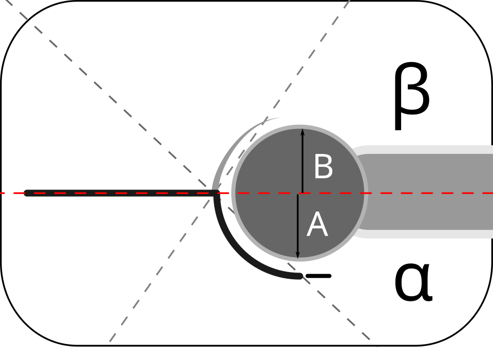
A schematic of the Graf hip measurement. The red baseline runs along the side of the pelvis. The alpha angle (α) measures the slope of the bony socket roof. The beta angle (β) measures the cartilage roof above it. A and B mark the lower and upper halves of the femoral head.
Ultrasound puts a number on how well a hip is formed. The standard way is the Graf method, named for the doctor who devised it. It rests on one clear view: a slice down the side of the hip that shows the socket, the ball, and the straight bony wall of the pelvis above. From that view a clinician measures the angles that define the hip. Holding that one plane steady is what the measurement depends on. Off that plane, the angle means nothing.
The main number is the alpha angle. It measures the slope of the bony socket roof against the straight wall of the pelvis. A well-formed socket gives a steep roof and a large angle. A review of ultrasound screening for hip dysplasia sets the normal at sixty degrees or more. An angle below sixty marks a socket too shallow to hold the ball well. The alpha angle is the single most useful number the scan gives. It puts one repeatable figure on a socket’s depth. A figure is something a screening programme can act on and audit.
The normal value depends on the baby’s age. The angle is allowed to sit a little lower in a newborn, reaching sixty degrees by about three months. A clinician reads each hip against the baby’s age, using the value Graf set out for each week. Reading the angle without the age behind it can mislead. A number read against the wrong age can call a fine hip abnormal.
Beyond the angle, the scan checks how the ball sits. A well-formed hip holds the ball deep in the socket, with the bony roof curving over more than half of it. A clinician looks at how much of the ball the socket covers, a quick check that backs up the angle. A healthy socket covers at least half of the ball. Good coverage and a good angle together say a hip is well formed.
The numbers turn a look into a measurement. Two clinicians scanning the same hip, using the Graf method, arrive at close to the same angle. That makes the scan something a unit can rely on and repeat. A measured angle, written down, can be set beside the next scan to see whether the hip is improving. That repeatability is what makes it fit for screening.
The Graf hip, by the numbers
Reading
What it means
Alpha angle (bony socket roof)
60° or more is normal
Alpha below 60°
a socket too shallow to hold the ball
Graf type I
a normal, well-formed hip
Graf type II
an immature or mildly shallow socket
Graf type III–IV
the ball pushed to the edge, or out of the socket
Best time to scan
the first months, before about 6 months
Chief risk factor
breech position before birth
The cartilage roof
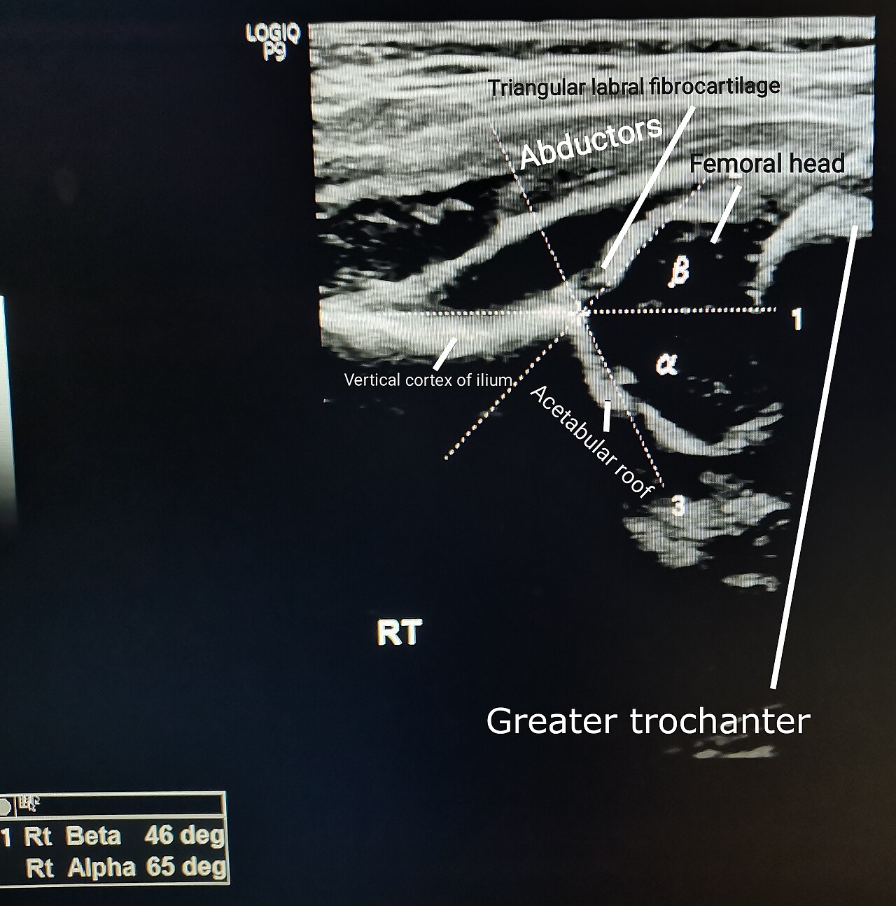
A coronal ultrasound of a normal infant hip. The bright line of the ilium forms the baseline. The femoral head sits in the socket, with the bony acetabular roof and the labral fibrocartilage above it. Here the alpha angle reads 65 degrees, within the normal range.
The cartilage part of the socket has its own measure. Above the bony roof, the rim of the socket is cartilage, ending in a small lip called the labrum. The beta angle measures the slope of that cartilage roof. Together, the alpha and the beta describe the socket fully, the bony part and the cartilage part.
The cartilage roof tells how the hip is coping. When a socket loosens, the cartilage rim rides up, pushed out of place by a ball that no longer sits deep. A clinician reads the cartilage roof alongside the bony angle, the two together giving the fuller picture. Read together, the alpha and the beta place a hip on the Graf scale. On the screen the labrum shows as a small bright triangle over the ball.
The dynamic exam
A sound hip needs two things: a well-shaped socket and a stable joint. The scan reads both. Beyond measuring the shape, it watches the joint move, to see whether the ball holds its place or slips in the socket. Looseness, as much as shape, decides whether a hip will dislocate.
The dynamic test gently stresses the joint. During the scan, a clinician moves the baby’s leg and presses gently, the same movements used in the hands-on newborn hip exam. On the screen, a clinician watches whether the ball stays seated under the push. A ball that slides toward the rim, or out over it, marks an unstable hip. The push shows in seconds what a still picture can hide.
Watching the movement adds what a measurement alone cannot. The scan reads both the shape and the stability in one study. That live read is something only ultrasound can give, since it watches the joint as it moves. Both readings come from one short scan.
Stability changes the plan. A borderline hip that holds firm may only need watching, with a repeat scan in a few weeks. A hip that slips needs treatment, whatever its angle. By watching the joint move, the scan flags the hips that need a harness, early enough to start. Shape and stability together decide what a hip needs. Stability is read in seconds, with the probe held steady on the joint.
Timing the scan
Timing is everything with a hip scan. The window for ultrasound is the first months of life, the months the hip is still mostly cartilage. Around six months the head of the thigh bone begins to turn to bone. That bone blocks the sound, casting a shadow over the socket behind it. After that, an X-ray takes over, since the bone now shows on a film. There is a lower limit too: in the first weeks, a normal hip can look a little loose, because the ligaments are still relaxed from the mother’s hormones. Screening is usually set at about six weeks of age, once that settles. The best window is roughly six weeks to six months, early enough that a shallow hip can still be guided to grow well. Scanning in that window is what lets a simple harness do the work that surgery would otherwise have to. Miss the window, and a simple harness gives way to an operation. Those few months are the whole reason DDH is screened so early.
Which babies are scanned
Not every baby needs a hip scan. Different countries scan different babies. Where screening is selective, the scan goes to the babies at higher risk of DDH. Some babies carry a clearly higher risk. Those are the ones a clinician scans first.
A handful of things raise the risk. The strongest is breech position before birth, a baby that lay bottom-down in the womb. A family history of DDH raises it too. So does being a first-born, and so does being a girl. A baby with any of these is sent for a scan, even after a normal hip exam at birth. A risk factor is only a reason to scan. Most scanned hips turn out normal.
The hands-on exam and the scan work together. A midwife or doctor checks every newborn’s hips by hand, feeling for a click or a slip. A hip that feels abnormal goes straight for a scan. The scan confirms what the hands found, and it catches the loose hips a hand can miss. The scan puts numbers on what the hand can only feel.
The Graf types
Graf sorted hips into types, from normal to dislocated. The types run on the alpha angle, with the cartilage roof and the dynamic test filling in the detail. A clinician reads a hip, measures it, and names its type. The type sets what happens next.
Type one is a fully formed hip. The alpha angle is sixty degrees or more, the socket deep, the ball well held. A type one hip needs nothing but a clean bill. Most hips scanned, even many sent for risk, turn out to be type one. It is the result a unit hopes to write most often. A type one hip is signed off and needs no return visit.
Type two is the immature or mildly shallow hip. Its angle sits below sixty, the socket a little too flat for the baby’s age. A type two hip may be slow to mature and catch up on its own, or it may need a harness to help the socket deepen. A repeat scan tells which way it is heading. Many type two hips simply need a few weeks and a repeat look.
Types three and four are the serious hips. In these the ball has been pushed to the edge of the socket, or right out of it, the cartilage rim folded over and the joint dislocated. These hips need treatment without delay. The scan names the type plainly, so the worst hips are sent for care first. These are the hips that screening exists to catch.
What the scan guides
The scan steers the treatment. A shallow or loose hip, caught early, is treated with a harness, a soft brace that holds the baby’s legs bent and turned out, so the ball sits deep in the socket. Held that way for some weeks, a shallow socket grows in around the ball. The harness is soft, worn under the baby’s clothes. Most babies settle into it within a few days.
The scan also follows the treatment. A baby in a harness is scanned again to check the ball is sitting deep and the angle is improving. If the hip is deepening, the harness stays on. The scan watches the socket grow over the weeks. It confirms when the hip is fixed and the harness can come off. A hip confirmed normal on a follow-up scan needs no more checks. Most hips caught early are normal again within a few months.
How the scan is done
The scan asks little of a baby. A clinician lays the baby on its side, settles the small probe on the side of the hip, and finds the standard Graf view. The baby can feed or sleep through it. The whole scan, both hips, takes only a few minutes. A calm baby gives the steadiest image.
Getting the right view is the skill. The Graf angles only hold true on one exact slice, the coronal view that shows the straight wall of the pelvis. A clinician learns to find that plane reliably, since a tilted view throws the angle off. With the plane right, the angle is quick to measure.
A handheld machine does the job well. A small micro-convex or high-frequency linear probe reads a baby’s shallow hip clearly. A pocket-sized scanner carries either. The scan can be done in a clinic, on a ward, or on a home visit, wherever a baby is seen. For a screening test run on many babies, that portability makes it easy to reach every one. A clinic, a ward round, a home visit: the scanner goes to all of them. The image and the angle save to the baby’s record for the next visit.
What ultrasound gives
For the infant hip, ultrasound is the tool the early months call for. It reads the soft cartilage that an X-ray cannot show. It measures how well the socket is formed, watches the joint for looseness, and names the type of hip, all from a few minutes’ scan. For finding DDH early, when it is still easy to treat, ultrasound has no equal in the first months. It turns a hidden problem into one a clinician can see and measure.
Its strength is reading cartilage and motion. The scan measures the bony socket with the Graf angle, reads the cartilage roof and the labrum for the soft part, and watches the ball under a gentle stress for looseness. From those, a clinician reads a hip fully. The whole joint is read in the cartilage months, before the easy fix is lost. No other test reads a cartilage joint this fully in a baby.
Screening is where this pays off. DDH gives no pain and often no outward sign in a newborn. A shallow hip can go unnoticed until the child limps. Ultrasound finds it at six weeks, when a harness can still set it right. A few minutes of scanning can spare a child an operation and a lifetime of trouble.
A baby’s hip is a small window that does not stay open long. For the months it is cartilage, ultrasound reads it fully, safely, at no cost to the baby. It measures the socket, watches the joint, and catches the hip that needs help, early enough for that help to stay simple. For the newborn hip, the scan is how trouble is found early, and set right in time. Found at six weeks, most of these hips never trouble the child again.
Common questions
Why is ultrasound used for a baby’s hip instead of an X-ray?
Because a baby’s hip is mostly cartilage. The ball of the joint, and much of the socket, are soft cartilage in the early months, turning to bone only later. Ultrasound reads cartilage clearly, so it pictures the whole joint from birth. An X-ray needs bone to show a joint, so it sees little of a hip this young. For the first months, ultrasound is the tool that shows the hip plainly.
What is the alpha angle, and what is normal?
The alpha angle measures how well the bony socket is formed. It is the angle between the straight wall of the pelvis and the slope of the bony socket roof. A larger angle means a deeper socket. Sixty degrees or more is normal for a hip past about six weeks of age. Below sixty, the socket is too shallow. The hip then needs following or treatment.
When should a baby’s hips be scanned?
In the first months, roughly six weeks to six months. Before about six weeks, a normal hip can look a little loose, because the ligaments are still relaxed from the mother’s hormones, so a very early scan can mislead. Around six months, the head of the thigh bone turns to bone, which blocks the sound and ends the window. Scanning between those points reads the hip in the months it is still cartilage. It is also the age when a shallow hip is easiest to treat.
Which babies need a hip scan?
The babies at higher risk, and any whose hip exam is abnormal. The strongest risk factor is breech position before birth. A family history of DDH, being a first-born, and being a girl each raise the risk too. Where screening is selective, these higher-risk babies are the ones picked out. A baby whose hips feel loose or click on the newborn exam is sent for a scan whatever its risk.
What happens if the scan finds DDH?
It is treated early, usually with a harness. A shallow or loose hip caught in the first months is treated with a soft harness that holds the baby’s legs bent and turned out, so the ball sits deep and the socket grows around it. Worn for some weeks, it guides most hips to normal. The scan follows the progress and confirms when the hip is fixed. Caught this early, the great majority of hips are put right without surgery.
About the Author
Julien Mercier
Senior R&D Engineer · Medical Ultrasound Transducer Development
Senior R&D Engineer with an M.S. in Applied Physics and over 15 years of experience in medical ultrasound transducer development, specializing in the design verification and performance testing of high-frequency imaging transducers. Currently leading the development and verification of the company’s next-generation high-frequency linear-array transducer, responsible for imaging performance evaluation and reliability analysis in preclinical testing. Brings extensive hands-on experience in piezoelectric element tuning, beamforming parameter optimization, and system-level performance testing.