Neonatal lung ultrasound reads a newborn’s lungs from the surface of the chest. A baby’s lung is full of air. Air normally turns sound back, so the scan does not picture the lung the way it pictures a solid organ. What it reads are the patterns that air and fluid throw up at the lung’s surface: bright lines, dark bands, and the slide of the lung against the chest wall. From those patterns a clinician can read a healthy lung, spot a wet one, find a collapse or an air leak, and sort out why a newborn is struggling to breathe, all at the cot.
What it is, and why
A newborn’s chest suits ultrasound well. The chest wall is thin. The ribs are soft, with wide gaps to scan between. The lung lies right beneath them. A small probe set between two ribs reads the lung surface directly, in fine detail. Between the ribs, the probe looks straight onto the lung.
The scan replaces a lot of X-rays. For years a breathless newborn meant a chest film, often several over days in intensive care. Lung ultrasound answers many of the same questions at the cot, in radiation-free minutes. A baby can be scanned again and again when its breathing changes, at no cost to a small body. Sparing those films spares a fragile baby a real dose over weeks of care.
It has grown into a frontline tool in the nursery. A clinician can scan a struggling newborn, read the cause, and act on it, the baby never leaving the cot. For sorting out why a baby cannot breathe well, lung ultrasound has become one of the first tools a clinician reaches for. It belongs in the same kit as the stethoscope on a busy round.
The lung gives up only a handful of signs. They are quick to learn. Bright horizontal lines, bright vertical lines, the slide of the surface, a liver-like patch, a pocket of dark fluid: each points to a particular state of the lung. Each of these signs has its own section below. Together they let a clinician read a newborn’s chest in minutes.
Reading an air-filled lung
Reading an air-filled lung takes a different eye. Sound cannot pass into healthy lung, because the air inside scatters it straight back. So the scan does not show the lung tissue itself. It reads the surface where the chest wall meets the lung, the pleural line, and the patterns that bounce off it. The surface is enough, because every sign a clinician needs forms right there.
The pleural line is where it all begins. It shows as a bright horizontal line a centimetre or so down, where the lung surface lies. Everything ultrasound reads about the lung comes off that line: the lines below it, the way it slides, the breaks in it. A clinician finds the pleural line first, then reads what it is doing. Finding it well is the first skill a learner picks up.
From that line, a clinician reads two things: the lines that hang off it, and the way it moves. The lines show how wet the lung is. The movement shows whether the lung is sliding. Together they build a picture of the lung underneath.
The normal lung
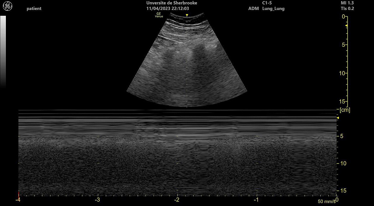
An M-mode lung ultrasound. The sand-like band below the still lines is normal lung sliding, the pattern called the seashore sign. The text around the image is the machine’s own settings.
A healthy lung gives a steady pattern that repeats. Below the bright pleural line run faint horizontal lines, evenly spaced, called A-lines. They are an echo of the pleural line itself, repeating down the screen. A-lines are the sign of an air-filled lung, the look of lungs working as they should. Their even spacing is the easiest normal sign to recognise.
The lung also moves. The scan catches that movement: with each breath, the lung surface slides back and forth against the chest wall. On the screen the pleural line shimmers along its length, a sign called lung sliding. Sliding tells a clinician that the lung is up against the chest wall, inflating and deflating as it should. Lost anywhere, that slide is itself a clue.
Frozen in time, the sliding shows up in another mode. Switched to M-mode, which plots one line of the picture against time, a sliding lung draws a grainy band like sand beneath the still lines of the chest wall. This is the seashore sign: still lines above, sandy band below. It is the M-mode signature of a lung sliding normally. M-mode freezes the motion that the live picture shows in real time.
Together, A-lines and lung sliding say the lung is healthy there. The pattern is quick to recognise. A clinician learns it first. Once the normal look is fixed in mind, the abnormal patterns stand out plainly. Most of lung ultrasound is reading how far a lung has drifted from this baseline. A scan that shows the normal pattern across the chest is reassuring in seconds.
The normal pattern also rules things out. A lung that slides, with clean A-lines, has no collapse and no air leak at that spot. Finding that pattern across the chest can settle a worry quickly. Sometimes the most useful thing the scan does is show that a breathless baby’s lungs look normal, and point the search elsewhere.
B-lines, the wet lung
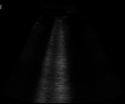
B-lines on a lung ultrasound: bright vertical bands falling from the pleural line. They come from fluid in the lung. A screen full of them means a wet lung, as in a newborn with RDS or transient tachypnea.
When fluid enters the lung, the pattern changes. Bright vertical lines appear, rising from the pleural line and running straight down the screen. These are B-lines. They come from fluid in the tiny spaces of the lung, which sends the sound echoing in a new way. The change is quick to see, even for an eye new to lung ultrasound.
B-lines tell how wet a lung is. The more of them a clinician counts, the more fluid the lung holds. A scatter of them can be normal in the first hours of life. Packed close between the ribs, they mean a lung heavy with fluid, a pattern called lung rockets. Merged into a solid white field, they mean a very wet lung, the look named white lung. Reading the density of the lines, a clinician gauges how heavy the fluid load is.
The breathless newborn
Two common conditions make a newborn’s lungs wet. The first is transient tachypnea, the wet lung of a baby whose lung fluid has been slow to clear after birth. On ultrasound it shows as B-lines, often heavier low in the chest, that fade over the first day or two once the fluid clears. The scan can watch that clearing, scan by scan. Watching the lines fade confirms the diagnosis without a single film.
The second is respiratory distress syndrome, the lung of a premature baby short of surfactant. Such a baby has stiff lungs that have not filled. On ultrasound they show heavy B-lines or white lung, with a pleural line that looks rough, its smooth edge broken up. The pattern covers both lungs evenly. Lung ultrasound picks up RDS early and grades how severe it is. Caught early, RDS can be treated before the baby tires.
Each condition has its own look and its own course. A clinician reads the pattern alongside the baby’s age and how its breathing unfolds. The scan points to the likely cause, and to the treatment that fits it. Reading it early can start the right care sooner. Naming the cause is the first step toward the right support. The table sets the common patterns side by side.
Common newborn lung patterns on ultrasound
Pattern
What it suggests
A-lines, with lung sliding
a normal, air-filled lung
A few B-lines, fading over a day
transient tachypnea, a wet lung clearing
Heavy B-lines or white lung, rough pleural line
respiratory distress syndrome
A patch like liver, with bright specks
consolidation, often pneumonia
No sliding, no B-lines, a lung point
pneumothorax, an air leak
Dark fluid above the diaphragm
pleural effusion
The scan also tracks the response. After surfactant is given, or once a wet lung dries, the B-lines ease and the A-lines return. A clinician scans again a few hours on and reads whether the lung is improving. That close follow-up, safe to repeat, is one of the scan’s real strengths in the nursery.
Consolidation
When a piece of lung fills with fluid or pus, it stops behaving like air and starts to look like a solid organ. On the scan that stretch of lung takes on the look of liver, a soft grey block where there should be bright lines, a change clinicians call hepatisation. Within it, bright dots and branches often show, the air still trapped in the small airways, a sign called air bronchograms. Consolidation like this is the mark of pneumonia, or of a stretch of lung that has gone airless. The scan shows how big the consolidated patch is and where it sits. A clinician follows it over days to see it clear or grow. For a baby with a fever and hard breathing, finding a consolidated patch can name the problem at the cot, with no film needed. A consolidation that shrinks scan by scan is a lung getting better. A patch that spreads points to an infection gaining ground.
Air leak
An air leak is the emergency the scan rules in or out fast. When air escapes into the space between the lung and the chest wall, a pneumothorax, it pushes the lung away from the wall. The lung no longer touches the chest wall there, so it no longer slides against it. On the scan the pleural line goes still. That stillness is the first thing to look for.
Absent sliding is the first clue. The lung point confirms it. Where air has leaked, the lung lies away from the wall, showing no sliding and no B-lines. At the edge of the leak sits a spot where sliding lung meets the still wall, the lung point, a sign specific to pneumothorax. A review of neonatal lung ultrasound notes that the scan reads pneumothorax more reliably than a plain film, and faster.
An air leak leaves little time to spare. A large pneumothorax can squeeze the lung and the heart. A baby can go downhill fast. Ultrasound gives the answer in seconds, at the cot, with no wait for a film. For a newborn suddenly worse, that quick read can be what gets the air drained in time. An answer that arrives in seconds can change what happens in the next minute.
Fluid and the diaphragm
Fluid can also gather outside the lung. When fluid collects in the space between the lung and the chest wall, a pleural effusion, it shows on ultrasound as a dark pocket above the diaphragm. Fluid lets sound through, so an effusion shows up clearly and directly. The scan reads its size and shape with ease.
Ultrasound is the best test for an effusion. It finds even a small pocket of fluid, measures how deep it is, and shows whether it is clear or full of debris. It marks the safest spot to put a needle, when the fluid needs draining. For finding and guiding the drainage of an effusion, the scan is the tool of choice. Draining under ultrasound guidance keeps the needle clear of the lung and the heart.
The diaphragm comes into the same view. The scan shows the sheet of muscle between the chest and the belly. It watches the muscle move with each breath. A diaphragm that lies still, paralysed, can leave a baby struggling for air. The scan catches that stillness, one more answer in a single study. One sweep of the chest reads the lung, the fluid, and the diaphragm together.
Effusion and the diaphragm round out the ABCDE the scan covers. A clinician runs through A-lines, B-lines, consolidation, the diaphragm, and effusion, in order, on each side of the chest. The set survey means no finding is missed. From a few minutes of scanning, the whole chest is read.
The probe and the sweep
A small probe does this work. A micro-convex probe, with its small curved face, fits between a newborn’s ribs and fans a wide view across the lung field, taking in several rib spaces at once. Its frequency reaches deep enough to read a consolidation or an effusion below the surface. For a quick survey of the whole chest, the micro-convex probe is well suited.
A high-frequency linear probe brings out the finest surface detail. Pressed over the ribs, it reads the pleural line and the B-lines coming off it with great clarity, which is why it is favoured for the surface signs. A clinician may sweep with the micro-convex, then switch to the linear for a close look at the pleura. Both small probes fit a newborn chest. A handheld machine can carry either.
Scanning again and again
Breathing changes fast in a newborn, so one scan is rarely the end. A baby with wet lungs may clear within hours, or worsen into something that needs more support. A scan repeated through the day follows how the lungs change. The signs read in sequence tell more than any single picture. A worsening trend on the lines often shows before the numbers on the monitor do.
Serial scanning suits the bedside tool. The scan uses no radiation, takes minutes, and asks nothing of a baby beyond a warm probe on the chest. A clinician can scan a struggling baby every few hours, watching the B-lines, the sliding, and the consolidation shift. That close watch lets a team match the support to the lung from hour to hour. Each repeat costs the baby nothing, so a clinician scans as freely as the case needs. No other chest test can be brought back so often.
Beyond the chest X-ray
For decades the chest X-ray was the test for a newborn’s lungs. It shows the lungs as light and shadow, the heart’s outline, and the place of every tube and line. It catches a large pneumothorax and a whited-out lung. The chest film keeps its place as a fast first look.
Lung ultrasound reads the surface more closely. It reads how wet a lung is from the lines, finds a small consolidation the film may miss, and reads a pneumothorax more surely. It does all this at the cot, in radiation-free minutes, on a baby who may need many looks. For the day-to-day picture of a newborn’s lungs, the scan reads finer and costs less. On a baby who needs daily looks, that adds up to many spared X-rays.
The two work side by side. Many units now reach for the scan first in a breathless baby. The film is kept for what it does best. Used together, the X-ray and the scan leave little about a newborn’s chest unread.
What lung ultrasound gives
For a newborn’s lungs, ultrasound has become a tool a unit reaches for early. It reads the chest from the surface, off the patterns that air and fluid throw up: the lines, the sliding, the fluid, the still patches. From those it reads a healthy lung, spots a wet one, finds a collapse or a leak, and names why a baby is breathless. All of it happens at the cot, in minutes.
Its strength is reading air, the very thing that once put the lung beyond ultrasound. A-lines and sliding mark a healthy lung. B-lines mark the fluid in a wet one. A liver-like patch marks consolidation. A still pleural line with a lung point marks an air leak. From a handful of signs, the scan reads the lungs of the whole nursery. A clinician carries that short list in mind and reads a chest fast.
The scan fits the smallest patients especially well. A premature baby in respiratory trouble is exactly the patient who cannot spare a trip to X-ray, or a dose of radiation, or a wait. Lung ultrasound comes to the cot, gives its answer in minutes, and can be repeated as often as the breathing demands.
A newborn’s breathing can turn within minutes, so seeing the lungs clearly, scan after scan, is what makes the difference. Lung ultrasound gives a team that view, safely, at the cot, as many times as a baby needs. For the breathless newborn, it has become one of the surest ways to read the lungs and act in time.
Common questions
How can ultrasound see a lung that is full of air?
It reads the patterns at the lung’s surface. It does not picture the lung tissue itself. Air turns sound back, so the scan cannot see deep into a healthy lung. What it reads are the lines and the movement at the pleural line, where the chest wall meets the lung. From those patterns, a dry lung, a wet one, a collapse, or an air leak each shows a look of its own. The air that blocks the view is the very thing the scan learns to read.
What do B-lines mean on a newborn’s lung scan?
They mean fluid in the lung. B-lines are bright vertical lines that rise from the pleural line and run down the screen, thrown up by fluid in the small spaces of the lung. A scatter of them can be normal in the first hours after birth. Packed close together, they mean a wet lung, as in transient tachypnea or respiratory distress syndrome. The more a clinician counts, the more fluid the lung holds.
Can lung ultrasound find a pneumothorax?
Yes, and quickly. When air leaks between the lung and the chest wall, the lung stops sliding against the wall there. On the scan the pleural line goes still. At the edge of the leak, a spot called the lung point shows where sliding lung meets the still wall, a sign specific to pneumothorax. Ultrasound reads this at the cot in seconds, more reliably than a plain film. For a newborn suddenly worse, that speed can be what gets the air drained in time.
Does it replace the chest X-ray in a newborn?
No, it works with it. The chest X-ray stays a fast overview. It shows the lines and tubes in a sick baby. Lung ultrasound reads the lung surface more closely, in radiation-free minutes at the cot: how wet the lung is, a small consolidation, a pneumothorax. Many units now scan first in a breathless baby and keep the film for what it does best. Side by side, the two give the fuller picture.
What probe is used for a newborn’s lungs?
A small one, the same kind used elsewhere on a newborn. A micro-convex probe fits between the ribs and sweeps a wide view across the lung field, deep enough to reach a consolidation or an effusion. A high-frequency linear probe reads the pleural line and the B-lines off it in the finest detail. A clinician often sweeps with the micro-convex, then switches to the linear for a close look at the surface. Both are small enough for a newborn chest and fit on a handheld machine.
About the Author
Julien Mercier
Senior R&D Engineer · Medical Ultrasound Transducer Development
Senior R&D Engineer with an M.S. in Applied Physics and over 15 years of experience in medical ultrasound transducer development, specializing in the design verification and performance testing of high-frequency imaging transducers. Currently leading the development and verification of the company’s next-generation high-frequency linear-array transducer, responsible for imaging performance evaluation and reliability analysis in preclinical testing. Brings extensive hands-on experience in piezoelectric element tuning, beamforming parameter optimization, and system-level performance testing.