What Ultrasound Probes Work For Neonates And Infants Handheld
The first choice in scanning a baby is the probe. A newborn carries small organs just under the skin, within easy reach of sound. The probe that suits them has a small footprint. It runs at a high frequency. A micro-convex probe handles most of the work, joined by a high-frequency linear probe for the most superficial scans and a sector probe for the heart. Picking the right one is where a good pediatric scan begins.
What decides the probe
Two things about a baby drive the choice. The body is small, so the probe must have a small face to fit the spaces it offers: a soft spot, a gap between ribs, a tiny limb. The organs lie shallow, within a few centimetres of the skin, so a high frequency can be used to draw fine detail. A small face and a high frequency: those two together name the probe for a newborn. A few centimetres of reach is all a newborn asks of it.
An adult probe gets neither right. Its face is too broad to sit on a newborn. Its frequency is set for organs deep in a grown body. A baby’s probe answers both at once: small enough to sit on a newborn, quick enough to sharpen a shallow organ. Pediatric scanning starts by reaching past the adult probes for one sized to a newborn.
Three probe shapes do the work, each with a job it fits. The micro-convex covers the general scan. The linear takes the most superficial targets. The sector reaches the heart. Each of the three has a shape and a frequency suited to a particular task. Each probe gets its own section below, with the scans it fits and the frequency it runs at.
The micro-convex workhorse
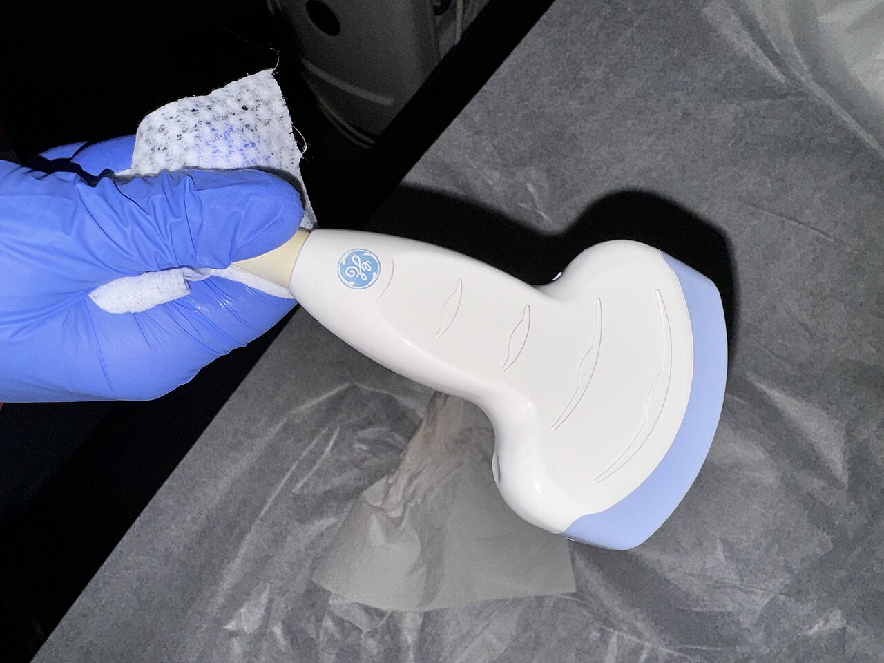
A curved-array ultrasound probe. The micro-convex probe used on a newborn is this shape shrunk to a small face, narrow enough to sit between two ribs or on a soft spot.
The micro-convex probe does most of the work in a nursery. It is a curved probe shrunk to a small footprint, with a face that curves over a short radius. That small curved face sits where a baby gives room: between two ribs, under the costal margin, on the soft spot of the skull. From a tiny patch of contact, it fans a wide view into the body below. The curved face throws a fan-shaped field that broadens below the skin. That fan suits a belly or a head, where a clinician wants a broad sweep from a small point of entry. The curve also throws sound around the gas in the bowel, reaching what lies behind it. That reach behind gas is why the curved shape suits a belly full of bowel.
Its frequency, around five to eight megahertz, suits the depth a baby’s organs sit at. The same probe reads the brain through the fontanelle, the bowel through a soft belly, and the kidneys and bladder below. A unit that owns one micro-convex probe can cover most of what a newborn needs scanned. It serves the vessels too, finding a large vein for a central line. For the bulk of a newborn’s scans, the curved probe is the one in a clinician’s hand. Most machines carry a neonatal preset for it, the depth and frequency already dialled for a small patient. It is the first probe to reach for and the last to put down.
The high-frequency linear probe
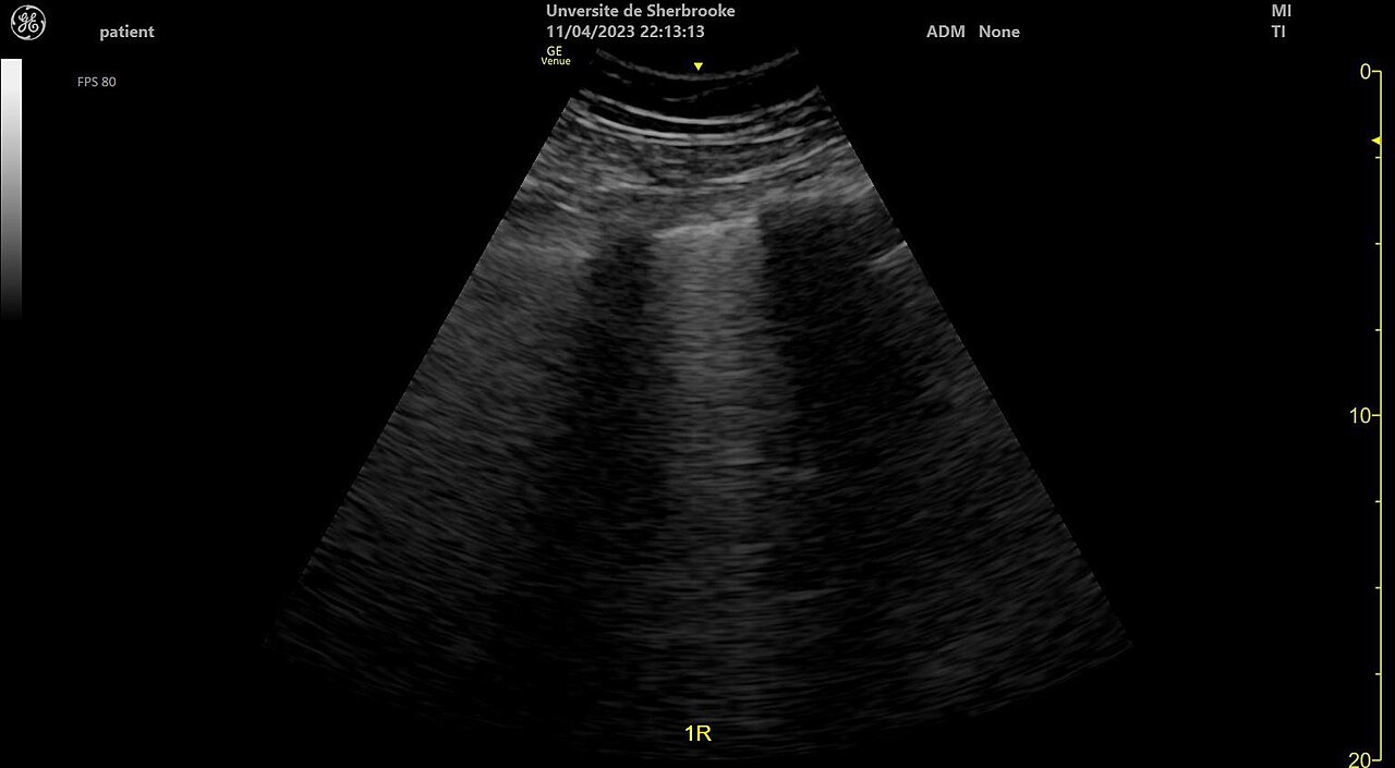
A lung ultrasound. The bright bands fanning down from the top are B-lines, the sign of fluid in the lung. A high-frequency probe brings the lining and these lines out most sharply.
Some scans call for fine detail in the shallowest layers. For these a high-frequency linear probe steps in, running anywhere from ten to eighteen megahertz. Its flat face lays a rectangular field on the skin and resolves the surface finely. The lung sits right under the chest wall. A linear probe reads its lining and the bright lines that come off it with the most clarity. A rectangular field shows the layers of the surface in their true width, side by side. Near the probe, where a baby’s organs live, the linear probe resolves detail no other shape can match.
The evidence backs the linear probe for the newborn lung. A comparison of probe types for neonatal lung ultrasound found the linear probe gave the clearest images, scoring well above the micro-convex on the same babies. For the pleura and the fine signs that read off it, the high-frequency linear is the probe of choice. The gain holds for babies of every weight and skin tone.
The infant hip is the other classic linear scan. A baby’s hip is shallow cartilage. A high-frequency linear probe shows its shape and the socket’s angle precisely. The flat face lines the bony rim up cleanly for the measurement. The angle, read against that straight edge, puts a number on how deep the socket is. A small hockey-stick linear probe, shorter than the standard one, suits the tiny hip of a newborn. For screening a hip in the first months, the linear probe is the standard tool.
The same probe serves the shallowest targets elsewhere. It maps the spine of a newborn for a dimple that might tether the cord. It finds a vein for a line and guides the needle in. Wherever the target sits a centimetre or two down, the linear probe brings back the sharpest picture. Soft tissue, nerve, and the wall of a vessel all come up crisp at these frequencies.
The sector probe and the heart
The heart needs a probe of a different shape. It beats behind the ribs and the lung, both of which block sound. A phased or sector probe has a very small face, small enough to aim between two ribs. From that narrow window it fans a wide sector. A single rib space opens a full view of the beating heart. The clinician works through a few set windows, below the ribs and beside the breastbone, angling the small face to catch each view.
Its frequency runs lower, often two to seven megahertz, to reach the depth the heart sits at and to keep up with its speed. A lower frequency draws a coarser picture, the price of that depth. For the heart, reach and frame rate come first. A sector probe gives both. Colour Doppler runs on the same probe, painting the flow through the valves and the great vessels.
A small footprint serves here too, as much as on the chest. The same narrow face that fits between ribs can sit on the fontanelle. Some clinicians read the newborn brain with a sector probe. For the heart, it is the one probe that does the job. A baby’s heart beats fast. The sector probe’s high frame rate keeps every beat in view. Neonatal echo runs on the sector probe.
Frequency and the patient’s size
Frequency is the dial that matches a probe to a patient’s size. The higher the frequency, the finer the detail it draws, and the shorter its reach into the body. The art is to use the highest frequency that still reaches the organ in view. On a baby, that frequency is high, since the organs sit so shallow.
A baby rewards a high frequency. The organs sit close, so the short reach of a high frequency costs nothing. The detail it brings out is exactly what a small structure needs. Nothing of interest sits deep enough to need a lower frequency. On a premature baby, the thinnest of patients, even the deepest organ sits within a high-frequency probe’s reach.
The smaller the baby, the higher the frequency goes. A premature newborn, tinier and thinner than a term baby, takes a higher frequency still. The lung of a tiny preterm reads best at the top of the linear probe’s range. On a bigger, older baby, the frequency comes down to reach the deeper organs of a larger body. A smaller patient simply allows a higher number on the dial.
Most handheld probes let the frequency be set within a range. A clinician picks the part of the range that fits the depth in view, sitting at the high end for the shallowest scans. The number is not fixed by the probe alone. It is tuned to the baby and the organ in front of it. A neonatal preset sets a sensible starting point. The clinician nudges it from there.
This is why a baby is, in a sense, the easy patient for ultrasound. Its organs sit shallow, in plain view of a high-frequency probe. The detail comes free. The lost depth is never needed. A small body suits ultrasound better than it suits almost any other tool.
The footprint must be small
Whatever the probe, its footprint has to be small to work on a baby. A newborn offers almost no flat ground: a fontanelle the size of a coin, rib spaces a finger wide, a hip no bigger than a thumb. Only a small face sits flush on ground like that. It holds full contact and angles into the narrow windows a baby’s anatomy leaves open. The smaller the face, the more of a tiny body it can reach. This is why every probe made for newborns, curved or flat or sector, comes with a small face. Gel fills the last gap between the small face and the curved skin, so the contact is complete. On a curved little limb, only a small face holds steady contact end to end. The footprint is the first thing that has to fit, ahead of frequency or anything else.
Matching the probe to the scan
Each scan has a probe that suits it best. The brain through the fontanelle goes to the micro-convex, the small curved face fitting the soft spot. The belly goes to the same micro-convex, which reaches the bowel and the organs behind it. These two scans, the commonest in a newborn, run on the one curved probe. One probe covers the two busiest scans of a nursery.
The superficial scans go to the linear probe. The lung, the hip, the spine, a vein for a line: each sits close to the skin, where the high-frequency linear draws the finest picture. A unit that does these scans keeps a linear probe alongside the micro-convex. Vascular access leans on it as well, the probe in one hand guiding the needle under its eye. The two cover between them almost everything a baby needs. Between the pair, a nursery is equipped for nearly every scan it meets.
The heart goes to the sector probe, the one scan the curved and flat probes cannot do well. A nursery that reads hearts adds a sector probe to the set. The table pairs each common scan with the probe that suits it, and the frequency that probe runs at.
Which probe for which newborn scan
Scan
Probe
Frequency
Brain, through the fontanelle
micro-convex
about 5–8 MHz
Belly and bowel (NEC)
micro-convex
about 5–8 MHz
Lung
high-frequency linear
about 10–18 MHz
Hip (DDH)
high-frequency linear
about 10–18 MHz
Heart (echo)
phased / sector
about 2–7.5 MHz
Why shallow organs read so well
It helps to see why a high frequency cannot also reach deep. Sound at a high frequency carries fine detail, since its short waves resolve small things. Those same short waves fade fast over distance, so a high frequency cannot reach far into the body. A rough rule ties the two together: the higher the megahertz, the finer the detail and the shallower the reach. The depth a probe can see is set by how fast its sound fades.
A newborn’s organs fall inside that short reach. They sit within a few centimetres of the skin, exactly where a high frequency is sharpest. A clinician can run the probe fast and lose nothing to depth. Every organ a baby has lies in the high-frequency zone.
This is why the detail comes free in a baby. The high frequency that resolves a small structure also reaches it, since the structure is shallow. No one setting has to be traded against another. The picture reaches the organ and stays sharp. A low frequency would only buy depth the baby has no use for.
This is the deep reason a baby is easy to scan. The probe is small because the body is small. The frequency is high because the organs are shallow. Both choices, forced by the baby’s size, happen to be the choices that give the best picture. The clinician spends no effort coaxing depth out of a shallow patient. Size works in the clinician’s favour from the start.
One probe or several
How many probes a unit needs depends on what it scans. A single micro-convex probe covers the brain, the belly, and the general work of a nursery. For many units that is enough. That single probe is where most units begin. Many never need more. It is the one probe a newborn service cannot do without.
A unit that scans lungs, hips, or hearts adds a linear or a sector probe alongside it. A full neonatal service ends up with all three, each reaching for its own scans. The cost of a second or third probe is small against the scans it opens up. Many handheld machines take more than one probe, or switch a single probe between modes, so a small device can still cover the range. A spare probe head costs far less than the scanner it plugs into.
On a handheld machine
On a handheld scanner, the probe is most of the machine. A pocket-sized device is often a probe with a screen attached, or a probe that sends to a phone or tablet. The probe is where the image is made. Its quality sets what the whole device can do. Choosing it well is choosing most of the scanner.
This puts a premium on the right probe for a baby. A handheld micro-convex probe puts the brain and the belly in a pocket. A handheld linear probe puts the lung and the hip there too. The same pocket form that suits the bedside carries the probe a baby needs. Slipped into a coat pocket, it goes from cot to cot through the unit.
Some handheld probes now cover more than one shape in a single head. A wide-band probe can switch in software from a curved format to a linear one. For a nursery short on space or budget, one such probe can stand in for two. A wireless probe sends its image to a tablet over the air, untethered at the cot. The trend puts the range of pediatric probes within reach of a single handheld device.
Why a baby needs its own probe
A baby’s probe is its own tool, chosen for a newborn’s small body and shallow organs. Its small face fits the spaces a newborn offers. Its high frequency draws the detail a shallow organ rewards. Both choices follow from one fact: a newborn is small. Sizing and tuning the probe to that fact is what makes a clean newborn scan possible. Everything downstream, the image and the reading, rests on that first fit.
Reaching for the right probe is the start of every pediatric scan. A clinician who picks the micro-convex for a belly, the linear for a lung, the sector for a heart, has already done half the work. The picture comes easily once the probe fits the patient and the scan. The habit forms fast. Within a few shifts the choice is automatic. The probe is the first decision. The rest depends on it.
Picking the probe
For a newborn, the probe is chosen for a small body and shallow organs. A small face to fit the body. A high frequency to read the organs in fine detail. Those two demands, both set by the baby’s size, settle most of the choice before a clinician thinks about it. The rest is detail, set once and left alone.
The micro-convex probe covers the bulk of the work. The linear probe covers the superficial detail. The sector probe covers the heart. Between them, these three small probes reach a newborn from head to hip.
None of this is complicated in practice. A unit settles on its probes once, fits them to its handheld machines, and reaches for the right one by habit. The probes live on the machine, wiped clean between babies and ready for the next. The choice that looks technical on paper becomes second nature at the cot. The thinking happened once, when the unit chose its probes. A clinician picks up the small probe and scans.
The probe is the quiet foundation of every newborn scan. Choose it well, and the brain, the lung, the belly, and the hip all come back clear on the screen. A small face on the skin and a high frequency in the sound: that is most of the secret of scanning the smallest patients. Everything else in pediatric ultrasound rests on the probe that fits the child.
Common questions
What kind of ultrasound probe is used on a newborn?
A small one. The everyday choice is a micro-convex probe, a curved probe with a small face that fits between ribs, on a fontanelle, or on a small limb, running around five to eight megahertz. For the most superficial scans, the lung and the hip, a high-frequency linear probe is used. The heart calls for a small sector probe. All three share one thing: a small face to fit a small body.
Why does a baby need a higher frequency than an adult?
Because a newborn’s organs lie shallow. A high frequency reads shallow structures in the finest detail. Its short reach costs nothing in a baby, since nothing of interest sits deep. The smaller the patient, the higher the frequency a clinician can use. A premature baby takes the highest of all.
Can one probe do all the newborn scans?
Often, yes. A single micro-convex probe covers the brain through the fontanelle, the belly and bowel, and the general work of a nursery. For many newborn units that one probe is enough. A unit that also scans lungs, hips, or hearts adds a linear or a sector probe alongside it. A full service uses all three, each for its own scans.
Which probe is used for the newborn lung?
A high-frequency linear probe. The lung sits just under the chest wall. A linear probe running at ten megahertz or more reads the lining and the signs that come off it most clearly. Studies of probe types in newborns put the linear probe ahead of the curved probes for the lung. Where no linear probe is on hand, a micro-convex probe still does the job, at a little less detail.
Does a handheld scanner have the right probe for a baby?
Yes. Many handheld scanners take a micro-convex probe, the one a nursery uses most. Some take a linear or a sector probe as well. A few use a single wide-band probe that switches between a curved and a linear format in software. On a handheld machine the probe is most of the device, so its quality sets what the scanner can do. A pocket scanner with a micro-convex probe covers the bulk of newborn work.
About the Author
Julien Mercier
Senior R&D Engineer · Medical Ultrasound Transducer Development
Senior R&D Engineer with an M.S. in Applied Physics and over 15 years of experience in medical ultrasound transducer development, specializing in the design verification and performance testing of high-frequency imaging transducers. Currently leading the development and verification of the company’s next-generation high-frequency linear-array transducer, responsible for imaging performance evaluation and reliability analysis in preclinical testing. Brings extensive hands-on experience in piezoelectric element tuning, beamforming parameter optimization, and system-level performance testing.