





































Vitreous Hemorrhage Ultrasound Assessment Handheld Ophthalmic Probe
Vitreous hemorrhage is blood loose in the clear gel that fills the back of the eye. The gel, a transparent jelly that takes up most of the eyeball, is normally as clear as water. Blood spreading through it clouds it red. Vision drops in proportion to how much has bled. The loss comes on without pain. A few red cells bring a sudden shower of floaters and fine cobwebs across the sight. A heavy bleed can black the eye out almost completely within minutes, leaving the patient able to tell light from dark and not much else. The blood comes from a torn or diseased vessel somewhere on the retina. The same blood that steals the sight also hides the retina from anyone trying to look in. A handheld ultrasound reads the eye through that blood and shows what lies behind it.
Blood in the clear gel
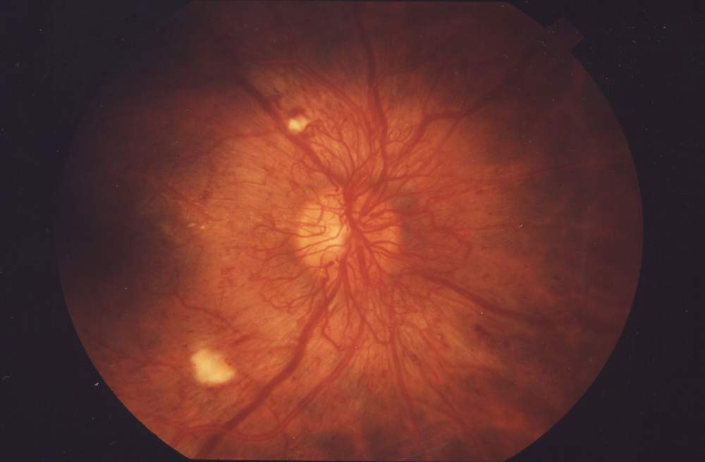
The reason for the scan is simple. A doctor looks into the eye with a bright light and a lens, reading the retina directly through the pupil. That direct look is the whole of an ordinary eye exam. It depends on a clear path from the pupil to the back of the eye. Blood in the vitreous turns that path to a red fog, or blanks it out altogether. The retina behind the blood goes unseen. So does any tear, detachment, or tumor that might have caused the bleed. Sound reads the eye a different way. It carries through blood as easily as through clear gel, scattering only a little off the red cells on its way. A probe laid on the closed lid sends its pulses past the blood, off the back wall, and home again. The machine builds the whole back of the eye into a grey picture. The view that the blood took away comes back on the screen, detachment and danger and all. In an emergency room or a clinic with no eye specialist on hand, that grey picture is often the only look anyone gets at the back of a blood-filled eye for days. No other tool at the bedside sees past the blood into the eye, which is why the handheld has become the first look anyone takes at a red-blind eye in an emergency room.
Where the blood came from matters as much as the blood itself. The commonest source by far is the diseased retina of advanced diabetes. Years of high sugar weaken the retinal vessels until the eye grows fragile new ones across its surface. These new vessels have flimsy walls, and they break at the slightest pull from the gel. A retinal tear or a detachment can tear a vessel, the bleed and the tear arriving together. A posterior vitreous detachment, the aging gel separating from the retina, can snap a small vessel on its way off. A blow to the eye ruptures vessels directly, blood filling the gel within minutes of the injury. Blood thinners and bleeding disorders make any of these worse. A spontaneous bleed in an older patient points first to diabetes or a vitreous detachment. A bleed right after an injury points to direct vessel damage. The patient’s story starts the search before the probe ever touches the lid. Each cause sends blood into the vitreous by its own route. Each carries its own urgency and its own treatment. Naming the source is half the work the scan is there to do.
A vitreous hemorrhage is rarely an emergency for the blood alone. Most of it clears on its own. The eye recovers its sight once the gel settles. The danger sits in what the blood may be hiding. A heavy bleed can cover a retinal detachment completely, blanking out every warning sign the detachment would normally give. The shadow, the curtain, the flashes are all lost behind the red. A detachment left unfound under blood spreads quietly for weeks. By the time the blood clears, the macula may be gone for good. The job of the scan is to see past the blood to that hidden danger, fast, on an eye no light can enter. A masked detachment caught in those first days is an eye saved. That catch is the whole reason a probe goes on the lid.
The blood and its cause together make the case. The blood itself, light or heavy, clears in most eyes given time. The danger is what may sit under it: a detachment or a diseased retina wearing the same red disguise as a harmless bleed. Both look alike from the outside: a red reflex gone dark, a vision dropped to hand movements, an eye no ophthalmoscope can read. The outside of the eye gives no hint which one it is. Telling the harmless bleed from the dangerous one, on an eye no one can see into, is exactly the work the handheld is there for. The whole value of the scan lies in that one piece of sorting.
Scanning through the blood
The scan is the same gentle technique used for any eye. Drops are not needed; the probe works over a closed, numbed lid cushioned by a thick layer of gel, resting light enough that it never presses the globe. The patient lies back and looks ahead under the lid. A linear probe, the high-frequency kind used on vessels and nerves, gives the fine detail the vitreous needs. The examiner fans the beam slowly across the whole globe, across and up and down, in more than one plane, so no pocket of the cavity goes unscanned. A bleed can pool anywhere, and a thin scatter in one corner is easy to miss on a quick look. Each view is held long enough to read it, and the bleed is mapped in two planes so its true extent on the back wall is clear.
One control matters more here than anywhere else: the gain. Gain is the machine’s sensitivity, how loudly it listens for the faint echoes coming back. Blood gives only faint echoes, far weaker than solid tissue, so its whole look on the screen depends on where the gain is set. The examiner runs the gain high to bring the blood out of the dark, then steps it down to watch how much of it fades, reading the bleed across the whole range of the dial. A scan locked at one gain setting can miss a light bleed entirely or mistake it for solid tissue. The high setting brings out the faintest scatter of cells. The low setting strips the soft blood echoes away and leaves only what is truly solid. That sweep of the gain, high to low and back, is the heart of reading a vitreous hemorrhage, and almost every sign that follows is read off it. The gain reads out in decibels on the screen, and a scanner soon knows the high setting that lifts a faint bleed out of the dark and the low one that strips it away.
What blood looks like on the scan
At first, on a fresh light bleed, the vitreous cavity is almost as dark as a healthy eye, marked only by a scatter of fine bright dots floating in the black. Each dot is a small clump of red cells catching the sound. Turn the gain up and more of them appear, filling the cavity with a soft snow that thickens toward the part of the eye where the blood has gathered. The dots have no shape and no anchor. They hang where the blood happens to sit, drifting a little with the slow currents of the gel, with none of the tethered line of a detached retina. A fresh bleed is mostly this: a haze of free dots, dense in places, thin in others, dark between them. Gravity pulls the heavier blood down, so the haze often lies deepest along the lower part of the cavity. Left a day, the same blood settles into a denser pool low in the globe. The look of one bleed can shift from one visit to the next for that reason alone.
The single feature that names blood is the way it answers the gain. At high gain the cavity crowds with bright specks, a snowstorm filling the black. Bring the gain down step by step and those specks dim and thin, melting from the screen until the cavity reads dark again. That fading at low gain is the surest single sign that a vitreous opacity is blood. The clinician sets the gain high to find the blood, then drops it low to confirm it, watching the snowstorm clear step by step. A finding that survives the low-gain sweep is something more solid than blood, and that is the finding worth hunting for. One mimic is worth knowing here: asteroid hyalosis, tiny calcium-fat bodies that hang in the gel as bright sparkling dots. They keep their brightness at every gain setting and settle back to the same spots each time. That steadiness through the gain is what marks them as the harmless lifelong finding they are.
Blood moves in its own telltale way. Ask the patient to move the eye and the cloud of dots churns through the cavity like silt stirred in a glass of water, rolling and folding on itself, settling back only long after the eye has stopped. Clinicians call it the washing-machine look. The heavier the bleed, the more it moves, a dense hemorrhage rolling in slow waves across the screen. This free, swirling, after-running motion is blood’s second signature, and it shows even when a still frame would leave the bleed looking like solid tissue. Movement read together with the gain settles what the opacity is. The two tests run in the same few seconds: drop the gain, then move the eye, and watch.
The amount and the age of the blood change the picture. A sparse fresh bleed is a thin scatter of dots, easy to miss at low gain. A dense one fills the cavity with a thick cloud that can hide the back wall entirely. Older blood begins to organize, its cells clumping into strands and sheets and sometimes settling under gravity into a layer along the floor of the cavity, a level that shifts when the head tips. Those organized strands can mimic a membrane. They give themselves away by staying softer and fading further at low gain. A long-standing hemorrhage can thicken into true membranes that pull on the retina in their own right. Reading the density and the age tells the clinician how fresh the bleed is and how much it stands in the way of the view behind it. A bleed that has begun to organize has been there a while, a clue in itself about what set it loose. Blood organized into thick grey sheets has usually been bleeding and rebleeding for months, the mark of an eye that keeps tearing its own new vessels, the way a diabetic eye does.
Put together, the three signs read a vitreous hemorrhage at a glance: faint dots and clouds that crowd at high gain, fade at low gain, and swirl freely when the eye moves. A clinician who has run the gain up and down on a few bleeds learns the pattern quickly. The dots that bloom at high gain and vanish at low, churning when the eye turns, are blood, wherever in the cavity they sit and however thick they lie. None of it asks for a perfect frozen frame; the bleed shows itself across the whole sweep and the whole movement. Once that pattern is clear, the scan moves on to the real question, which is what the blood is hiding and what set it loose.
How a bleed clears
Time clears most of the blood on its own. The eye reabsorbs a vitreous hemorrhage slowly, the cells carried off by the eye’s own scavenger cells at a rate of roughly one part in a hundred a day, a light one clearing over weeks and a heavy one over months. Each repeat scan over those weeks reads a clearer cavity, the snowstorm thinning toward the dark of a healthy eye. That slow clearing is itself useful to watch. A bleed that thins on schedule needs only patience and a repeat look. A bleed that stays thick after weeks of waiting, or grows, raises a suspicion that something keeps feeding it, a vessel still bleeding or a detachment still pulling at the retina. A hemorrhage that refuses to clear becomes a reason to act, taken to surgery to wash the gel clear and treat whatever lies beneath. Diabetes, a young dense bleed, and a bleed that keeps recurring all clear more slowly, and they pull the decision toward operating sooner. An eye that cannot afford a long wait, a child’s eye or a patient’s only seeing eye, is taken to early surgery to clear the gel and read the retina at last. The scan, repeated over weeks, follows a settling bleed down to nothing or flags the eye that is not clearing on its own. The repeat scan is quick, a minute at a follow-up visit, and it turns a worrying blind eye into a tracked one with a clear trend on paper.
Finding what bled
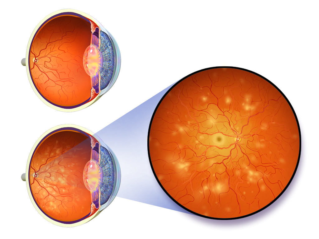
The most important thing the scan does is look for what caused the bleed. Behind a heavy hemorrhage the examiner hunts above all for a retinal detachment, the one finding that turns a wait into surgery. The trick is the gain again. Run it low to clear away the soft blood echoes, and any structure left standing bright on the screen is something more solid than blood: a detached retina, a thick band of scar, a sheet of organized tissue. A bright line that survives the low-gain sweep, tethered at the optic disc and swaying stiffly when the eye moves, is a detachment hiding under the blood. A detachment under blood is pulled into tented peaks by diabetic scar or lifted in a smooth fold by a tear, both tethered at the disc and both bright at low gain. Finding either one on a blind, blood-filled eye changes everything about the hours that follow. Without that finding the same eye might be sent home to wait. The hidden detachment would spread unseen for a week before anyone looked again.
What the scan finds points back to the source of the bleed. A funnel-shaped membrane tethered at the disc is a detachment, often dragging the very tear that bled. A short flap lifting from the far edge of the retina can be the tear itself. A thin line swinging free of the disc is a posterior vitreous detachment, the gentle and common kind of bleed that usually clears on its own. Heaped tissue growing off the retina, with abnormal strands reaching forward into the gel and tugging the retina up into peaks, is the neovascular disease of diabetes, the most common reason an eye fills with blood. Each of these patterns carries its own plan. The scan reads them through blood that hides them from every other test. The same low-gain sweep that confirms the blood is what brings these solid findings out from under it.
The cause sets the clock on the eye. A bleed from a posterior vitreous detachment is watched and left to clear, with a repeat scan to be sure nothing worse sits under it. A bleed hiding a fresh retinal detachment goes to surgery within days, before the detachment can spread across the macula and take the central sight for good. A bleed from diabetic disease is planned for laser or a vitrectomy once it clears enough to work through, or sooner if a detachment is found beneath it. A bleed from trauma is watched alongside the other injuries of the hit. On the scan, that one red fog sorts into very different paths, each with its own plan. The scan is what does that sorting, on an eye no one else can see into. Without it, every one of these eyes would look the same from the front and wait the same anxious days for the blood to clear before anyone knew which was which.
| Item | Figure | Note |
|---|---|---|
| Vitreous, share of eye volume | about 80% | the gel filling the back, ~4 mL |
| Probe frequency | about 10–15 MHz | linear probe on the closed lid |
| Leading cause | diabetic retinopathy | the fragile new vessels that bleed |
| Spontaneous reabsorption | roughly 1% per day | a slow clear over weeks to months |
| Gain to confirm blood | run high, then low | blood fades out at low gain |
| B-scan for a hidden detachment | high, about 90%+ | the danger the scan hunts |
| When to suspect a hidden cause | a bleed not clearing in weeks | the trigger for surgery |
What the scan settles
A handheld scan answers the two questions that matter at the bedside. Is this blood, and is anything dangerous hiding behind it. For the first, the gain test and the swirling motion settle it in seconds: a haze that blooms at high gain, fades at low, and churns when the eye turns is blood. For the second, the low-gain hunt for a tethered membrane finds or rules out the detachment that would change the whole plan. Studies of bedside scanners put ultrasound’s read of a vitreous hemorrhage high for catching a detachment behind the blood, good enough to act on a positive finding. A scan clean of any membrane points the plan toward watching and rescanning, with the worst danger ruled out for the moment. Those two answers are enough to sort a frightening red-blind eye into a wait, a referral, or an operation. The scan turns a blank, unseeable eye into a picture a clinician can act on, in a minute, at the chair or the bedside.
What the scan does not give is the fine detail of the retina itself. It shows a detachment as a bright line. The small tear that started it stays below the scan’s resolution. Grading the diabetic disease and planning the laser are beyond it too. Those belong to a dilated exam and the retinal specialist, once the blood has cleared enough to see through. The handheld does its work earlier, in the blind hours when the eye is full of blood and no one can look in. It reads the blood, finds the danger under it, and starts the patient down the right path days before the view comes back. A bleed that would otherwise sit unread for weeks gets sorted on the first night it is seen. The eye behind it gets its chance. On a busy night, that one early sort is what gets the right eye to the surgeon in time.
Common questions about ultrasound for vitreous hemorrhage
How does vitreous hemorrhage look on ultrasound?
As faint dots, strands, and clouds floating in the dark vitreous, brightest at high gain. The defining sign is gain-dependence: turn the gain down and the blood dims and clears, until the cavity reads dark again. The blood also swirls freely when the eye moves, churning through the cavity and settling only long after the eye has stopped. The amount of blood sets how thick it looks, from a thin scatter of dots to a dense cloud that can hide the back wall.
Why use ultrasound when the eye is full of blood?
Because the probe reads straight through the blood. Sound crosses a blood-filled vitreous as easily as a clear one and draws the retina on the screen behind it. The direct view with a light fails once blood clouds the gel, so the scan is what sees in. It works in seconds at the bedside, on an eye no light can enter, and shows whatever the blood is hiding.
How is blood told apart from a retinal detachment on the scan?
By the gain and by the motion. Blood fades at low gain and swirls loosely when the eye moves. A detached retina holds its brightness at low gain and stays tethered at the optic disc, swaying stiffly. Running the gain low is the quickest test of the two: the soft blood echoes melt away and leave any solid membrane standing on the screen.
Does vitreous hemorrhage clear on its own?
Often, yes. The eye reabsorbs the blood slowly, over weeks to months depending on how much there is. Vision returns once the cavity clears. A bleed that thins on each repeat scan needs only patience. A bleed that stays thick after weeks, or grows, suggests something is still feeding it, and earns a closer look for a tear or a detachment underneath. A hemorrhage that will not clear can be cleared surgically.
Can ultrasound find the cause of the bleed?
It can find the dangerous ones. With the blood echoes cleared away at low gain, the scan shows a retinal detachment as a tethered bright line, a tear as a short lifting flap, and the heaped neovascular tissue of diabetes as strands reaching off the retina. The smallest tears and the fine grading of diabetic disease stay beyond it, waiting for a dilated exam once the blood clears. What the scan does at the bedside is catch the detachment that cannot wait.

About the Author
Julien Mercier
Senior R&D Engineer · Medical Ultrasound Transducer Development
Senior R&D Engineer with an M.S. in Applied Physics and over 15 years of experience in medical ultrasound transducer development, specializing in the design verification and performance testing of high-frequency imaging transducers. Currently leading the development and verification of the company’s next-generation high-frequency linear-array transducer, responsible for imaging performance evaluation and reliability analysis in preclinical testing. Brings extensive hands-on experience in piezoelectric element tuning, beamforming parameter optimization, and system-level performance testing.