





































Axial Length AL Measurement Before Cataract Surgery Handheld Ophthalmic Ultrasound
Axial length is the distance from the front of the cornea to the retina at the back, the through-and-through length of the eyeball. It is the first measurement taken when an eye is booked for cataract surgery. On a normal adult eye that length runs to a little under twenty-four millimetres. The cloudy natural lens is about to come out. A clear plastic lens will take over its focusing job. That replacement comes in a long ladder of powers, from weak to strong. Once it is in, it stays for good. The length of the eye decides which rung on the ladder lands the focus cleanly on the retina.
Why a cataract operation needs a length
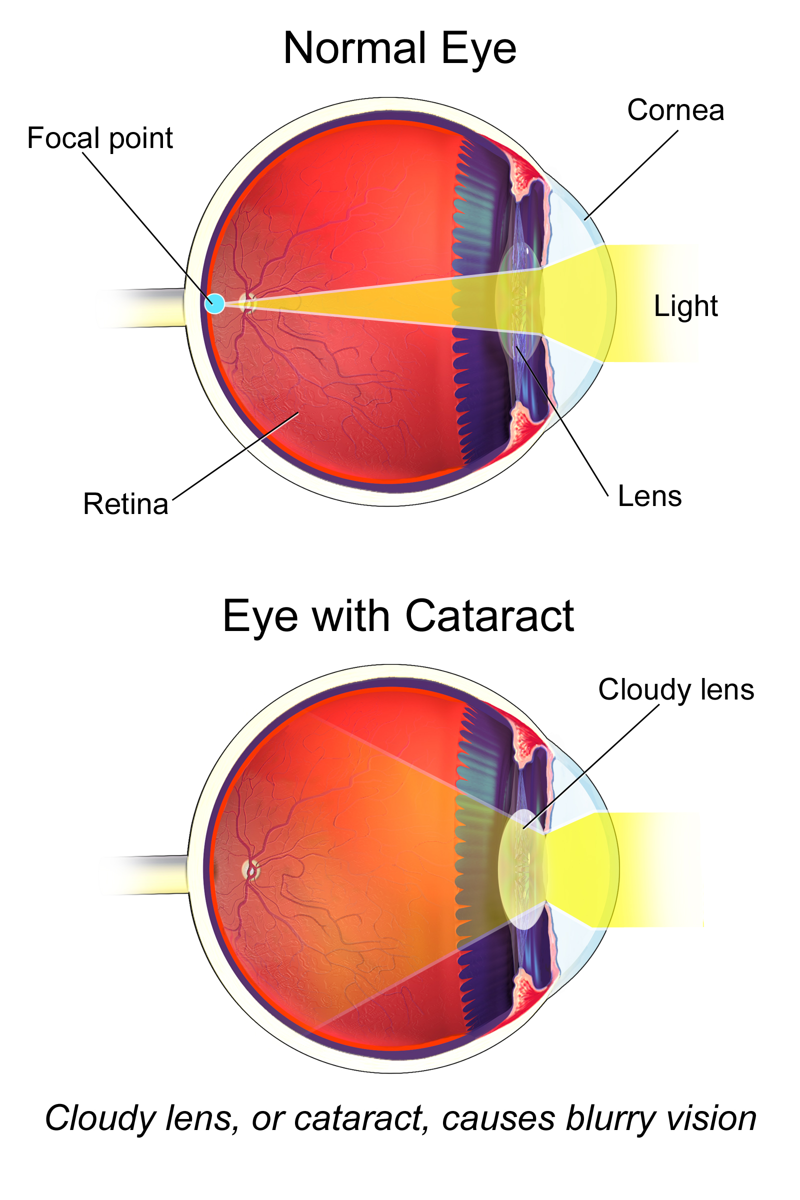
That makes the length the largest single number in the whole calculation. A formula takes it together with the curve of the cornea and the depth of the front chamber and returns a lens power in dioptres. Of those inputs the length carries the most weight, because the eye is a long optical tube. A small change in length, run down so long a tube, moves the focus a long way. An error of a single millimetre pushes the result by roughly two and a half to three dioptres, a miss large enough to leave the eye needing strong glasses for the rest of a life. The surgeon gets one chance to choose the lens. The power is set before the operation. After surgery it cannot be dialled up or down like a pair of spectacles. A wrong choice means living with the blur, or a second operation to exchange the lens. The surgeon also picks a target before measuring. Usually the aim is sharp distance vision. Sometimes the aim is set a little near, for the patient who would rather read without glasses. Whatever the target, the length still has to be right, or the focus lands where nobody chose.
For most of the history of cataract surgery this length was taken with ultrasound. On the cloudiest eyes it still is. A probe sends a pulse of sound into the eye. The sound bounces off each surface it crosses. The time each echo takes to return is turned into a distance. The whole measurement takes only seconds. It asks nothing of the patient beyond holding still. A plastic lens chosen on a length that is half a millimetre wrong will sit in the eye for the rest of the patient’s life, focusing a little in front of or behind the retina the whole time. Half a millimetre is about the width of a pencil line. It is worth taking slowly.
Both the picture and the measurement of the eye come from the same family of probes. A clinic that can scan the eye can also measure it. The length is the part with a number on it, the part a surgeon plans an operation around. A small hospital with one portable machine can work up an eye for surgery, with no need to send the patient elsewhere. All of it comes back to that one figure.
Measuring the length with sound
Drops numb the surface of the eye first. A numbed eye lets the probe rest against it while the patient holds still. The probe is lined up along the eye’s own axis, looking straight down the middle from the cornea through the pupil to the macula, the small pit at the centre of the retina. Most probes carry a tiny fixation light. The patient stares straight into it. That alone lines the eye up on its axis. Aim that is even slightly off the axis, crossing the eye at a slant, reads a shorter or skewed path and gives a wrong length. The axis matters more than almost anything else in the reading.
On the screen the returning echoes stand up as a row of spikes, one for each surface the sound crossed. The first marks the cornea. The next two mark the front and back of the lens. The tall spike toward the right of the trace marks the retina, the end of the journey. Between them the trace stays low across the clear vitreous, a part of the eye that returns almost no echo of its own. The distance the machine reports is taken between the first spike and that retinal spike, the cornea-to-retina length. A trained eye takes in the shape of the whole row at a glance and knows whether the sound went where it should. The gain can be turned down to sharpen the spikes, or up to pull a faint retinal echo out of a noisy trace. Setting it well is part of the craft.
The retinal spike is the one that decides whether the reading is any good. It has to stand tall, rising almost straight up from the baseline, which happens only when the sound has struck the retina square-on at the macula. A spike that leans, or sits low, means the sound met the retinal wall at an angle, off the macula. The length under such a spike cannot be trusted. The examiner watches for that clean, vertical retinal spike and throws out the traces without it. Just behind the retinal spike, smaller spikes from the sclera and the orbital fat often follow. The length is taken to the leading edge of the retinal spike, the point where the retina itself begins.
One measurement is never enough. The examiner takes several, watching that the retinal spike stays clean and that the length holds steady from one to the next, then lets the machine average them. A spread of more than a tenth of a millimetre across the measurements is a sign to start again. Both eyes are measured at the same sitting, even when only one carries the cataract being treated, because the second eye is the natural check on the first. Lengths that hold steady across several readings, close between the two eyes, are the ones a surgeon will plan on.
Contact and immersion
There are two ways to bring the sound to the eye. The plain one is contact: the probe tip is set directly on the numbed cornea to read the length through it. Contact is quick, needs no extra kit, and works at the bedside, which is why it is the common method. The cost of contact is the touch itself. A probe pressed even lightly dents the cornea inward. The eye then reads shorter than it truly is, by a fraction of a millimetre. A tenth or two of a millimetre lost this way turns into a quarter or half a dioptre of error in the lens. Taken with a heavy hand, the contact length runs short. The lens picked from a short length sits a little too strong. The honest way around it is the lightest possible touch, the probe barely meeting the cornea.
The steadier way is immersion. A small cup or shell holds a pool of fluid against the eye. The probe floats in that pool a few millimetres off the cornea, with nothing pressing on the surface. The sound crosses the fluid and enters the eye with no dent at all, so the length comes back truer. The fluid also gives the corneal spike a clean shape, since the probe no longer sits on top of it. Immersion takes a little more setup and a cooperative patient lying back, which is why a clinic keeps it for the eyes that need the most exact figure, the long and the short ones especially. Many surgeons trust the immersion number first when the two methods disagree.
When sound is the only way in
For an eye with a clear path through it, the length today is usually read with light. An optical biometer shines a fine low-coherence laser the length of the eye and reads the reflection off the retina, with no probe touching the surface. It reports the length to about a hundredth of a millimetre, finer than sound can manage, in a few seconds per eye. The same machine usually reads the corneal curve and the chamber depth at the same sitting, so one device hands over most of what the formula needs. Most cataract eyes are measured this way now, and the patient barely feels it. Over the last twenty years the optical method has become the first choice wherever a clinic can afford the machine. It is faster for the staff and easier on the patient.
The limit is in the word optical. The laser reads by sending light to the retina and catching it on the way back, so the light has to make the round trip through clear ocular media: a clear cornea, a clear lens, a clear vitreous. The light is a fine beam, easily scattered. It needs all three reasonably clear to complete the trip. The figure is only as good as the eye’s clarity allows.
A cataract dense enough to need surgery is, by definition, a lens that has stopped being clear. A thick brown or white cataract scatters the laser light so badly that little or none of it reaches the retina and comes back. The densest brown cataracts and the swollen white ones are the worst offenders. A bleed in the vitreous does the same. So does a cornea clouded with old scar tissue. On those eyes the optical biometer gives no usable length: the screen reports a failure, or a number the machine itself flags as unreliable. Trying the laser a second time changes nothing.
Sound does not depend on a clear path. An ultrasound pulse crosses a cloudy lens and a blood-filled vitreous almost as readily as a clear one, reflects off the retina, and comes back to the probe. The machine times that round trip and reports the length. The dense brown lens that stops the laser still lets sound through. A wave of sound passes through a brown cataract much as it passes through any soft tissue, slowed a little, scattered a little, still strong enough to return a clean retinal echo.
So ultrasound biometry holds a settled place in the modern clinic. It is the method used when light cannot measure the eye: the dense cataract, the vitreous bleed, the scarred cornea. When one of those defeats the laser, the length still has to come from somewhere. Sound supplies it. A brown cataract that would otherwise stall the whole operation gets its number this way. In a clinic with both machines, the probe waits in reserve for the eyes the laser cannot measure.. That reserve role keeps the skill alive in the clinic. A team that never touches the probe loses the feel for it, so the dense-cataract patient is exactly the one who needs a hand that still knows the trace.
The speed the machine assumes
Sound does not travel at one speed through the eye. It runs faster through the dense lens than through the watery aqueous and vitreous around it. The machine cannot see those parts apart on a simple trace, so it converts each echo time into a length using one assumed average speed for the whole eye, a single figure that suits a normal eye with its natural lens in place. That assumption breaks the moment the eye stops being normal inside. An eye already emptied of its lens carries no dense lens to speed the sound, so its average runs slower. An eye filled with silicone oil after retinal surgery slows the sound far more. Each of those eyes needs the machine told what is inside it, set to a different speed, or the length comes out long or short by millimetres. The setting is a small switch with a large consequence. Forgetting it is a classic way to ruin a good reading.
The error that reaches the glasses
The reason all this care is spent on a few millimetres is that the eye magnifies the error. Run the numbers through a standard lens formula and a length wrong by one millimetre shifts the eye’s final focus by roughly two and a half to three dioptres. Three dioptres is not a subtle miss. It is enough to move an eye from reading a wall clock across the room to needing glasses to see it at all. On a long ladder of lens powers, one wrong millimetre has stepped the choice several rungs off. The cornea and the chamber depth carry weight in the formula too. For sheer leverage over the result, the length still leads them both by a wide gap. An error in the corneal reading adds its own shift on top, so the two can stack into a larger miss than either alone. The length is the one most worth getting right.
The error does not fall evenly across all eyes. The longest, most myopic eyes and the shortest, far-sighted ones both sit far from the average the formulas are tuned around. A small length error on those eyes throws the focus further than the same error on an average eye. Those eyes are measured with the most care, with immersion preferred, several formulas compared, and the figure double-checked, because they are the least forgiving of a careless millimetre. A short eye packs a strong lens into a small space. A fraction of a millimetre there moves the focus a long way. The long eye carries its own traps in the shape of its back wall.
From length to lens power
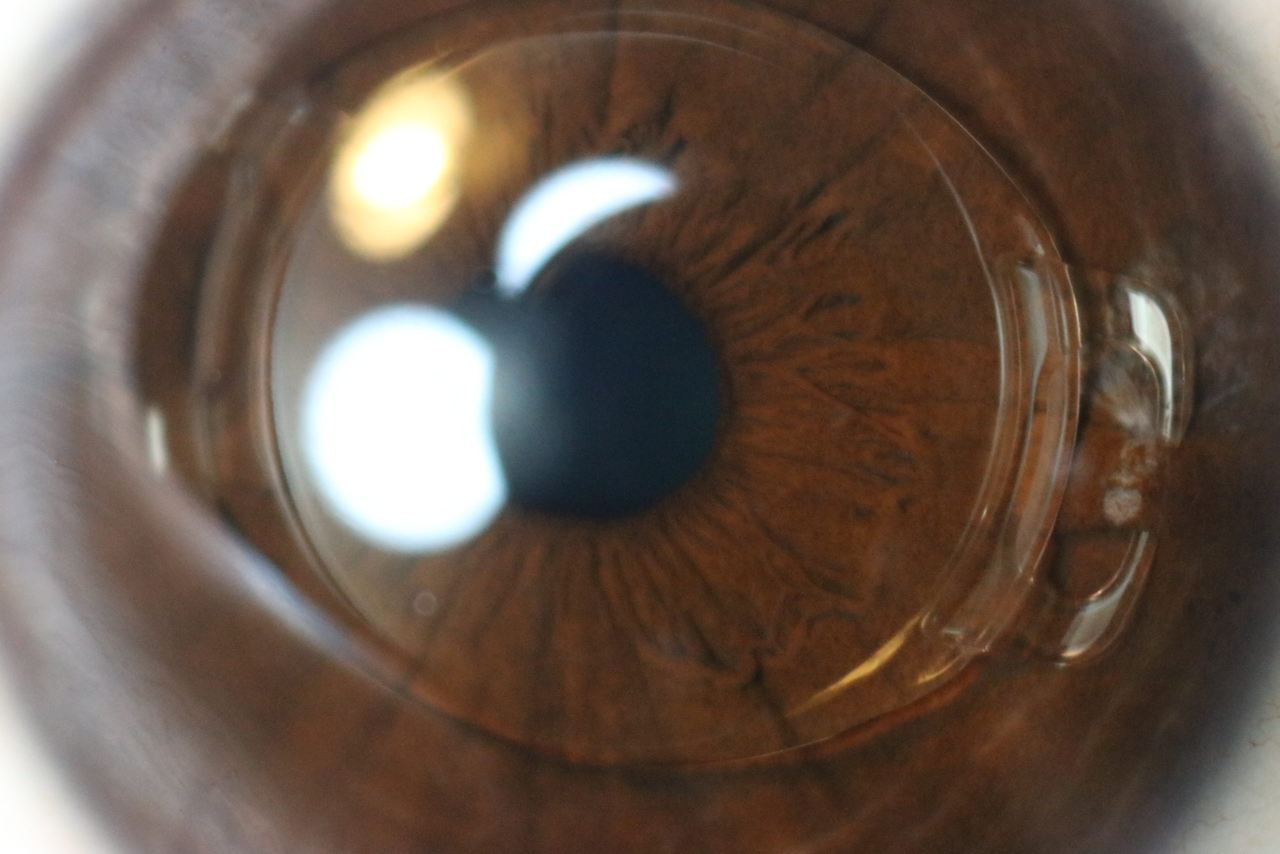
The length on its own does not name a lens. It is fed into a formula together with two other readings: the curve of the front of the cornea, which says how strongly the cornea already bends light, and the depth of the front chamber, the gap between cornea and lens. The formula weighs the three and returns the power, in dioptres, of the implant that should land the focus on the retina. The hardest part of the sum is guessing where the new lens will finally sit once the eye has healed. The length feeds straight into that guess. The corneal curve sets how the eye bends light before the implant is in place. A steeper cornea calls for a weaker lens behind it. The chamber depth hints at how far back the new lens will rest. The length frames all of it.
Modern formulas carry the names of the people who built them and differ in how they handle the unusual eye. Each lens model also brings a constant of its own, a number that tells the formula how deep in the eye that particular lens will end up sitting. A surgeon’s own past results sharpen that constant over time. What every formula shares is a heavy reliance on the length. Feed in a length half a millimetre off and each one, old or new, hands back the wrong lens. The formula cannot know the number it was given is wrong. Good measurement is what the whole calculation stands on. The published reference on intraocular lens power calculation lays out how each formula leans on the length.
The two eyes are compared again at this stage, now as lengths and lens powers side by side. Most people’s eyes are close to each other in length, within about a third of a millimetre. The powers picked for them sit close as well. A wide gap between the eyes is treated as a warning that one reading may be off, unless a known difference in the patient’s vision explains it. That eye is measured again before anyone plans a lens on it. A patient who has always worn a much stronger glasses lens on one side is the exception the comparison expects.
One anatomical trap is watched for in the long eye. A highly myopic eye can carry a staphyloma, a localised bulge where the back wall has stretched outward into a pouch, often a little to one side of the macula. Sound aimed at the deepest part of the pouch reads too long, because the floor of the bulge lies deeper than the macula. The length that counts is the one measured along the axis through the macula itself. On a known staphyloma the examiner works carefully to land the reading on the macula, sometimes guided by a B-scan picture of the back wall, so the length belongs to the right point on the retina. This is one place where the picture and the measurement work side by side.
| Item | Figure | Note |
|---|---|---|
| Mean axial length, adult eye | about 23.5 mm | the through length; most eyes 22–25 mm |
| A-scan (ultrasound) precision | about 0.1 mm | the length sound can resolve |
| Optical biometry precision | about 0.01 mm | the finer reading, used when the eye is clear |
| Error per 1 mm of length | about 2.5–3.0 D | shift in the eye’s focus at the glasses |
| Sound speed in the lens | about 1,640 m/s | faster than the watery parts |
| Sound speed in aqueous and vitreous | about 1,530 m/s | the fluid-filled aqueous and vitreous |
| Assumed average speed, normal eye | about 1,550 m/s | the single figure a normal eye uses |
| Two-eye length agreement | within about 0.3 mm | a wider gap prompts a recheck |
Sound at the cataract clinic
A handheld ultrasound unit puts this whole measurement in a portable box. The probe is small. The screen reads out the length on the spot. The same machine that took a B-scan of a cloudy eye can switch to the A-scan trace and measure it. Nothing has to leave the room. For a clinic without a laser biometer, or for the eye too cloudy for one, the handheld is the tool that still returns a length. In much of the world it is the only biometer a cataract service has. It carries the whole workload alone.
What it asks in return is skill. A contact figure is only as good as the lightness of the hand holding the probe; lean on the eye and the length shortens. An immersion figure is more exact, harder to set up freehand, and slower. Both demand a steady axis through the macula and a clean retinal spike before the number means anything. The machine reports a length whatever the aim. Whether that length can be trusted is for the examiner to judge. Training shows in the readings: a practised hand brings back lengths that agree time after time, on eye after eye. A careful clinic builds in quiet checks: a repeat on any odd eye, a second pair of hands on the hard ones, a glance back at the patient’s old glasses when a number looks strange. None of it is fancy. All of it guards the one figure the operation rests on.
So the length that decides which lens a cataract patient receives, and how sharply they see for years afterward, often begins as a row of spikes on a handheld trace. The measurement rewards patience: a numbed eye held steady, an axis kept true to the macula, several echoes averaged, the speed set to match the eye in front of the examiner. Get those right and the sound probe hands the surgeon a number to build an operation on, even on the cloudiest eye, the one no laser could measure. The smallest careful measurement at the start is what lets a patient read a page again at the end.
Common questions about measuring the eye for a lens
Why is the eye’s length measured before cataract surgery?
The cloudy lens is removed. A clear plastic lens is put in. That implant comes in many powers. The length of the eye is the largest factor in which power focuses light on the retina. A length measured well lets the surgeon pick the lens that puts the eye in sharp focus.
How accurate does the length have to be?
To about a tenth of a millimetre. A length wrong by a full millimetre shifts the eye’s focus by roughly two and a half to three dioptres, the difference between sharp distance vision and needing a strong correction. Several measurements are averaged for each eye. The two eyes are then checked against each other to keep the figure tight.
When does the cataract clinic use ultrasound for the length?
When the eye is so cloudy that light cannot cross it. The everyday tool is an optical scanner that takes the length with a laser, which needs a clear path to the retina. A dense cataract, a vitreous bleed, or a scarred cornea blocks that light. Sound carries through the murk and still times an echo off the retina, so the length can be measured when the laser returns nothing.
Does the probe touch the eye?
It can. With the contact method the probe rests on the numbed cornea for a moment. The immersion method floats the probe in a small fluid bath, with nothing pressing on the eye. Drops numb the surface either way. The immersion figure runs a little truer, so it is the one used on eyes that need the most exact length.
Can a length be wrong even when the scan looks fine?
Yes. A length can read short if the probe pressed on the cornea. It can read long if the eye has a staphyloma, a back-wall bulge that sits off the macula. In both cases the machine still reports a number and does not flag it. This is why the retinal spike is studied for height and shape, why both eyes are compared, and why an odd length is measured again before a lens is chosen on it.

About the Author
Julien Mercier
Senior R&D Engineer · Medical Ultrasound Transducer Development
Senior R&D Engineer with an M.S. in Applied Physics and over 15 years of experience in medical ultrasound transducer development, specializing in the design verification and performance testing of high-frequency imaging transducers. Currently leading the development and verification of the company’s next-generation high-frequency linear-array transducer, responsible for imaging performance evaluation and reliability analysis in preclinical testing. Brings extensive hands-on experience in piezoelectric element tuning, beamforming parameter optimization, and system-level performance testing.