Bladder Scanner Alternative Handheld Multipurpose Ultrasound
What a single-purpose bladder scanner is
A bladder scanner is a small device with one job. It measures the volume of urine in the bladder. A nurse presses it to the lower belly, aims it at the pelvis, and takes a number off the screen. The device fires sound into the pelvis from several angles. It finds the dark pool of the urine, builds a rough three-dimensional shape, and works out the volume on its own. It prints the figure in milliliters in a second or two. Many models show an aiming guide first, a crosshair that centers the bladder before the measurement. A nurse lines up the crosshair, presses the button, and notes the result. A figure near zero means an empty bladder. A figure in the hundreds means urine left behind. The dedicated scanner does this one job in seconds. It runs on a ward round with almost no training. The device asks only for a steady hand and a button.
The dedicated scanner lives on hospital wards and in clinics. A nurse checks the bladder after surgery, after a birth, or after a catheter comes out. The device flags a bladder that holds urine after a void. It saves a person from a needless catheter when the bladder is already empty. It calls for a catheter when the volume climbs too high. The scanner runs all day on a ward without a doctor present. A team trusts it for a quick number at the bedside. It carries the routine bladder check on a busy ward. A unit clips to a wall charger between uses. A nurse wipes the probe head and moves to the next bed. The whole check takes under a minute from start to figure. A ward built around that routine runs smoothly on the simple box. The device handles a high load of post-void checks every shift.
The dedicated scanner gives a number. The screen shows a figure alone. The device works out the volume from echoes a person never sees. A nurse notes the milliliters and trusts the box. The scanner cannot show why the bladder holds that volume. It cannot show a stone, a mass, or a catheter balloon inside. It cannot show the kidneys above or the prostate below. It cannot tell a blocked bladder from one that just sits full. The number stands alone. The reasons behind it stay dark. A nurse who sees a high figure knows the volume. The cause stays a mystery. The box answers its one question and no other. The figure travels into the chart as a bare number with no picture behind it.
The scanner counts any pelvic fluid as urine. The device hunts for a dark pocket and treats it as bladder. An ovarian cyst, a fibroid, or a belly full of ascites can fill that pocket. The box then prints a large volume from fluid that is not urine at all. One report describes a false reading in a patient with ascites and no urine to drain. A false high number can send a person to a needless catheter. The dedicated scanner cannot tell urine from other fluid, since it never shows the operator the pocket it measured. A number from a box carries that risk in a swollen belly. The same trap catches a large cyst in a woman and a loop of fluid-filled bowel. A clinician who trusts the figure alone acts on a measurement of the wrong thing. The box gives no warning, since it sees the dark and reports a volume either way.
The handheld that does more
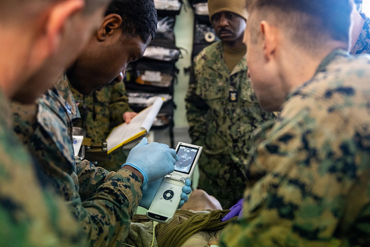
A clinician uses a handheld ultrasound at the bedside. The whole scanner fits in one hand, with the live picture on its own small screen. A probe like this measures the bladder and reaches much of the rest of the body.
A multipurpose handheld scanner is a full ultrasound shrunk to the size of a phone probe. It connects to a tablet or a phone and draws a live picture of whatever sits under it. A clinician points it at the bladder and sees the urine, the wall, and the shape of the pocket. The same probe measures the volume with a built-in tool. The clinician traces the bladder in two planes. The device works out the milliliters from width, depth, and height. The figure lands on the screen beside the picture that produced it. Some handhelds now run an automatic bladder measurement of their own, a tap that finds the bladder and prints the volume like the dedicated box. The handheld covers the bladder scanner’s one job. It carries the work much further across the body. One device holds a curved probe for the deep belly and a linear probe for shallow parts in the same housing. A clinician picks a bladder preset and sets the depth to fill the screen with the pelvis. A turn of a dial brightens the picture in a dark belly.
The picture changes what the bladder check can find. A clinician sees a full bladder fill the screen as a clean dark pocket. A stone settles to the floor as a bright spot. A mass stands up from the wall. A catheter balloon sits as a round bubble inside. A clot drifts as a loose clump in the urine. The same sweep climbs to the kidneys and checks for a backup. The same probe drops to the prostate and gauges its bulge into the bladder. One pass covers the whole lower tract. The clinician saves a clip of the scan to the chart. A specialist far away opens that clip later and checks it again. The picture becomes a record in the file. A scan kept as an image lets a second eye confirm the call.
What the picture adds at the bladder
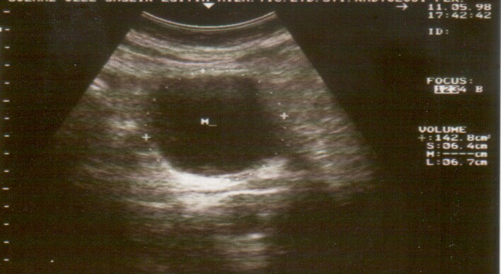
A bladder measured on ultrasound. The small crosses are calipers set on the bladder in two directions. The machine works the volume out from those lengths and prints it on the right, here about 143 milliliters.
A high volume marks a full bladder. The picture behind it shows why. A clinician sees retention from a blocked outlet as a smoothly stretched bladder. A clinician sees a tumor as a lump on the wall. A stone shows as a bright spot with a shadow. A wall thickened into ridges speaks of years of straining against a block. The same high number can come from any of these. The picture sorts them in a glance. A clinician takes in the volume and the cause in the same look. A picture points straight at the problem behind the volume. A clinician sees the cause without a guess. The bladder check grows from a single figure into a full study of the lower tract. A clinician leaves the bedside with the size, the cause, and a plan.
A catheter complicates the result. The box cannot explain that change. A balloon holds the catheter in place inside the bladder. The balloon shows on the picture as a round bubble with a bright rim. A blocked catheter leaves the bladder full around that bubble. A clinician sees the full bladder and the blocked catheter together in one image. A kinked tube shows the same full bladder for a different reason. The picture tells the two apart at a glance. The dedicated scanner reports a volume and stops there. The picture shows the clinician the balloon, the urine around it, and the block at once. A clinician acts on the cause behind the number. A nurse flushes a blocked catheter and watches the bladder empty on the screen. The image guides the fix and confirms it worked. A clinician documents the blocked catheter with a saved image. The picture proves the cause for the next person who opens the chart.
A multipurpose handheld reaches far past the bladder. At the bladder it gives the volume and the picture behind it. It shows the wall for thickening or a mass. It shows a stone on the floor or a clot in the urine. The same probe climbs to the kidneys and checks each one for the swelling of a backed-up tract. It drops to the prostate and measures the bulge that blocks an older man. One device covers the whole lower urinary tract in a single visit. The same handheld then turns to the rest of the body. It checks the lungs for the lines of fluid in heart failure or the slide of a healthy edge. It scans the four corners of the belly for free fluid after an injury. It presses the leg veins to hunt a clot behind a swollen calf. It measures the depth of fluid around a lung in a breathless patient. It checks the big vein by the heart for a read on the fluid state. It guides a needle into a vein, a joint, or an abscess. It dates a pregnancy, checks the lie of the baby, and finds the heartbeat. It looks at the heart, the gallbladder, the aorta, and the soft tissue under a swelling. A clinic buys one handheld and runs a dozen exams on it. A nurse measures a post-void residual on it in seconds. A doctor scans a lung or a vein on the same probe an hour later. A student learns anatomy on it between patients. A midwife checks a pregnancy in a far village with the same tool. A multipurpose probe stays busy across the whole clinic all day. The bladder volume becomes one task among many on a single device. A clinician who owns the picture owns far more than a number.
The picture settles a puzzling case. A frail person shows a high volume on the box. A clinician scans the same belly and finds a large ovarian cyst. The bladder behind it sits empty. A catheter then drains nothing. The cyst stands explained. The picture spares that person a fruitless catheter and a wasted procedure. A swollen belly from ascites tells the same story. The picture shows the bladder lying empty behind the fluid. A clinician sees the real state and holds the catheter. A confused number on the box becomes a clear answer on the screen. The image turns a wrong number into the right call. A clinician who scans the belly avoids a needless catheter in a hard case.
The handheld measures the volume from the picture itself. A clinician traces the bladder across its widest width, depth, and height. The device multiplies the three by a set factor and prints the milliliters. The factor, zero point five two, turns the box of measurements into the rounded shape of a bladder. The number rests on a bladder the clinician can see and check. A clinician who doubts a figure scrolls back through the picture and measures again. The handheld puts the whole measurement in plain view. A clinician trusts a volume drawn on a bladder in full sight. A clinician centers the bladder, freezes the clearest frame, and sets each caliper by hand. A careful trace lands close to the true volume. The clinician checks the trace against the picture and corrects any drift.
One probe, many jobs
A multipurpose handheld does the bladder scanner’s job and many more besides. One probe carries the work of a whole shelf of machines.
Where the dedicated scanner still fits
The dedicated scanner still has its place. A busy ward runs dozens of post-void checks a day. A nurse needs a number in seconds with one button. The dedicated scanner gives exactly that. It needs no image and no training. A ward with a steady stream of routine checks leans on that speed. The scanner asks for almost no thought at the bedside. A team puts it in any nurse’s hands on the first day. For pure volume at high speed, the single-purpose box holds its ground. A new staff member learns it in one short demonstration. A scanner survives a busy ward better than a delicate probe and tablet. The box takes a knock and keeps working. A ward that wants one simple task done fast finds the dedicated tool hard to beat. A dedicated scanner needs no gel station and no image archive. A ward plugs it in and forgets it between rounds.
A clinic weighs that speed against the skill a picture asks for. A multipurpose handheld asks a clinician to learn the image. The picture takes a little training and a steady eye. A ward with no trained sonographer leans on the simple box. The dedicated scanner fits a team built around one routine task. A clinic that needs only post-void numbers can stop at the single-purpose tool. The choice rests on who holds the probe and what the clinic needs from it. A nurse-led bladder service runs well on a one-button box. A team with a trained eye outgrows the box quickly. The training to learn an image takes hours of practice and a mentor at the elbow. A clinic without that support leans on the tool that needs none.
Bladder volume by device: the numbers
What is measured
Value
Note
Bladder scanner accuracy, on the label
within 15% and 15 mL
of the true volume
Bladder scanner error, in studies
about 20% on average
often an underestimate
Detects 100 mL or more
90% sensitive
81% specific
Ellipsoid volume on a handheld
W × D × H × 0.52
shown on the screen
Post-void residual to act on
over 200 mL
retention, look closer
False high volume from other fluid
about 1 in 11 scans
a cyst, ascites, or fibroid
Accuracy and the false readings
Both tools measure volume to about the same accuracy. A dedicated scanner claims a figure within fifteen percent of the true volume. A handheld traces the bladder and reaches a similar range with the ellipsoid formula. Studies put the real error of a bladder scanner near twenty percent on an average bladder. Neither tool hits the tight five percent a lab test would demand. A clinician treats a bladder volume as a good estimate of what the bladder holds. The number guides a decision about a catheter. A rough figure serves that decision well. A bladder of a hundred milliliters or a thousand shows clearly enough on either tool. The exact figure matters less than the broad band it falls in. A clinician acts on a high volume or a low one. A rough number answers that question. A clinician rounds the figure to a band and moves on. A bladder near four hundred milliliters calls for action whatever the exact count.
A bladder scanner tends to come in low. The box underestimates the true volume on many bladders. The error grows on a small volume and on a heavy build. A scanner can call a bladder empty when a little urine still sits inside. A clinician who needs a true low number turns to the picture. The image counts even a thin layer of urine. A handheld holds its accuracy down to a small volume. A study of women after birth found the box low in more than four cases in five. The box works through fat and scar with less certainty. A heavy belly, a recent section scar, or a low-lying womb each throws the automatic count. A clinician who sees the bladder on the screen looks past those traps.
The worst trap fills the box with the wrong fluid. A scanner reports a high volume from a cyst, a fibroid, or ascites in the pelvis. The box treats any dark pocket as urine. A false high number sends a person toward a catheter that drains nothing. The picture closes that trap at once. A clinician sees a cyst as a round walled sac apart from the bladder. Ascites shows as fluid spread around the bowel loops. The real bladder sits off to one side, holding little. The image names the fluid for what it is. A reported false-positive rate of about one scan in eleven traces back to this confusion. A clinician with the picture avoids the needless catheter that follows a false high count. The eye on the screen sees the thing it measures.
The hard cases call for the picture. A frail person, a swollen belly, a body heavy with fluid all defeat the box. A clinician reaches for the image when the number looks wrong. The picture turns a doubtful number into a clear finding. A clinician confirms a real retention before a catheter goes in. A clinician holds the catheter when the bladder sits empty behind a cyst. The picture guards against a needless procedure in exactly the cases the box gets wrong. A catheter carries its own risk of infection and injury. A scan that cancels a needless catheter spares the person that risk. A clinician who can see the bladder catheterizes only the people who need it. The picture does its best work in the patients the box handles worst. A clinician explains the held catheter to the team with the image on the screen.
Choosing one for the clinic
A clinic choosing a device weighs cost against reach. A multipurpose handheld buys the bladder volume and dozens of other exams besides. A single handheld serves urology, the wards, the emergency room, and the clinic out in a village. One purchase covers the work of several machines. A clinic short of money stretches a single probe across every need. The handheld turns a tight budget into wide coverage. A basic handheld now sells for about the price of a dedicated scanner. The handheld then does the work of many machines for that one price. A clinic that counts every purchase gains more from a tool that does many jobs. One device on a shelf answers the bladder, the lung, the belly, and the pregnancy that walk through the door.
A handheld fits a clinic far from a hospital. A rural clinic runs on one device and a phone. A clinician scans a bladder, a pregnancy, a lung, and an injured belly on the same probe in a day. A clinic with no radiology department leans on that one tool for every scan it needs. The handheld carries a department’s range to a place with a single room. A worker checks a bladder there in the morning and a baby in the afternoon. One probe stands in for a shelf of machines a small clinic could never house. A clinician sends a saved clip to a specialist in the city for a second opinion. A device that runs off a phone battery works where the power comes and goes. A handheld asks for no gel cart, no print paper, and no service contract on a heavy machine. A small clinic owns its imaging for the first time with one probe in a drawer. A handheld lives on a charger and waits for the next call. A worker grabs it for a scan and returns it to the shelf.
The choice comes down to the work ahead. A clinic that needs only fast post-void numbers can stop at a dedicated scanner. A clinic that wants more reaches for the handheld. The handheld answers the bladder volume and a hundred other questions besides. A clinic that buys the picture buys the bladder scanner’s job inside a far larger tool. The handheld measures the volume, shows the bladder, and stays set for whatever the next patient brings. A bladder volume is where many clinics start with ultrasound. The same probe then grows into the lung, the heart, and the pregnancy as the staff learn. One purchase opens a door to a whole practice of imaging. A clinic that picks the picture picks room to grow.
Common questions
Can a handheld ultrasound replace a bladder scanner?
Yes. A multipurpose handheld measures the bladder volume the same way a dedicated scanner does. It shows the bladder on screen as well. The handheld traces the bladder in two planes and works out the milliliters from the size. It then serves the lungs, the abdomen, the vessels, and a pregnancy on the same probe. A clinic gains the bladder scanner’s job and a whole department’s range in one device.
Is a handheld as accurate as a dedicated bladder scanner?
Both measure volume to about the same range, within roughly fifteen to twenty percent of the true amount. A dedicated scanner works the volume out automatically. A handheld reaches a similar figure from the picture with the ellipsoid formula. The handheld holds its accuracy on a small volume and on a hard body, where the automatic box tends to slip. A clinician treats either number as a solid estimate for a catheter decision.
Why does a bladder scanner sometimes give a wrong result?
A bladder scanner counts any dark pocket of fluid in the pelvis as urine. An ovarian cyst, a fibroid, or ascites can fill that pocket and drive the number too high. The box cannot tell urine from other fluid, since it shows no picture. A false high number can send a person to a catheter that drains nothing. A multipurpose handheld shows the fluid on screen and names it, so a clinician sees a cyst or ascites for what it is.
When is a dedicated bladder scanner still the better choice?
A dedicated scanner suits a ward that runs many post-void checks a day with little training. A nurse presses one button and gets a number in seconds. The box needs no eye for an image. A team built around fast routine volumes runs well on the simple tool. A clinic that wants the picture, the reasons, and the rest of the body chooses the multipurpose handheld.
About the Author
Julien Mercier
Senior R&D Engineer · Medical Ultrasound Transducer Development
Senior R&D Engineer with an M.S. in Applied Physics and over 15 years of experience in medical ultrasound transducer development, specializing in the design verification and performance testing of high-frequency imaging transducers. Currently leading the development and verification of the company’s next-generation high-frequency linear-array transducer, responsible for imaging performance evaluation and reliability analysis in preclinical testing. Brings extensive hands-on experience in piezoelectric element tuning, beamforming parameter optimization, and system-level performance testing.