





































ESC Valvular Heart Disease Guidelines Using Continuous Wave Doppler
The ESC guideline on valvular heart disease settles its calls on a measured number, the severity grade of a tight or a leaking valve. Continuous-wave Doppler is the beam that reads that number. It turns the speed of the blood through the valve into a pressure the guideline grades. A handheld carries that read to the bedside in minutes. A clinician grades the valve against the guideline’s lines and carries the grade into the plan for the patient.
What the ESC guideline grades a valve by
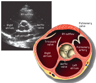
The ESC guideline reads a valve in stages. A clinician first names the lesion, a narrowing of the opening or a leak back through it. A clinician then grades how far the lesion has gone, from mild through moderate to severe. The grade decides the next step for the patient. The whole pathway hangs on that one word, severe. A measured number stands behind the word for every valve. The guideline spells out, valve by valve, what counts as severe, so two clinicians reading the same heart land on the same grade. A clinician reads the lesion, the grade, and the number as one chain that ends in a decision. The guideline names a mild, a moderate, and a severe band for each lesion, so the same valve falls in the same band for any reader who measures it.
Severe is a measured thing in this guideline. A clinician does not call a valve severe by eye alone. The guideline fixes a velocity, a pressure gradient, or an opening area for each lesion. The number crosses a stated line. The valve earns its grade from where the number falls. Continuous-wave Doppler supplies the velocity and the gradient behind nearly every one of those lines. A clinician reads the number against the line the guideline draws for that valve. The opening area on the left side often follows from the same Doppler trace through a short calculation the handheld can run. A clinician keeps the raw number in the report so the next reader sees the basis for the grade.
The number drives the decision more than the eye does. A clinician weighs a severe grade against the patient’s complaints and the strength of the heart. The guideline ties each grade to an action down the line. A severe valve in a breathless patient points toward repair or replacement. A clinician reads the grade and the symptoms as one picture of the patient. The guideline turns a valve read into a plan a clinician can act on the same day. A clear number at the bedside moves the patient along the pathway without a wait for the lab. A clinician who grades the valve in clinic spares the patient a second trip and gives the team a number to plan around.
What continuous wave reads at a valve
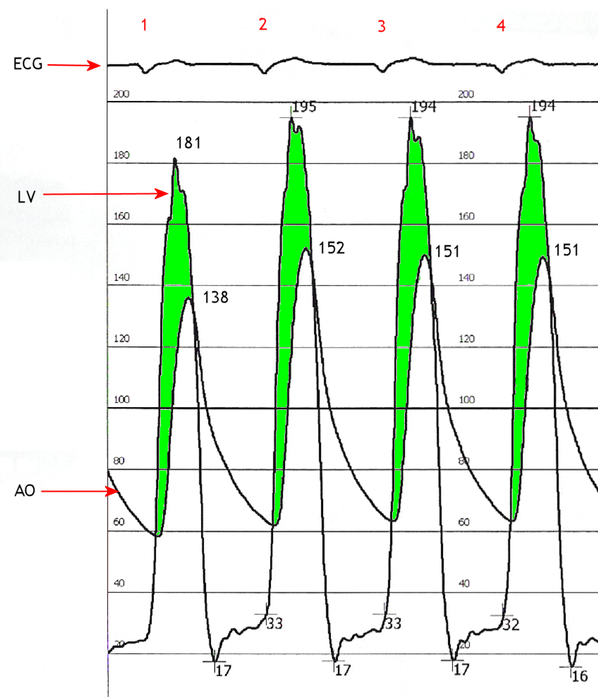
Continuous-wave Doppler reads the speed of the blood through a valve. A clinician lines the beam along the jet the valve throws. The mode catches the fastest blood anywhere on that line and prints it as a trace. A clinician reads the peak of the trace in metres a second. A narrowed valve or a leaking valve drives a fast jet a clinician can catch from the surface. The speed is the raw figure the whole grade rests on. A clinician reads the peak the instant the blood races through the valve. A clean peak from a well-aimed beam holds the speed the guideline grades, so a clinician spends the time to aim it. A clinician keeps the gain low enough that only the true jet writes on the trace, since a hot gain paints a fuzzy edge that reads the peak too high.
The speed turns into a pressure by a simple rule. A clinician squares the peak speed and multiplies by four. The result is the gradient across the valve in millimetres of mercury. A jet at four metres a second works out to a sixty-four millimetre gradient at its peak. The guideline grades the aortic valve on that gradient. A clinician reads the peak gradient and the mean gradient off the same trace. The mean comes from a tracing drawn around the whole envelope of the beat. A clinician records both numbers so the grade rests on the mean the guideline leans on for the aortic valve. A clinician also reads the ratio of the speed below the valve to the speed across it. A ratio under a quarter marks a severe aortic valve on its own, clear of the patient’s size and the angle of the beam. A clinician leans on that ratio when the gradient and the area disagree.
The shape of the trace carries its own message. A dense envelope that fills the trace marks a clean read straight down the jet. A clinician angles the probe in small steps to fill the envelope and sharpen the peak. A clean trace is the sign the number can be trusted. A thin signal reads the speed low and grades a tight valve too kindly. A clinician chases the fullest envelope so the peak reads honest. A clinician who grades a valve off a faint trace risks calling a severe valve only moderate. A clinician repeats the sweep from each window until the trace looks its fullest on the screen.
A clinician reads the jet from the window that lines up best with the flow. The aortic jet reads from the apex and from the right edge of the sternum. A clinician swings the probe through small arcs and keeps the highest clean peak. The highest peak from the best window holds the true speed of the jet. A clinician marks the window in the notes for the next study to match. The read assumes the beam sits near the line of the jet, so a clinician hunts the angle that fills the trace. A small dedicated probe head reads the jet from a tight rib space a larger footprint cannot reach.
The severe line
The guideline draws the severe line at a set of numbers for each valve. A clinician reads the peak velocity and the gradient against those lines. A number past the line names the valve severe. A clinician reads the velocity first, since the velocity gives the gradient directly. The table below sets out the lines the guideline draws across the common valves. A clinician reads each valve against its own line. The same beam reads them all, so a clinician moves from valve to valve with one tool.
A clinician reads the numbers together for one grade. The velocity, the gradient, and the opening area agree in the usual valve. A clinician gains confidence when the three line up on the same valve. A number out of step points to a measure to repeat or a flow problem to weigh. A clinician reads a valve severe when the lines it crosses hold together. A single number on its own carries less weight than three that agree. A clinician reads a lone high velocity with care, since a leak nearby or a high flow can lift it past the line on its own.
| Lesion | Continuous-wave measure | Severe | Highest grade |
|---|---|---|---|
| Aortic stenosis | Peak jet velocity | 4.0 m/s and up | 5.0 m/s and up |
| Aortic stenosis | Mean gradient | 40 mmHg and up | 60 mmHg and up |
| Aortic stenosis | Valve area | 1.0 cm² and under | – |
| Aortic regurgitation | Jet pressure half-time | under 200 ms | – |
| Mitral stenosis | Valve area from half-time | 1.5 cm² and under | 1.0 cm² and under |
The aortic valve and the gradient that calls for action
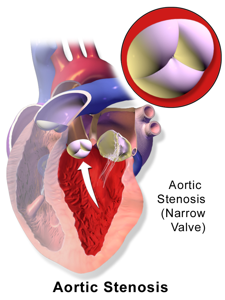
The aortic valve shows the gradient calling for action at its plainest. A clinician reads a tight aortic valve from the speed of the jet it drives in systole. The guideline draws the severe line at a peak velocity of four metres a second, a mean gradient of forty millimetres of mercury, and a valve area at or under one square centimetre. A clinician reads the three together as one picture of a tight valve. The 2021 ESC valve-disease guideline sets the lines a clinician grades against. A clinician keeps all three numbers in the report so the grade stands on more than the velocity alone. A clinician indexes the valve area to the patient’s body when the build runs small or large. The guideline draws the indexed severe line at six-tenths of a square centimetre for each square metre of body. A clinician reads a leaking aortic valve by how fast its backward jet fades on the trace. A clinician times that fall, since a steep drop under a fifth of a second marks a severe leak.
A steeper line marks the highest grade the guideline names. The guideline draws it at a peak velocity of five metres a second or a mean gradient of sixty millimetres of mercury. That grade earns the closest follow-up and the soonest referral. A weak heart can hide a tight valve behind a low number, so a clinician reads the gradient against the strength of the ventricle. A failing ventricle cannot drive the blood fast, so the gradient stays low even where the valve sits well past the line. The guideline names this low-flow, low-gradient stenosis. A clinician leans on the valve area here, since a weak heart can hold the gradient down below its true mark. The full lab settles the case with a low-dose stress study the handheld cannot run. A clinician who spots the mismatch at the bedside sends the patient on for that study. The guideline ties the severe grade to the symptoms before it acts. A symptomatic severe valve points toward a new valve, by open surgery or a catheter route. A clinician reads the gradient, the area, and the patient’s breathlessness as one call. A clinician who finds a clear severe gradient in a breathless patient has found a plain reason to refer.
The gradient turns into a decision at the bedside. A clinician who finds a severe gradient in a patient with chest tightness, faintness, or breathlessness has found a reason to refer that day. The guideline points symptomatic severe stenosis toward a new valve. A clinician carries the grade and the symptoms together to the heart team. A handheld brings the read forward to the day the patient is first seen. A clinician dates the first symptom in the note as the marker the team acts on.
A clinician follows a severe valve without symptoms on a close watch. The guideline sets a clock for the next study by the grade and the velocity. A clinician reads the velocity again at each visit to catch a valve on the move. A velocity climbing past three-tenths of a metre a second in a year marks a valve heading for surgery. A clinician shortens the interval the moment a symptom arrives. A patient on a watch list learns the symptoms that bring them back early. The guideline allows a treadmill test to draw out symptoms a patient plays down at rest. A clinician who sees the blood pressure drop or a symptom appear on exertion reads the valve as ready for the team.
A clinician reads the aortic valve from more than one window, keeping the highest true peak. The apex, the right sternal edge, and the suprasternal notch each open the jet to the beam. A clinician keeps the highest clean peak from the whole set of windows. A foreshortened view or an off-axis beam calls the valve milder than it is. A clinician chases the fullest envelope from each window so the grade comes out honest. A dedicated continuous-wave probe with a small face slips between the ribs the apex hides behind.
The mitral and the right-sided valves
The mitral valve answers to the same beam from the apex. A clinician reads a tight mitral valve by the mean gradient across it and by the pressure half-time of the inflow. The guideline grades a severe mitral stenosis at a valve area at or under one and a half square centimetres. A clinician reads the valve area from the half-time through a fixed formula the handheld runs on the trace. A clinician reads a leaking mitral valve by the density of its backward jet and by the width of the leak at its neck. The guideline grades a severe leak by the size of the orifice and the volume that crosses it each beat. A tight mitral mouth lets the pressure equalise slowly, so the inflow speed takes longer to fall away. A clinician reads a longer half-time as a tighter valve with a smaller mouth.
The right-sided valves carry their own grades. A clinician reads the tricuspid jet to find the pressure in the lungs, since the speed of the backward jet gives the pressure in the right heart. The guideline reads a severe tricuspid leak by the width of the jet at its neck and the strain it lays on the right heart. A clinician reads the pulmonary valve the same way, by the speed of the blood across it. The right side rounds out the valve study a clinician carries from one window to the next. A handheld reads the tricuspid jet from the same apical window that read the mitral. The peak speed of that backward jet, squared and multiplied by four, gives the pressure the right heart works against, with the pressure in the right atrium added on. A clinician reads a high lung pressure as a sign a left-sided valve has begun to load the right heart.
A clinician grades each valve against its own line in the guideline. Each valve carries its own line, so a clinician keeps the right number for the right valve. A clinician reads the number the valve gives against the number the guideline sets for it. The grade names the severity the same way across two readers and two visits. A clinician carries each grade into the plan the guideline draws for that lesion. One probe and one beam read the whole set, so a clinician grades the heart valve by valve in a single study.
What a handheld can grade
A handheld with continuous wave grades the numbers the guideline keys its decisions to. A clinician reads the aortic peak and gradient, the mitral half-time, and the tricuspid jet at the bedside in one sitting. Those readings cover the valves behind the everyday decisions. A clinician sends the hard case on for the full valve area by planimetry or the low-dose stress study the lab runs.
Where the number fits the ESC pathway
The guideline reads the number against the symptom every time. A clinician pairs a severe grade with what the patient feels day to day. A severe valve that has begun to cause breathlessness gives a plain reason to send the patient on for a new valve. Before that day, a clinician keeps the same severe valve on a close watch and a set clock. A clinician and the guideline act on the number and the symptom together. The grade opens the conversation the heart team finishes. A clinician times the referral to the symptom, since a severe valve that has turned symptomatic calls for the team within weeks.
The grade sets the speed of the next step. A clinician moves the highest grade forward faster than a borderline one. A fresh severe grade in a sick patient can trigger a same-day call to the heart team. The guideline matches the urgency of the move to the size of the number and the depth of the symptom. A clinician reads a bigger number as a quicker move down the pathway. A clinician who waits on a clear severe grade owes the patient a reason for the wait.
A clinician records the number plainly for the next reader to match. The report gives the velocity, the gradient, and the area, each with the window it came from. A clinician ties the grade to the symptoms in the same note. The next clinician repeats the study and compares like with like. A pathway runs on numbers that hold their meaning from one visit to the next. A clinician who writes the window down saves the next reader a guess.
A clinician brings the bedside grade to the heart team. The team weighs the grade, the symptoms, the heart’s strength, and the other imaging together. A clinician reads the handheld grade as the opening move of that decision. The guideline holds the whole team to one set of lines so the call travels from clinic to clinic. A clinician carries the number into the room where the call is made. A grade measured to the guideline at the bedside fits the terms the team already uses.
Reading the same valve over time
Valve disease moves slowly, so the guideline leans on repeat studies over years. A clinician follows a moderate aortic stenosis with a study every year or two by the grade. A hardening valve pushes the peak velocity up from one study to the next. A clinician marks the climb across the studies on one chart. A jump in the gradient brings the next decision forward in time. A clinician reads the run of numbers across the visits.
The same windows and the same beam keep a trend honest. A clinician returns to the apex or the sternal edge that read best last time. The beam runs along the jet the same way each study. A number measured the same way each time shows a true change in the valve. A clinician keeps to one method across visits so the grades compare cleanly. A change measured two different ways tells a clinician little. A clinician notes the heart rate alongside the gradient, since a racing heart lifts the mean gradient at the same valve.
A clinician reads the grade against the symptoms at each visit. A new breathlessness on a severe valve brings the referral forward at once. A clinician asks the patient about chest tightness, faintness, and breathlessness every time. The first symptom on a severe valve changes the plan that day. A clinician dates that symptom as the marker for action. A patient who knows the symptoms reports them before the next booked study.
A clinician records each study with the grade, the windows, and the symptoms in one place. A clinician saves the trace and a clip of the valve for the next reader. The record lets a clinician plot the velocity over the years on a single line. A rising line warns of a valve heading for surgery well before the day arrives. A clinician trusts the trend across studies above any single number. A clinician dates each study so the rate of change reads true across the years. A valve that gains speed fast needs a shorter gap between studies.
The ESC valve-disease guideline turns a valve read into a plan. A clinician grades the valve by the number, weighs the number against the symptom, and carries the pair to the team. A handheld with continuous wave brings that grade to the bedside in minutes. A clinician reads the heart where the patient sits and carries the hard case forward to the lab. A guideline number measured at the bedside starts the patient down the right pathway sooner.
Common questions about ESC valve grading with continuous wave
How does the ESC guideline grade a valve?
The ESC guideline grades a valve by a measured number, a velocity, a gradient, or an opening area. A clinician reads the number against the severe line the guideline draws for each valve. Continuous-wave Doppler supplies the velocity and the gradient. A clinician carries the grade into the decision to watch the valve or to refer the patient.
What gradient marks a severe aortic stenosis?
The ESC guideline draws the severe line at a peak velocity of four metres a second or a mean gradient of forty millimetres of mercury, with a valve area at or under one square centimetre. A reading past five metres a second or sixty millimetres marks the highest grade the guideline names. A clinician reads the three numbers together. A symptomatic severe valve points toward a new valve.
Can a handheld grade a valve to the guideline?
Yes. A handheld with continuous wave reads the peak velocity and the gradient the guideline keys its decisions to. A clinician grades an aortic valve, reads a mitral half-time, and estimates the lung pressure at the bedside. A clinician sends the hard case on for the full valve area or the stress study the lab runs.
Does a single number decide on surgery?
No. The guideline pairs the severity grade with the symptoms and the heart’s function. A severe valve points toward repair or replacement once the patient feels it. Until that day, the same valve stays on a close watch. The clinical picture and the heart team make the call together.
How often is a valve rescanned?
A clinician sets the interval by the grade and the speed of the change. The higher the grade and the faster the climb, the closer the next study. A moderate valve that holds steady can stretch to a year or two. A clinician scans sooner the moment a symptom appears, since the first symptom of a severe valve changes the plan.

About the Author
Julien Mercier
Senior R&D Engineer · Medical Ultrasound Transducer Development
Senior R&D Engineer with an M.S. in Applied Physics and over 15 years of experience in medical ultrasound transducer development, specializing in the design verification and performance testing of high-frequency imaging transducers. Currently leading the development and verification of the company’s next-generation high-frequency linear-array transducer, responsible for imaging performance evaluation and reliability analysis in preclinical testing. Brings extensive hands-on experience in piezoelectric element tuning, beamforming parameter optimization, and system-level performance testing.