Automated bladder volume calculation turns a picture of the bladder into a number in millilitres. The machine measures the bladder across three directions on screen and runs the three figures through one fixed formula. A volume lands beside the image a moment later. A handheld does the same work at the bedside, off a battery, the probe sweeping across the lower belly. The figure it returns is the urine the bladder holds at that moment, found with no catheter and no trip to a scanning room. A clinician reaches for it to settle a plain question. How full is this bladder right now. A hand on the belly cannot answer that. A guess from how a person feels misses badly in both directions. The scan puts a figure where the hand had none, the same figure in any hand that holds the probe. That number is only as honest as the steps that build it. Those steps start with a formula older than any handheld.
The figure counts because a bladder gives the hand almost nothing to go on. A bladder swollen to twice its size and one freshly emptied can press the belly wall with the same soft give. It sits low behind the pubic bone, out of reach of a clean press. A heavy belly buries it further. The volume cuts through all of that. It lifts the urine straight off the image, in cubic centimetres, into a figure a clinician can write down and compare. One scan before a person voids, one after, the drop between them showing how well the bladder emptied. A single scan on a person who cannot pass urine at all says how hard the bladder needs draining. The same probe covers the kidneys above in the same sitting, since a bladder that backs up pushes its trouble upstream. One device, one short scan, and the lower tract gives up a number that once took a catheter to find. A person feels nothing past the cool gel and a light press, the scan repeating as often as the care asks with no fresh risk each time. The bladder is the easiest organ to reach in the belly, a dark sac that all but jumps off the screen. The volume comes within reach of a light touch and a steady frame, no deep skill needed to land it.
The ellipsoid sum
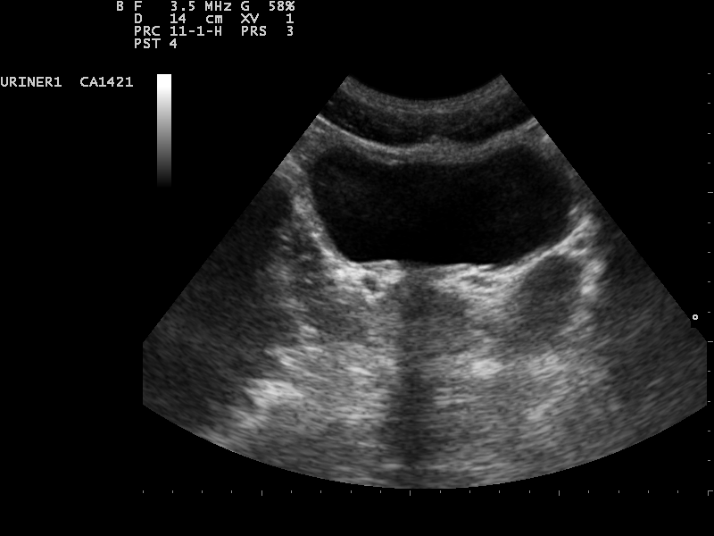
A full bladder in cross-section. A scanner outlines the dark pool and measures its width and depth on a view like this, then its height on a second, lengthwise view.
The formula treats the bladder as an ellipsoid, a ball stretched along its three axes. A full bladder rounds out close to that shape, near enough to the model to trust. The sum needs three numbers off the bladder, the width across, the depth front to back, the height top to bottom. Multiplied together, the three give the volume of the box that would just hold the bladder. A bladder does not fill its box, its corners being round, so the sum scales the box down by a fixed share for the rounding. That share is the factor 0.52. A clinician marks width, depth, and height on screen. The machine carries the 0.52 from there. The whole calculation is three measured spans and one constant, run in the instant after the last caliper drops. Nothing in it shifts from one bladder to the next, which is what lets two figures stand side by side and mean the same thing. The sum holds no surprises and no judgement of its own. Width, depth, height, and a constant go in; a volume comes out, the same way every time on every machine that runs it.
The factor falls straight out of geometry. The volume of an ellipsoid is four-thirds of pi, times each of the three half-axes multiplied together. Full widths come off the screen, so each axis carries a factor of one half. Three halves multiplied give one eighth. Four-thirds of pi, divided by eight, comes to 0.5236. Rounded, that is the 0.52 the machine holds, pi over six, the share of its bounding box any ellipsoid fills. The arithmetic never reaches a clinician, since the constant lives in the software. Some machines carry 0.7 instead, a looser factor for a bladder that fills out past the clean ellipsoid shape. The choice of factor shifts every volume a machine reports, so two scanners can size one bladder and print numbers a fifth apart. Knowing which factor a machine uses puts its numbers in the right light. Comparing two machines means checking they run the same constant before trusting the gap.
The three spans come off two passes of the probe. The first view, the probe held across the belly, cuts the bladder in section; width runs side to side there, depth front to back on the same frozen frame. A quarter-turn brings the second view, a cut down the bladder’s length, with height running top to bottom. Three calipers drop onto the two frames, one pair to each span. The machine takes the distance between each pair and feeds the three into the formula. A careful hand catches each span at the bladder’s widest on its plane, since a caliper set short of the edge drops the whole volume below the truth. Squaring the probe to the bladder before the freeze keeps the cut true, since a slanted cut crosses the bladder askew and shaves every span too short. The bladder can shift a little between views if a person breathes deep or stirs, so the two spans come in quick succession, catching the one bladder at the one size. The two views take seconds, the volume following with no pause.
The ellipsoid sum, term by term
Part of the sum
What it is
Width
the bladder’s span side to side, off the cross view
Depth
its span front to back, off the same view
Height
its span top to bottom, off the long view
0.52
the share of its box a rounded bladder fills (pi over six)
Volume
width – depth – height – 0.52, read in millilitres
The answer arrives in cubic centimetres, a cubic centimetre being a millilitre to the clinician. A bladder measured at eight by six by seven centimetres comes to three hundred and thirty-six cubic centimetres before the factor. Scaled by 0.52, it lands near one hundred and seventy-five millilitres. That figure on the screen is the urine in the bladder, ready to act on. The same clean formula keeps printing whatever the bladder’s shape, even on one scarred, slack, or shoved out of round, with no hint that its footing has gone. The arithmetic cannot feel the bladder broke its own assumption. Only the picture shows that, which is why the shape on the screen comes before the figure the shape was forced into.
The same sum in every machine
A dedicated bladder scanner and a handheld reach the volume along the same road. Both point the beam at the bladder, take its measure across, and run the ellipsoid sum on what they find. The arithmetic is one and the same. A volume is width times depth times height times a constant, whether a nurse thumbs a scanner in a ward or a clinician sweeps a handheld in a clinic. The number a scanner flashes and the number a handheld shows come off the identical formula. Learn the sum on one device and the other holds no mystery under its shell. A figure from either carries the same meaning, once both run the same factor. The device sets how fast the volume comes and how much of the working shows. It does not touch the sum that finds it. A figure means the same thing whichever box it came out of, once the factor behind it is known.
What the machine traces
The word automated points at the part a clinician once carried by hand. Every caliper went on alone, the frame frozen, each edge of the bladder marked by eye. The machine now finds those edges for itself. It traces a line around the dark pool of the bladder, picks out the widest span on the view, and drops the calipers where they belong. The whole measure happens in the breath after the probe steadies. A clinician shifts from placing the marks to checking them, watching whether the traced line sits on the true wall. This is the heart of what automation moved. The judgement of where the bladder ends, once a skill built over hundreds of scans, now arrives as the machine’s first guess. Experience goes into confirming or fixing that guess. The first stroke on a blank screen now belongs to the machine. A beginner reaches a usable volume far sooner, the hardest part of the measure coming pre-drawn. The skill that once took a hundred scans to build now comes loaded into the software. The saved effort goes onto judging the outline, the one part the machine cannot vouch for.
A dedicated bladder scanner reaches the same volume by a different route, one that sidesteps the three-line measure. The scanner freezes no single picture. It fans the beam through the bladder in a slow sweep, lifting a stack of thin slices from one wall across to the other. It outlines the bladder on each slice and adds those outlines into one volume, the way the size of a loaf comes from adding its cut pieces. The sweep loosens the grip of the ellipsoid assumption, a stack of real slices standing in for the guess that the bladder is a clean ellipsoid. A handheld with an automated volume mode borrows the same trick, building its outline frame by frame, each tilt of the probe adding a stroke to the line. The newest tools hand the outline to a trained network. A program studies the look of a bladder across thousands of labelled scans, learns where a wall tends to run, and then draws the boundary on a fresh scan in a fraction of a second. One study built such a tool, training a network to spot the bladder and measure its diameters off two-dimensional images. The automated volume it produced flagged a residual above one hundred millilitres at a sensitivity near 88 percent and a specificity of 100 percent, a match for the figures a sonographer reached by hand on the same scans. The payoff is speed and a steady result. A nurse on a ward round sizes a dozen bladders in the span a manual measure once gave to three. The figure comes out the same in any hand, the machine laying the calipers with no eye in the loop to vary them. The mode also stores the frame and outline the number came from, so a later check sees what was measured and weighs it with no rescan.
A clinician still owns the final line.
The two device types hand a clinician a different amount to see. A dedicated scanner shows almost nothing of its working. It clears the screen and prints a volume, no frame left behind to weigh. The figure goes on trust, with no way on the device to catch an outline that strayed, as good as the machine’s guess and no better. On a handheld the bladder stays in plain view through the whole measure. The traced outline lies over the picture, the frame held for a closer look. Outline and number come together as one thing. A line gone astray shows itself against the picture at once. The gap bites hardest on a hard bladder, where the outline is the part likeliest to slip. A clean bladder leaves almost no room to get it wrong. A difficult one turns that same outline into the whole question. Seeing the line, a clinician can fix it. Handed a bare number, all a clinician can do is believe it.
Automation leans on a full bladder more than a hand ever did. A bladder at a full, tight stretch gives the machine a clean edge to trace, the trained line snapping onto it with little to argue about. The same edge wanders on a bladder left half full, the creases throwing shadows the outline takes for walls and the traced line drifting onto a fold, sizing the bladder smaller than it stands. A scan that has to count wants a full bladder first, a person asked to hold on or to come with a comfortable load. A slack bladder earns a lighter trust, the automated edge standing on thin ground. Bowel gas calls for the same care, the machine losing the wall in the haze and the outline guessing through it. A press of the probe pushes the gas aside and clears the edge before the machine draws. The cleaner the bladder shows, the less there is to undo. A full bladder is the one thing a clinician can set up in advance, a glass of water and a short wait turning a hard scan into an easy one.
Where a clean number misleads
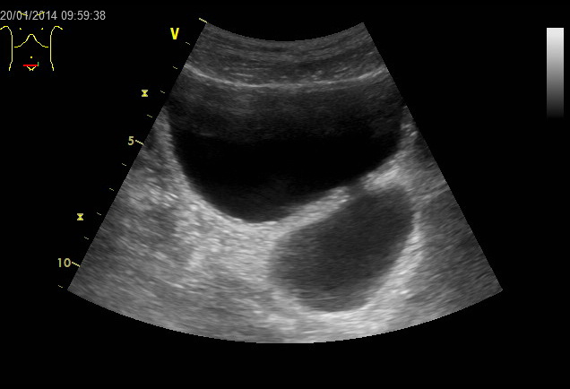
A bladder with a diverticulum, the rounded pouch below and to the right. A volume measured across the main bladder leaves out the urine the pouch holds.
A volume prints out to a tenth of a millilitre, a fine figure wearing a confidence the estimate behind it has not earned. The formula rests on one belief, that the bladder holds a clean ellipsoid shape. A long-blocked bladder breaks that belief. Years of pushing against a tight outlet thicken the wall and pull it into ridges and pouches, a shape the geometry never pictured. A bladder scarred by past surgery sits crooked in its bed. A fibroid or a full bowel can shove a bladder out of round, leaning where the model assumes it stays even. The sum runs across any of these without complaint, printing a number in the same clean font as ever. The arithmetic has no way to feel the shape has gone wrong; it measures three lines and trusts them, whatever shape strings them together. A clinician carries the doubt the machine cannot, weighing the figure against the shape on the screen. A bladder far from a tidy oval earns a number held loosely, checked with a second look before it goes on the record. The danger sits in the neatness of it. A figure to the tenth of a millilitre wears a precision the wrong bladder never gave it.
A pouch budding off the bladder fools the sum past any shape error. A diverticulum pushes out through a weak point in the wall and swells into a chamber of its own, filling with urine through the same opening it grew from. The ellipsoid measure spans the main bladder and stops dead at the main wall, the pouch sitting outside the three lines, uncounted. A bladder that looks near empty on the screen can hold a small cupful in a diverticulum the sum never crossed. A person voids, the main bladder flattens, the scanner prints a low number. The pouch keeps its own load out of sight of that number. A clinician sweeps wide of the bladder for a second dark chamber before calling the bladder drained. A handheld matters here, the picture showing the pouch a bare number would bury. Spotting the diverticulum, a clinician measures it on its own and adds it back, or at least notes that the printed volume is the main bladder alone. A large pouch can hold as much urine as the bladder it hangs off, the share the sum missed matching the share it counted. Urine trapped in a diverticulum breeds infection in a pool that never drains, one more reason to sweep wide before trusting a low count.
Smaller errors stack below the gross ones. A probe held off square slices the bladder on a slant, crosses it short of its true width, and hands the formula three spans all a touch too small, the volume coming out low for no reason but a tilted hand. A bladder caught partway through filling gives a number honest only for that minute, the kidneys feeding it and the level still climbing. An automated edge brings fresh trouble, latching now and then onto the wrong border. It can grab a loop of bowel, a cyst beside the bladder, or free fluid pooled around a sick belly, folding that dark patch into the bladder it was hunting. The volume jumps on a false edge as fast as it drops on a tilt. A catheter balloon left sitting in the bladder throws its own bright echo into the picture. The automated line can wrap that balloon into the count it was meant to leave out. A deep belly works the other way, pushing the bladder past easy reach into faint depths where the machine can only guess at its edge. Some settings tax the machine harder than others. A fresh postpartum bladder comes out low by tens of millilitres in published series, the swollen, slack tissue of the days after birth sitting far from the model. A clinician checks the outline against the picture every time and drags it back onto the true wall when the machine has wandered off. No automated outline is final until a human eye has signed off on where it sits. The eye stays the last check on the line, the same way it always was, signing off the outline the machine drew.
Weighing the number
A clinician treats the volume as an estimate in the dress of a measurement. On a bladder scarred, slack, pouched, or caught on a slant the figure calls for doubt, the kind earning a second look. A second look costs little, the probe re-squared, a slack bladder refilled, the measure run again in two clean planes by hand. A manual measure on a good pair of views often steadies a number the automated mode left shaky. Weighing the picture behind the figure is what makes the figure mean anything. The record notes which number was kept, the automated one or the hand measure, so the next clinician on the chart knows what it stands on. The number rewards a clinician who knows where it bends. The shape on the screen tells how far to trust the digits beneath it. That judgement is the one no automation has taken over.
The volume does its real work in the decision that comes next. A high number left after a void marks a bladder that cannot empty and turns a clinician toward the cause and the fix. A low number after a void closes the question in one scan. A number rising across a week of checks tracks a bladder losing ground, or a treatment failing to hold it. None of those calls is safe on a figure no one has weighed. A decision rests on the number, the number on the outline, the outline on a bladder that may or may not have kept the shape the formula assumed. Working down that chain comes before acting at the top of it. The machine has taken over the drawing and the arithmetic. What it hands back is the time to do the one thing it cannot, judging whether the tidy number tells the truth about the bladder it came from. That last judgement is human work, left to a person once the machine has done its measuring. The number is a tool a clinician picks up, sets against the bladder on the screen, and acts on once it holds up.
Common questions
What formula gives the bladder volume?
A scanner treats the bladder as an ellipsoid and multiplies its width, depth, and height by 0.52. The 0.52 is pi over six, the share of its bounding box a rounded shape fills. The result comes in millilitres, a cubic centimetre and a millilitre being the same amount.
Is the automated number accurate?
The automated figure drifts on an odd-shaped, scarred, or near-empty bladder, where the ellipsoid shape no longer fits. The picture settles which bladder is in front of the clinician before the digits get trusted.
How many measurements does the volume need?
Three, the width, the depth, and the height of the bladder. Width and depth come off one cross view, height off a long view. The machine multiplies the three and scales them by 0.52 to land on the millilitres.
Why does an automated volume come out low on a real bladder?
A probe held off the bladder’s widest line, a fill caught too early, or a pouch off the bladder can each drop the count. The sum measures the main cavity and trusts it to be a simple round shape. Squaring the probe and sweeping wide for a pouch comes before trusting a low number.
About the Author
Julien Mercier
Senior R&D Engineer · Medical Ultrasound Transducer Development
Senior R&D Engineer with an M.S. in Applied Physics and over 15 years of experience in medical ultrasound transducer development, specializing in the design verification and performance testing of high-frequency imaging transducers. Currently leading the development and verification of the company’s next-generation high-frequency linear-array transducer, responsible for imaging performance evaluation and reliability analysis in preclinical testing. Brings extensive hands-on experience in piezoelectric element tuning, beamforming parameter optimization, and system-level performance testing.