





































Pulse Repetition Frequency PRF Optimization in CW Doppler Handheld
Pulse repetition frequency is how often a pulsed Doppler mode sends a pulse. It sets the top speed the mode can read. A clinician tunes it to fit the flow on the screen and keep the trace from folding over. A handheld puts that control a tap away at the bedside. The right setting reads a flow clean, from a slow vein to a brisk inflow.
What pulse repetition frequency is
Pulse repetition frequency counts the pulses a mode sends each second. A pulsed mode sends a pulse and waits for its echo before the next. The wait fixes the depth the mode reads. The rate of those pulses is the pulse repetition frequency. A clinician sets it through the scale on the screen. A wider scale runs a higher rate and reads a faster flow. The mode reads the shift in the echo’s pitch, the Doppler shift, to find the speed. A faster flow shifts the pitch more, so the mode needs more samples to catch it. The pulse rate is how many samples a second the mode takes. A clinician sets the rate to match the speed of the flow. The scale on the screen shows that rate as the speed it can read. A clinician reads a brisk carotid flow near a metre a second. A mitral inflow runs near a metre a second as well, and an aortic outflow near the same.
The rate ties straight to the speed the mode can read. The mode samples the flow once per pulse, like frames of a film. A fast flow needs many samples a beat to trace its shape. A slow rate takes too few samples and loses the fast parts. The rate sets how fast a flow the mode can follow before it stumbles. A clinician matches the rate to the flow in front of them. The shift grows with the speed and with the angle of the beam to the flow. A beam along the flow reads the full shift, so a clinician lines it up. The mode turns the shift into a speed and draws it on the trace. A clinician keeps the beam near the line of the flow for an honest number. The cosine of the angle scales the speed the mode reads. A clinician keeps the angle small to hold that loss near nothing.
The depth of the reading caps the rate. A deep gate needs a long wait for its echo to return. The long wait holds the rate down, since the next pulse waits for the last echo. A shallow gate clears its echo fast and frees a higher rate. A clinician catches a fast flow more easily at a shallow depth. The depth and the rate pull against each other on the screen. The pulse must travel down to the gate and back before the next goes out. Sound runs near 1540 metres a second through tissue. A deeper gate adds travel time and forces a lower rate. A clinician treats the trade as a ceiling the depth sets on the rate. The shallowest gate that reaches the flow frees the highest rate. A deep mitral inflow runs at a lower rate than a shallow carotid. The depth on the screen tells the rate the mode can run. A clinician reads a deep flow at a lower rate and accepts the lower ceiling.
The screen scale shows the rate in plain speed. A clinician takes the top and bottom of the scale as the fastest the mode can show. A flow within the scale draws clean on the trace. A flow past the scale has nowhere to go and folds back. A clinician sets the scale to hold the whole flow with a little room to spare. The scale runs in centimetres a second on the screen edge. The number at the top edge is the ceiling the rate sets. The scale climbs when a clinician raises the rate. A flow that fits the scale draws its whole shape on the trace. A clinician leaves a margin above the peak so a beat-to-beat rise has room. A clinician edges the scale up when a flow grows through the study. The trace stays whole when the scale tracks the flow.
The rate is a pulsed-mode setting from end to end. A continuous mode sends sound without pause and never waits, so it carries no pulse rate at all. A clinician meets the pulse rate on pulsed wave and on colour flow. The setting governs every mode that samples in pulses. A clinician learns it once and uses it across those modes. Colour flow samples the same way, so it carries the same rate and the same limit. A clinician sets the colour scale as the colour version of the rate. The spectral trace and the colour map share the one setting in spirit. A clinician turns the rate up on either when a fast flow folds over. The pulsed family lives by the pulse rate from end to end. A clinician hears the audio of the flow alongside the trace. A faster flow gives a higher pitch, a second cue beside the picture. The mode builds the trace from the mix of speeds in the gate, sorted by a fast Fourier transform. A turbulent flow broadens that trace with a spread of speeds.
The Nyquist limit and aliasing
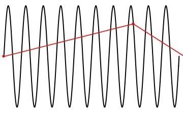
Half the pulse rate is the fastest speed the mode can read straight. This ceiling is the Nyquist limit. A flow below the limit draws clean. A flow above it cannot be sampled fast enough to trace. The mode then misreads the fast flow as a slow one in the wrong direction. This misread is aliasing. The rule sets the ceiling at exactly half the pulse rate. A rate of four thousand pulses a second reads a shift up to two thousand cycles a second. That shift turns into a speed by the Doppler rule. A clinician finds the Nyquist limit off the scale as the top speed on show. A higher rate lifts the limit and reads a faster flow clean. The half-rate ceiling is the Nyquist sampling limit, the same rule that sets the bit rate of recorded sound. A flow that crosses that half-rate folds at once. A clinician lifts the rate to push the half-rate higher.
Aliasing shows on the trace as a wrap. The peak runs off the top of the scale and reappears at the bottom. A clinician sees a signal that folds around the baseline. The wrap is the sign the flow has outrun the rate. The faster the flow runs past the limit, the more tangled the wrap looks. The mode samples too slowly to follow the fast flow, like a film that misses the spokes of a turning wheel. The wheel can seem to crawl or turn backward on the film. The fast flow seems slow or reversed on the trace in the same way. A clinician confirms a wrap by raising the scale and watching it unfold. A true fast flow keeps its peak when the scale climbs. A clinician treats the wrap as a sampling failure. The fix is more samples a second or a mode that never samples. A clinician knows the wrap by its look on both sides of the baseline at once. The signal seems to run two ways in the one beat.
A clinician takes a wrap as a flag to act on. The folded trace hides the true peak behind the fold. A clinician who takes the wrapped number at face value reads the speed low. The fix raises the rate or moves to a mode with no limit. A clean trace, whole within the scale, is the goal before any number comes off it. A wrapped peak reads lower than the true speed. A clinician who trusts it undercalls a tight valve or a fast leak. The wrap also muddies the shape a clinician reads for timing. A clinician clears the wrap before marking a peak or tracing an envelope. The clean trace is the rule to follow. A clinician marks a number only after the trace sits clean. A clinician raises the scale to tell a true peak from a wrap.
Why continuous wave escapes the limit
Continuous wave sends and listens without a pause, so it never samples in pulses. The steady stream leaves no gap to set a rate, so the mode meets no Nyquist limit. It reads a jet at any speed, from a gentle flow to a six-metre stenotic jet. The trade is place: the mode reads the whole beam at once. The speed it gives sits somewhere on that line, unplaced. The continuous mode runs two crystals, one sending and one listening without a break. The steady send and receive leave no gap to time an echo, so no depth and no rate exist. The mode reads the fastest blood anywhere on the line, which suits a high-speed jet. A clinician accepts the loss of place for the gain of unlimited speed. A clinician fixes the place with the image and reads the speed with the beam. A clinician turns to continuous wave for a fast flow and settles the place with the image.
Tuning the settings on a handheld
A clinician has a few moves against a wrap. Raising the pulse rate lifts the Nyquist limit and reads a faster flow. The move costs a little depth, since a higher rate reaches less far. A clinician lifts the rate first when a flow aliases. The scale climbs with the rate and shows the faster speed. The higher rate samples the flow more often each beat. The extra samples catch a faster flow before it folds. A clinician pushes the rate as far as the depth allows. The fuller scale then holds the flow with room above the peak. A clinician steps the rate up and watches the wrap clear. The trace settles whole once the rate fits the flow. A clinician reads the scale number after each step up. The flow sits clean once the scale tops the peak.
Shifting the baseline hands more of the scale to one direction. A flow that runs one way needs little room the other way. A clinician slides the baseline down to give an upward flow the whole scale. The move buys headroom without a faster rate. A clinician traces a one-way flow clean with the baseline set right. A flow that runs toward the probe fills only the upward half of the scale. A clinician slides the baseline down and hands that whole range to the upward flow. The move doubles the speed the mode shows in one direction. The rate stays the same through the shift. A clinician uses the baseline first for a flow that runs one way. A clinician centres the baseline again for a flow that runs both ways. The setting follows the flow’s direction each time.
Dropping to a lower transmit frequency reads a higher speed before the wrap. The lower frequency shifts less for the same speed, so it aliases later. A clinician trades some fineness of the image for that headroom. A clinician picks the frequency that fits the depth and the speed together. A lower frequency returns a smaller shift for the same speed. The smaller shift sits under the Nyquist limit at a higher speed. The lower frequency also reaches deeper into the chest. The frequency knob serves the depth and the limit at once. A clinician matches the frequency to the depth of the heart in front of them. A deeper heart takes a lower frequency for the reach.
A clinician reads the table for the move that fits the flow. The settings stack: a higher rate, a shifted baseline, and a lower frequency add their headroom together. The depth of the gate sets the ceiling on the rate, so a clinician keeps the gate as shallow as the flow allows. A jet far past the limit outruns every setting, and a clinician then switches to continuous wave. The wall filter clears the slow thud of moving tissue from the trace. A clinician sets the wall filter to match the flow, higher for a brisk artery. The sweep speed spreads each beat across the screen for a clear shape. A high-pulse-rate mode adds gates along the line to read a faster flow. The added gates blur which depth the speed came from, a trade a clinician accepts for the speed. A tissue Doppler reads the slow heavy motion of the muscle at a low rate. A wide colour box drops the frame rate, since each scan line needs its own pulses. The ASE recommendations on quantitative Doppler set out these settings and their limits.
| Setting | Move | Effect |
|---|---|---|
| Pulse rate (PRF) | Raise it | Lifts the Nyquist limit and reads a faster flow |
| Baseline | Shift it | Hands more scale to one direction |
| Transmit frequency | Lower it | Reads a higher speed before aliasing |
| Sample depth | Bring it shallower | Frees a higher pulse rate |
| Mode | Switch to continuous wave | Removes the speed limit |
Reading a clean spectral trace
A clean trace is the aim of every setting. A clinician fills the envelope, clears the wrap, and sharpens the edge before reading a number. The gain sets the brightness of the trace and keeps the noise down. The scale holds the whole flow with a little room above the peak. The baseline sits where the flow has the room it needs. A clinician reads the peak off a trace that sits whole within the scale. A clinician fills the envelope so the fastest blood shows at the edge. The gain sits high enough to draw the trace and low enough to keep the noise down. A clinician steadies the probe on the line of the flow for the fullest signal. A clinician reads the spectral window beneath the trace for turbulence. A clean trace reads a number a clinician can trust, beat after beat. A clinician keeps the settings steady across visits for a fair compare.
When the flow outruns the rate
A flow can run faster than any pulse rate the mode allows. A stenotic jet or a tight regurgitation can pass five metres a second. No pulsed setting holds such a jet on the scale. A clinician reads the wrap, tries the easy fixes, and finds them spent. The flow has outrun the pulsed mode for good. A stenotic valve drives blood through a tight gap at a high speed. A tight leak fires a jet back at a high speed as well. A clinician tries a higher rate and a shifted baseline first. The fast jet runs past the room those moves buy. A clinician reads the spent settings as the sign to change mode. A clinician notes the rate already at its top. The flow needs the continuous mode from there. A high-pulse-rate mode trades the certainty of one depth for a higher speed. A clinician weighs that trade before leaving the pulsed mode. A clinician keeps the gate shallow to hold the rate as high as it goes.
The continuous mode answers a flow the pulsed mode cannot hold. A clinician switches to it with a tap and reads the speed whole. The mode reads the fast jet with no ceiling to fold it over. A clinician reads the peak off a clean continuous trace at once. The fast jet that broke the pulsed scale reads clean on the continuous one. A clinician aims the beam down the jet and reads the peak at the edge. The mode reads a six-metre jet as cleanly as a two-metre one. A clinician marks the peak and turns it into a gradient. The continuous mode reaches the speed the pulsed mode could not hold. The number the pulsed mode missed comes off the continuous trace.
Colour flow shows the same wrap as a patch of the wrong colour. A clinician sees the colour flip where the flow speeds past the limit. The same fixes lift the colour limit: a higher rate, a shifted baseline, a shallower view. A clinician reads the colour wrap as the same flag the spectral wrap raises. The one idea covers the colour map and the spectral trace alike. The colour scale carries its own Nyquist limit, half the colour pulse rate. A flow past that limit flips colour in a bright mosaic patch. A clinician lifts the colour scale to clear the mosaic. A narrow colour box and a shallow view both lift the colour rate. A clinician takes the mosaic as the colour form of a wrapped trace. A clinician judges the spread of colour as the speed across the vessel. A clean colour fill marks a flow within the limit. A clinician narrows the colour box to lift both the colour rate and the frame rate.
The controls on a handheld
A handheld puts the scale, the baseline, and the gain under a finger on the screen. A clinician taps the scale up when a flow aliases. A clinician drags the baseline to free the room a flow needs. The controls answer at once, so a clinician tunes the trace and reads it in the same breath. The gain rides a slider beside the trace. A clinician sets the depth and the gate by eye on the live image. The whole set of controls sits under a thumb on the screen. A clinician tunes the trace and reads it without looking away. The controls and the image share the one screen.
A clinician carries the whole set of moves in a pocket probe. The scale, the baseline, the frequency, and the mode switch all sit on the screen. A clinician reads a clean flow at the chair without a cart. A clinician learns the normal speed of each flow to spot one that runs too fast. A tricuspid jet past two and a half metres a second points to a raised lung pressure. The handheld runs the rate, the scale, and the filter the cart machine runs. A clinician reads a clean carotid flow and a clean valve jet on the one device. The setting that fits the flow falls to hand with practice. A clinician needs no second machine for the read. A clinician runs the whole spectral study on the one pocket device at the chair. The right setting turns a folded mess into a number a clinician can trust.
Common questions about PRF and aliasing
What is pulse repetition frequency in Doppler ultrasound?
Pulse repetition frequency is how often a pulsed Doppler mode sends a pulse each second. The mode sends a pulse and waits for its echo before the next. The rate sets the top speed the mode can read, half the rate being the Nyquist limit. A clinician sets it through the scale on the screen.
What is the Nyquist limit?
The Nyquist limit is the fastest speed a pulsed mode can read straight, half the pulse rate. A flow below the limit draws clean on the trace. A flow above it cannot be sampled fast enough and folds over, the aliasing a clinician sees as a wrap. A clinician raises the rate to lift the limit.
How does a clinician fix aliasing?
A clinician raises the pulse rate to lift the Nyquist limit first. A shifted baseline hands more scale to the flow’s direction. A lower transmit frequency reads a higher speed before the wrap. A jet that outruns every setting calls for continuous wave, which carries no limit.
Why does continuous wave have no Nyquist limit?
Continuous wave sends and listens without a pause, so it never samples in pulses. The steady stream leaves no gap to set a rate, so no Nyquist limit applies. The mode reads a jet at any speed off the line of the beam. A clinician settles where that speed sits with the two-dimensional image.
Can a handheld scanner adjust the pulse repetition frequency?
Yes. A handheld puts the scale, the baseline, and the gain on the screen under a finger. A clinician taps the scale up to raise the rate when a flow aliases. The controls answer at once at the bedside. A clinician tunes the trace and reads a clean flow in the same study.

About the Author
Julien Mercier
Senior R&D Engineer · Medical Ultrasound Transducer Development
Senior R&D Engineer with an M.S. in Applied Physics and over 15 years of experience in medical ultrasound transducer development, specializing in the design verification and performance testing of high-frequency imaging transducers. Currently leading the development and verification of the company’s next-generation high-frequency linear-array transducer, responsible for imaging performance evaluation and reliability analysis in preclinical testing. Brings extensive hands-on experience in piezoelectric element tuning, beamforming parameter optimization, and system-level performance testing.