





































VetBLUE Veterinary Lung Ultrasound Handheld Protocol
VetBLUE is a lung scan run at a set of windows across both sides of the chest. A vet lays the probe at each window and reads one thing: how much fluid the lung holds. The lung reads as wet or dry at every spot. The spread of the wet across the windows points to the cause. A vet runs it cage-side on a handheld, on a patient that struggles to breathe.
What vetBLUE reads
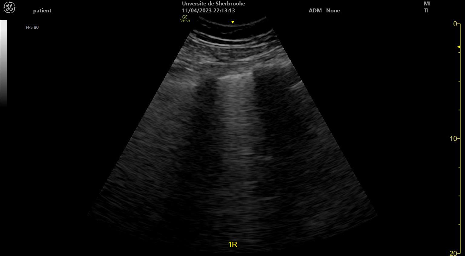
VetBLUE reads the lung for fluid. A healthy lung is full of air. Air throws up one ultrasound pattern on the screen. Fluid in the lung throws up another. The scan reads which pattern is there at each window. The pattern is what tells the vet how wet the lung is. The ultrasound cannot see into air. A lung full of air gives a surface and little else. Fluid changes what comes back to the probe. The scan reads the lung through what the air and the fluid send back.
The scan names each window wet or dry. A vet works along the windows one by one. They call each one before moving to the next. The reading builds a map of the lung across the chest. The map is the scan’s result. A vet does not read one window alone. The reading is the whole set of eight. A window is one point on the map. The map of all eight is what the vet reads.
The whole scan is a read of fluid in the lung. A vet runs the windows and counts the wet ones. The number and the place of the wet windows make the finding. Many wet windows mean a lung full of fluid. A clear scan has none. The count of wet windows is a quick measure. A vet runs the eight and tallies the wet. The tally says how much of the lung is involved. A rising tally on a repeat scan reads a lung filling up. The count is a number a vet can write down. It travels in a note better than a picture does.
The windows of the scan
VetBLUE runs at a set of named windows. The scan uses four windows on each side of the chest. That makes eight windows in all. Each window has its own spot on the chest wall. A vet lays the probe at each in turn. The set windows are what make the scan the same every time. A window is a fixed spot on the chest. A vet returns to the same eight on every patient. The fixed spots let two scans line up. The set windows are the backbone of the protocol.
The protocol was named and tested by its first study. The study that named the vetBLUE exam set out the regional approach. It read four lung regions on each side. It counted the lung rockets, the B-lines that mark a wet lung. A vet runs the windows that study laid down. The scan reads the same in any clinic that follows them.
The windows are named for where they sit. One reads the caudodorsal lung, up at the back. One reads the perihilar lung. One reads the middle lung. One reads the cranial lung, down at the front. A vet runs the four down each side of the chest. The four windows cover the lung top to bottom. A vet reads the back and the front of each side. The eight together sample the whole lung. No region is left unread.
The order of the windows holds the scan together. A vet runs the same four on each side, in the same order, on every patient. No part of the lung is missed in the rush. A vet who runs the order by habit covers the chest without a checklist. The set route is what makes one vet’s scan another’s. A vet learns the route as a fixed order. The same eight come in the same sequence. The habit covers the chest without a list. The order is what keeps a window from being skipped.
Wet lung and dry lung
The scan turns on one read at each window. A dry lung is a lung full of air. A wet lung is a lung holding fluid. The vet reads the sign at each window and calls it. The table below sets out the signs vetBLUE reads. The read at a window is quick. A vet sets the probe, looks, and calls the sign. The call takes a few seconds. The scan is the sum of eight quick calls. A vet does not linger at a window. The sign is there or it is not. A quick look settles it. The scan moves window to window without delay.
The signs are a short list a vet learns by heart. The table gives each sign, its look on the screen, and what it means. A vet reads a window, matches it to a sign, and moves on. The signs are the words the scan is written in. A vet who knows the signs reads a window at a glance. The signs are few enough to hold in the head. A new vet learns them in a sitting. The list is short by design.
| Sign | Look on screen | What it means |
|---|---|---|
| Dry lung | A-lines with a gliding lung | Normal, air-filled lung |
| Wet lung | B-lines, the “lung rockets” | Fluid in the lung: edema, contusion, or pneumonia |
| Shred sign | A ragged edge into the lung | Consolidation, as in pneumonia |
| Tissue sign | Lung that looks like liver | A larger area of consolidation |
| Nodule sign | A rounded soft-tissue spot | A mass or nodule in the lung |
| No glide sign | A lung that does not slide | Possible pneumothorax |
Reading the pattern
The place of the wet windows tells the vet the cause. A vet reads all eight windows and notes which ones are wet. The pattern across the chest points to what is filling the lung. Heart failure floods the lung from the back and the bottom first. Its wet windows sit in the caudodorsal and perihilar spots on both sides. Pneumonia patches the lung in one region or two. Its wet windows often fall on one side, with dry lung around them. Lung contusions from a knock sit under the spot that was hit. A vet reads the map of wet windows and asks what shape makes it. The spread of the rockets is the scan’s working diagnosis. A vet reaches a likely cause from the shape of the wet alone, before any other test. The reading takes a minute at the cage-side. The shape of the wet is the diagnosis the scan hands the vet. A vet reads it and names a likely cause on the spot.
The pattern reads the cause without a picture of the heart. A lung wet from heart failure looks one way on the windows. A vet reads that shape and thinks of the heart. The scan points to a cause the chest alone would not show on a film. A vet acts on the pattern while the next test is still being set up. The scan reads the fluid at the cage in a minute. A vet has an answer before the patient is moved. A working diagnosis comes without a trip to the x-ray room. The speed is half of what the scan is for.
The pattern changes as the patient is treated. A lung wet from heart failure dries out once the fluid clears. A vet scans again and counts fewer wet windows. The falling count reads the treatment working. The scan tracks the lung from wet to dry through the treatment. A vet does not guess whether a treatment is working. The repeat scan shows it in the count. Fewer wet windows read a lung clearing. The scan measures the response a vet would otherwise judge by feel.
The pattern is read fast and read again. A vet runs the eight windows in a minute or two. The scan is quick enough to repeat through a treatment. A vet scans on arrival, then again an hour on. The two scans read the change in the lung. The repeat is part of how vetBLUE earns its place. A first scan sets the baseline. Each scan after reads against it. The count of wet windows trends up or down. A vet reads the trend as the patient’s course.
The pattern is only the start of the workup. The scan points to a likely cause. A vet confirms it with the rest of the exam. The wet pattern of heart failure sends a vet to the heart scan next. The vetBLUE read opens the workup. It does not close it. VetBLUE narrows the field fast. A vet carries its pattern into the next tests. The scan is the opening of the workup. The rest of the exam settles the cause.
Dry is air, wet is fluid
VetBLUE asks one question at every window. It asks how much fluid the lung holds there.
The signs of consolidation
VetBLUE reads more than wet and dry. A lung packed solid with fluid or cells stops looking like lung at all. The scan shows it as a patch of tissue. These patches are the signs of consolidation. A vet reads them past the simple wet-or-dry call. Consolidation is lung gone solid. The air is pushed out of it. The scan shows it as a block of tissue. A vet reads it as a deeper change than the rockets.
The consolidation signs each have a name. The shred sign is a ragged edge where solid lung meets air. The tissue sign is lung that looks like liver. The nodule sign is a rounded soft-tissue spot. A vet reads which sign is there and what it points to. The signs read a lung gone solid in stages. A shred is a small patch turning. A tissue sign is a larger one. A nodule is a rounded growth. A vet matches the look to the name.
The consolidation signs point to the deeper trouble. A shred or a tissue sign reads pneumonia or a drowned lung lobe. A nodule reads a mass. The signs take vetBLUE past fluid alone and into the solid changes a lung can show. A vet reads the sign and follows it to the cause. A shred sign on one lobe reads a pneumonia. A tissue sign reads a larger patch gone solid. The scan tells the vet a lobe is involved. A vet works the cause up from there.
Pneumothorax and effusion
VetBLUE reads the space around the lung too. A healthy lung slides against the chest wall. The scan shows that slide as the glide sign. A vet looks for the glide at each window. The glide is the sign a lung is up against the wall where it should be. The glide is easy to see once a vet knows it. The lung surface shimmers along the wall. A still surface stands out at once. A vet checks the glide at every window.
A lost glide points to air in the chest. Air between the lung and the wall stops the slide. The lung sits still against the wall. A vet who finds no glide at a window thinks of a pneumothorax. The absent glide is the scan’s read for free air in the chest. A vet pairs the missing glide with the rest of the picture. Air in the chest is a finding to act on fast. The scan flags it at the window. A vet follows it up at once.
VetBLUE reads free fluid around the lung as well. Fluid in the chest pushes the lung up off the wall. The scan shows a dark space where the fluid sits. A vet reads that space as a pleural effusion. The lung floats in the fluid above it. A small effusion shows as a thin dark rim. A larger one lifts the lung well off the wall. A vet reads the depth of the dark space. The scan finds the fluid around the lung.
The space signs round out the scan. A vet reads the lung for wet and dry. A vet reads the space for air and fluid. The two reads together cover the chest. A vet runs vetBLUE and comes away with the lung and the space around it both read. A vet finishes vetBLUE with a full read of the chest. The lung is mapped for fluid. The space is checked for air and effusion. The chest is covered in one short scan.
The breathless patient
VetBLUE is built for the patient that cannot breathe. A dog or cat in respiratory distress cannot lie on its back for a scan. VetBLUE is run with the patient sternal or standing. The probe comes to the patient in the position it can bear. The scan asks nothing that makes the breathing worse. A vet does not lay the patient down for vetBLUE. The dog or cat stays in the pose it has found to breathe. The probe meets it there. A struggling animal is left as it is.
The scan is quick on a struggling patient. A vet runs the eight windows in a minute or two. The patient is handled little and stressed less. A breathless patient gets its working diagnosis fast, with no trip to a back room or an x-ray table. The scan is done where the patient sits. A trip to an x-ray table is hard on a breathless animal. VetBLUE asks for no such trip. The scan comes to the cage. A patient in distress is read where it lies. A vet handles a distressed patient as little as it takes. The probe does the rest from the surface. The scan adds no stress to a hard moment.
What vetBLUE leaves to other scans
VetBLUE reads the lungs and the chest space. It does not read the heart itself. A wet lung pattern points to the heart. The heart’s own scan reads it, set out in its own place. A vet runs vetBLUE for the lungs and turns to the echo for the heart. VetBLUE points at the heart without picturing it. A wet pattern from heart failure sends the vet to the echo. The lung scan raises the question. The heart scan answers it.
The belly and the rest of the body have their own scans. The abdominal scan reads the belly for free fluid. The chest scan reads for air and effusion around the heart. Each is a scan of its own, set out where it is covered. VetBLUE is the lung’s own part of the wider point-of-care exam. A vet who needs the heart turns to the echo. A vet who needs the belly turns to the abdominal scan. Each has its own windows and its own findings. VetBLUE keeps to the lung.
The full picture comes from the scans together. A vet runs vetBLUE for the lungs, the abdominal scan for the belly, the chest scan for the heart’s space. The reads add up to a whole-patient look. VetBLUE is one part of that look, the part that reads the lung. The other parts are set out in their own pages. A vet runs the scans that the case calls for. A breathless patient gets vetBLUE first. A bleeding one gets the belly scan. Each scan reads its own part of the patient.
VetBLUE on a handheld
VetBLUE was made for a handheld. The scan reads broad patterns. It does not need fine detail. A small screen shows a B-line as clearly as a big one. A vet runs the eight windows on a probe in one hand. The handheld is the machine vetBLUE was built around.
The handheld goes to the breathless patient. A patient in distress is not carried to a machine. The machine comes to the patient in its cage or its run. A vet scans it where it sits, in the position it can bear. The handheld is what lets the scan reach the patient that cannot move.
The scan is quick to set up and quick to run. A vet wakes the handheld and is scanning in seconds. The eight windows take a minute or two. The whole read is done before a cart machine would be plugged in. The speed is part of why vetBLUE suits the emergency. A breathless patient cannot wait for a machine to boot. A vet has the handheld reading in seconds. The scan keeps pace with the emergency. Speed is part of why the scan fits the crash.
The handheld put the lung scan in every clinic. A scan that needed a big machine can run off a probe in a pocket now. A small clinic reads a breathless patient’s lungs the same as a referral hospital. The standard scan reaches the everyday vet on a handheld. VetBLUE travels wherever the probe does. A vet carries the lung scan in a bag to a barn or a back room. The protocol runs the same wherever the probe lands. A small clinic reads a crashing patient lungs in minutes. The handheld put the scan within every vet reach.
The handheld and the protocol fit each other. VetBLUE asks for a quick pattern read at the patient’s side. The handheld is built for exactly that. The two together made the lung scan an everyday tool. A vet reads a breathless patient’s lungs cage-side, on a machine in one hand. The protocol and the machine grew up together. VetBLUE was written for a bedside read. The handheld made the bedside read easy. A vet runs the one on the other as a matter of course.
What vetBLUE gives
VetBLUE gives the breathless patient a fast read of the lungs. A vet runs eight windows in a minute. The scan reads each window for fluid. The pattern across them points to the cause. A struggling patient gets a working diagnosis cage-side, before any other test. The scan hands the vet a head start. A working diagnosis comes in the first minute. A vet starts treatment while the rest of the workup runs. The early read is the scan’s gift to the crashing patient.
The scan turns a hard call into a read a vet can make at the cage. A breathless dog or cat is one of the hardest patients to work up. VetBLUE gives the vet a quick look inside the chest, safe for a patient that cannot lie flat. The lung is read where the patient sits, on a handheld, in the minute it takes to run the windows. The scan reads the same in any clinic that runs it. The scan reads the lung where the patient is. A breathless animal needs no trip and no wait. A vet runs the windows and reads the cause. VetBLUE made the lung a fast read for the hardest patients.
Common questions about vetBLUE lung ultrasound
What is vetBLUE?
A lung scan run at set windows across the chest. VetBLUE stands for the veterinary bedside lung ultrasound exam. A vet reads four windows on each side, eight in all. Each window is read for fluid in the lung. The scan names each one wet or dry. The pattern across the windows points to the cause.
What are B-lines or lung rockets?
The sign of a wet lung. A B-line is a bright line that runs straight down the screen from the lung’s surface. It comes from fluid sitting next to air in the lung. A few B-lines at a window mark a little fluid. Many mark a lung full of it. A vet counts them to score how wet the lung is.
What can vetBLUE find?
Fluid, air, and solid changes in and around the lung. The scan reads a wet lung from edema, pneumonia, or contusion. It reads consolidation as the shred, tissue, and nodule signs. It reads a lost glide as a sign of pneumothorax. It reads a dark space as a pleural effusion. The table on this page sets out the signs.
Why run vetBLUE on a breathless patient?
Because it is quick. It asks little of a struggling patient. A patient in respiratory distress cannot lie on its back for a long scan. VetBLUE is run with the patient sternal or standing, in a minute or two. The scan gives a working diagnosis at the cage-side.
Can a handheld run vetBLUE?
Yes, and it suits the scan well. VetBLUE reads broad patterns, the kind a handheld shows clearly. A vet runs the eight windows on a probe in one hand, at the patient’s side. The handheld comes to a patient that cannot be moved. VetBLUE was built for a scan done where the patient sits.

About the Author
Julien Mercier
Senior R&D Engineer · Medical Ultrasound Transducer Development
Senior R&D Engineer with an M.S. in Applied Physics and over 15 years of experience in medical ultrasound transducer development, specializing in the design verification and performance testing of high-frequency imaging transducers. Currently leading the development and verification of the company’s next-generation high-frequency linear-array transducer, responsible for imaging performance evaluation and reliability analysis in preclinical testing. Brings extensive hands-on experience in piezoelectric element tuning, beamforming parameter optimization, and system-level performance testing.