





































AAEP American Association Equine Practitioners Ultrasound Standards
AAEP is the body for the vets who treat horses. It standardizes the work through the lameness exam. The association lays out the order a lameness is worked through. It sets a scale that grades how lame a horse is. It fixes the place the scan takes in the workup. A vet localizes the trouble with nerve and joint blocks first. The scan is then sent to read the spot the blocking has named. Find where the horse hurts, then scan to find what is wrong there. That order is AAEP’s gift to the equine scan.
The body for horse vets
AAEP is the body for the vets who treat horses. The American Association of Equine Practitioners draws the horse vets of its field into one body. It is a voice for equine practice. It sets the standards the field works to. A small-animal body speaks for the cat-and-dog vet. AAEP speaks for the horse vet in the same way. A horse vet who joins it gains a voice across a country and beyond. The association is the equine profession speaking as one. AAEP gathers the equine vets of its field. It gives them one standard to work to. A vet in a small practice shares it with a teaching hospital. The horse vet speaks through the body across the field.
The body’s work runs across equine practice. AAEP writes guidelines. It runs a yearly convention where horse vets meet and learn. It sets the standards for the lameness exam and the prepurchase. A horse vet scans, examines, and reports in a field the association has helped shape. The body’s standards reach the everyday vet as much as the referral hospital. A vet works to the AAEP standard whether they attend its convention or not.
Ultrasound sits inside the equine work. It matters above all in the lameness exam. AAEP is no imaging body. The scan is one tool in the workup it standardizes. The lameness guidance sets the place ultrasound takes. The probe is given its order in the run of the exam. AAEP says when the scan comes and what it is for. The reading of the picture is left to the vet’s own eye. AAEP keeps to the level of the standard. It names when a scan is called for. It names what a good one covers. The hand on the probe stays the vet’s own.
The lameness exam is the standard
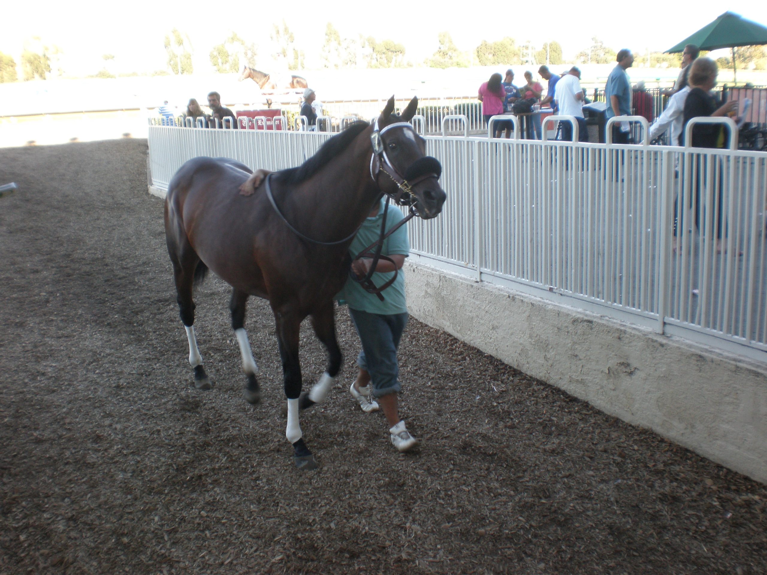
The work that brings a horse to the scan is usually a lameness. The lameness exam is where AAEP has set its clearest standard. The association lays out the workup as a set order of steps. The order is the same on every lame horse. It runs from the history to the standing exam. It goes on to the horse watched in movement. It goes on to the flexion tests. It comes to the nerve and joint blocks that find where the pain sits. It ends with the imaging that reads what is there. The scan is the last of those steps. The probe is laid on the spot the earlier steps have named. AAEP’s resources on understanding lameness hold that ultrasound is reached for in the face of a swelling, a wound, an effusion, or a wasting of the muscle. A lameness with none of those signs is pinned down with nerve and joint blocks first. The standard is the order. A vet finds where the horse hurts. Then a vet scans to find what is wrong there. A vet who works a lameness this way brings the scan to a named spot.
The order keeps the scan from being a guess. A probe laid on a lame leg with no localizing reads a dozen structures. It cannot say which one lames the horse. A scan run before the blocking can turn up an old half-healed injury. That finding has nothing to do with today’s lameness. The order holds the scan back until the blocking says where to look. The eye is easy to fool on a lame leg. A vet can see a finding and read it as the cause. The blocking is the check on that. It points the probe at the real trouble.
The standard lives in the sequence as much as in any one step. AAEP fixes the order a lameness is worked through. The history comes before the exam. The blocks come before the image. Each step narrows the trouble for the next. The flexion tests come before the blocks. A vet holds a joint bent for a moment, then trots the horse off. A worse lameness points the vet toward the part to block. The order matters as much as the steps. A vet runs each step in its place. Each one sets up the step after it. The workup tightens around the trouble.
The grade puts a number on how lame the horse is. The vet grades the lameness on a scale the association set, before the blocks and the scan. The number says how bad the lameness is. It gives every vet the same word for it. The grade is the first standardized thing the exam produces. A vet who knows the scale reads the grade the same way.
The whole exam reads the same from one vet to the next. A lameness is graded on the scale. It is localized by the blocks. It is imaged at the end. The record comes out a thing another vet reads without seeing the horse. A referral letter gives the grade, the blocks that worked, and the scan’s finding. That letter tells the next vet the whole of the lameness. A grade, a block result, and a scan finding fill a record. Another vet reads that record cold. They picture the lameness without the horse. The standard is what makes the record clear.
Grading the lameness
The grade is read on a scale of zero to five. AAEP set a scale that runs from a horse with no lameness to one that will barely bear weight. Each step is a level of how lame the horse is. A vet grades the lameness against the scale. The number is a word every equine vet knows. The grade is called early, before the leg is ever blocked. A vet watches the horse trot and puts a number on the lameness. The grade comes from watching the horse move. A vet trots it up and reads how it goes. The number marks the severity in one figure. Every equine vet reads that figure the same.
The scale gives the lameness a number any vet can read. The table below sets out the grades from zero to five. It gives the look of the lameness at each grade. A vet who grades a horse to the scale records a lameness another vet reads the same. A vet finds the grade in the table. They match the horse to a level. The grade is set before the leg is touched. It opens the record the workup fills.
| Grade | What it means |
|---|---|
| 0 | No lameness perceptible under any circumstance |
| 1 | Hard to see; not consistently apparent |
| 2 | Hard to see at a straight-line trot; consistent under certain conditions (a circle, hard ground, weight-carrying) |
| 3 | Consistently seen at a trot in all circumstances |
| 4 | Obvious at a walk |
| 5 | Minimal weight-bearing in motion or at rest, or unable to move |
Block first, then scan
AAEP’s standard puts the block before the scan. A vet localizes the lameness with nerve and joint blocks. The leg is numbed in stages until the horse goes sound. The block that takes the lameness away names the spot the pain sits in. Only then is the probe laid on. The blocks run from the foot up. The lowest part is numbed first. The vet works higher until the lameness lifts. A vet numbs the foot and trots the horse. The lameness may stay. The vet numbs higher and trots again. The leg gives up its sore spot block by block.
A block finds where a horse hurts. The scan, laid on that spot, finds what is wrong there. A nerve block that takes the lameness away names the part the pain sits in. The scan reads that part for what is wrong with it. It reads a torn tendon, a worn joint, or a chip of bone. The block and the scan together run a lameness to its cause.
The standard saves the scan from reading the wrong thing. A lame leg holds many structures the probe could read. A scan run before the blocking might read a finding that has nothing to do with the lameness. The block before the image sends the probe to the structure the lameness sits in. A horse can carry an old bow in one tendon and a fresh strain in another. The blocking says which one lames the horse today. The order points the scan at the live trouble. An old injury can sit quiet in a leg for years. A vet does not want the probe drawn to it. The blocking names the spot that lames the horse now. The scan reads that one structure.
The blocks are the equine way into the scan. A horse cannot say where it hurts. The blocking is the only way to ask. The leg is numbed part by part until the lameness lifts. A horse drops its head on one diagonal. It shortens a stride. It swings a leg wide. These signs tell the vet it hurts. The where of it is past the horse’s telling. The blocks are how a vet asks a question the horse cannot answer in words.
The block finds where, the scan finds what
AAEP fixes the order of the lameness exam. The blocks find the spot, then the scan reads it.
Where ultrasound fits the workup
Ultrasound is the soft-tissue eye of the lameness exam. The scan reads the tendons, the ligaments, and the joint surfaces. It reads the soft parts a lame leg can go wrong in. These are the things an x-ray cannot show. A torn tendon, a strained ligament, an inflamed joint lining, a pocket of fluid: the scan reads each one well. Where a lameness sits in soft tissue, the scan is the test that names it.
AAEP names when the scan is reached for first. A swollen leg shows soft tissue plainly. A wound, an effusion, or a wasted muscle shows it too. The vet scans those early, the cause there on the surface to follow inward. A quiet lameness with a clean-looking leg gives the eye nothing. The blocks are then the only way to find where to scan.
The detail of each soft-tissue scan lives in its own place. How a tendon, a ligament, or a joint is read on a horse is set out where each is covered. AAEP sets the order they sit in. A vet learns the scan of the equine leg from the clinical pages. The reading of a torn fibre or a core lesion belongs to that clinical work. AAEP’s part is to send the probe there. A torn tendon shows as broken fibres on the scan. A vet reads that on the clinical pages. AAEP sets where the scan sits in the workup. The reading of the picture is the vet’s own skill.
The prepurchase exam
The prepurchase exam is the other place AAEP’s standard meets the scan. A horse for sale is worked up the way a lame one is. The buyer is warned of a trouble before the money moves. AAEP sets out how the exam is done. The scan is among the tools a vet reaches for. A horse can look sound and still carry a quiet trouble in a tendon or a joint. The scan turns it up before the sale. A sound-looking horse can hide a weak tendon. The scan finds it on the table. A buyer reads the legs before money changes hands. The exam is the buyer’s look inside.
The imaging falls on the places that decide a horse’s soundness. A vet reads the front feet, the fetlocks, the hocks, and the stifles. These joints carry a horse’s long-term soundness. The scan reads the soft tissue of those spots. The x-ray reads the bone. The two together make the picture a buyer leans on.
The standard keeps a prepurchase honest. A buyer leans on the exam to know what they are getting. The standard is what makes one vet’s prepurchase another’s. A horse passed to the AAEP standard is read the same by any equine vet. The buyer leans on a method the whole field stands behind. They can trust it past the one vet who ran it.
The scan does its part in the soft tissue. A tendon can hold an old healed core. A suspensory can hide a quiet strain. A joint can carry a little fluid. Each shows on the scan in a prepurchase. A buyer would want to know any of them before the deal. The scan reads the soft warning a horse’s legs carry. A buyer wants the legs read before the deal. The scan shows a quiet strain a trot might hide. A finding read now can change the price. The exam puts the legs on the record.
The shared language of lameness
The grade is a language vets share. One vet calls a horse a grade-three lame. The next vet reads grade three and knows the same thing. The number means the same in any equine practice. The grade saves a page of describing. A single number stands in for a paragraph on how the horse moved. A vet reads grade three and knows the lameness without the words. A grade is a small word with a fixed meaning. A vet writes it once in a chart. The next reader knows the lameness at a glance. The number carries the picture across a field.
The shared scale lets a lameness be followed. A horse is graded over weeks. The number is marked down when the leg mends. It is marked up when the leg worsens. The scale itself does not move. A vet reads a horse’s grade against the last one. A horse that went from a grade three to a grade one shows its recovery in the two numbers. The grade tracks a horse’s soundness over a season the way a temperature tracks a fever. A vet grades a horse week by week. A mending leg earns a lower number. A reader sees the recovery in the chart. The scale makes a course of healing easy to follow.
What AAEP leaves to the clinical pages
AAEP sets the standard of the workup. The reading of each scan is left to the clinical work. How a tendon, a joint, or a foot is read on the screen is set out where each scan is covered. AAEP’s part is the order and the grade they sit in. A vet learns the scan itself elsewhere. A vet learns the way to work a lameness from AAEP.
The named protocols and the clinical detail belong to their own places. The point-of-care scans a horse vet runs are set out where they are covered. The reading of a tendon or a joint is set out there too. AAEP gives the frame around them. The body works at the level of the order and the grade. The doing of the scan is the vet’s own.
The standard on a handheld
The lameness scan runs on a handheld at the yard. A horse is lame where it lives. It stands in a stable or a field far from any imaging room. The scan is needed at the horse. The handheld goes to the lame leg. The standard workup is done where the horse stands.
AAEP’s standard runs the same on a handheld as on any machine. The standard lives in the method. It lives in the order, the grade, and the place of the scan. No part of it is tied to the size of the machine. A handheld and a set of blocks are all a vet needs. The whole exam is carried in a bag. The standard asks for a method. Any probe can run it. A vet sets up a handheld in a minute. The standard scan runs on it the same. The machine fits in a bag with the blocks. The whole workup travels to the horse.
The handheld put the lameness scan in the field vet’s bag. A vet carries a probe to the horse now. The soft-tissue read is taken at the yard the lameness was found in. The standard workup runs end to end at the horse. A field vet carries the blocks, the needles, and the handheld in one bag. The horse is worked from grade to block to scan without leaving its stable.
The field is where the equine scan belongs. A horse is hard to move. It is best read where it is. A vet works the lameness in the stable or the school the trouble showed in. The handheld brought the last step of the workup out to the yard. The first steps already happened there. The whole exam is kept in one place. A horse stays in its stable for the whole workup. The vet brings the grade, the blocks, and the scan to it. Nothing is shipped to a clinic. The standard exam happens at the yard gate.
AAEP’s order says how a lameness is worked. The handheld puts the scan to do its last step in the vet’s hand. The two together make the standard workup a thing done at the yard. A horse anywhere is worked to the same standard. The field is no longer cut off from the scan.
What AAEP gives the equine scan
AAEP gives the equine scan its place in the lameness exam. The body’s standard sets the order a lameness is worked. It sets the grade that measures it. It sets the spot the scan is sent to read. The probe is sent to a named spot. The whole leg is no longer a thing to fish in. A scan with a question to answer reads cleaner than one cast over a whole leg. AAEP gave the equine scan a target.
The grade and the order are the body’s gift to the work. AAEP’s scale puts a number on a lameness any vet can read. Its order runs the workup the same from one vet to the next. The scan is given its place at the end of the localizing. The equine field works a lameness as one. A lameness graded, blocked, and scanned to the AAEP order comes out the same in any vet’s hands.
The horse vet anywhere works a lameness to the same standard. The grade, the blocks, and the scan are each done the way AAEP set out. The workup reads the same in any equine practice. The scan takes its place in a lameness exam the whole field shares. The standard is what gives the probe its order. A vet anywhere runs the same steps. They grade, they block, they scan. The workup reads the same in every practice. The standard ties the equine field into one method.
Common questions about AAEP and equine ultrasound
What does AAEP say about ultrasound?
AAEP sets ultrasound’s place in the lameness exam. Its guidance reaches for the scan early when a leg is swollen, wounded, effused, or wasted. A lameness with none of those signs is localized with nerve and joint blocks first. The standard is the order. A vet finds where the horse hurts with the blocks. Then a vet scans to find what is wrong there. The scan is the soft-tissue eye of the workup.
What is the AAEP lameness scale?
A grading of lameness from zero to five. It runs from a horse sound under any test up to one barely able to bear weight or move. Each step is a level of how plainly the lameness shows. A low grade is hard to see. A high grade is obvious at a walk. The scale gives every equine vet the same word for how lame a horse is, set out in the table on this page.
Why block a horse before scanning it?
Because a horse cannot say where it hurts. AAEP’s standard localizes the lameness with nerve and joint blocks first. The leg is numbed in stages until the horse goes sound. The block that takes the lameness away names the spot. The scan is then laid on that one spot. It reads the structure for what is wrong. That is why the block comes first.
Is AAEP for horses only?
Yes, the horse is its field. The American Association of Equine Practitioners speaks for the vets who treat horses. The standards it sets are shaped for equine work. The cat and the dog have their own body. The farm animal has another. Each species has its standard-setter. AAEP’s standard is the equine one.
What is the prepurchase exam?
The workup of a horse before it is bought. A vet examines a horse for sale the way a lame one is worked up. The vet images the front feet, the fetlocks, the hocks, and the stifles. These spots carry a horse’s soundness. The scan reads their soft tissue. The buyer learns what the legs hold before the money moves. AAEP sets out how the exam is done.

About the Author
Julien Mercier
Senior R&D Engineer · Medical Ultrasound Transducer Development
Senior R&D Engineer with an M.S. in Applied Physics and over 15 years of experience in medical ultrasound transducer development, specializing in the design verification and performance testing of high-frequency imaging transducers. Currently leading the development and verification of the company’s next-generation high-frequency linear-array transducer, responsible for imaging performance evaluation and reliability analysis in preclinical testing. Brings extensive hands-on experience in piezoelectric element tuning, beamforming parameter optimization, and system-level performance testing.