Placental Position and Grading Handheld Ultrasound Assessment
A placental scan reads where the placenta sits in the womb and how it is built, the two things that shape how a pregnancy is carried and how the baby is born. A handheld probe finds the placenta against the wall of the womb, judges whether it lies clear of the way out, and reads its grain for the signs of age. The position settles the largest question: a placenta over the neck of the womb blocks the baby’s path and bleeds. The grade and the look add the rest, the placenta read as the organ the baby’s life hangs on.
Finding the placenta
The placenta plants itself somewhere on the wall of the womb early in the pregnancy, and the scan names the spot. It may sit on the front wall, the back wall, the top, or the side, each a normal home with no bearing on the pregnancy beyond where a needle would go. A reader names the wall and moves to the part that matters, how low the placenta reaches. The reader sweeps the whole womb for the placenta, no corner of the wall left unread before its edge is placed. The front-wall placenta sits between the probe and the baby, a soft cushion the scan reads straight through, its cushion muffling the kicks a mother feels and giving a reader cause to read less into a quiet day. The back-wall placenta sits behind the baby, harder to see when the baby lies against it, a reader shifting the probe to read around the spine for its lower edge. A reader notes it, since a needle for a sample must pass around it. A reader names the placenta’s wall plainly in the report, front or back, high or low, the word a guide to whoever scans or operates next. The wall and the edge are the two things a report always gives, the words that tell the next reader where the placenta lies.
The cord enters the placenta near its middle in nearly all pregnancies, the vessels fanning out from there across the disc. A placenta can grow a second small lobe set apart from the main one, joined by a thread of vessels across the membranes, a reader who misses the extra lobe missing a piece that can be left behind at the birth. A cord that joins at the edge, or runs free through the membranes before it reaches the placenta, is read with care, the free vessels at risk of tearing when the waters break. Vessels running free across the membranes near the neck are the rare danger called vasa praevia, a thread of cord the baby’s own blood runs through that can tear as the waters break, a reader who traces the cord to its landing the one who catches it.
The low edge of the placenta is the part a reader watches. A placenta planted high on the womb sits well clear of the neck, the way out open beneath it. The top of the womb, the fundal seat, is the textbook home, well clear of the neck, a reader noting a fundal placenta and reading on. The body of the placenta can hold dark pools of slow blood, the placental lakes, a normal pooling a reader names and passes by. A reader measures from the placenta’s lower edge to the neck of the womb, the gap between them the number that matters. A clear gap of a few centimetres puts the question of praevia to rest for good. A narrow gap brings the placenta under a closer eye. A reader follows the lower placenta down to the neck and reads the two together, an inside scan giving the surest view of the gap. The placenta holds the wall it implanted on, its lower edge the only part that moves. A placenta found and placed early sets the ground for every scan that follows, the wall and the edge a fixed point a reader returns to. The neck is read with the bladder a little full, the fluid lifting the lower womb into view, a bladder emptied or overfull able to fake a praevia and so kept half-full. The placenta low and to one side, its edge reaching the neck from the corner, is read as carefully as one dead-centre, the cover from any angle the thing that matters.
The placenta in the way
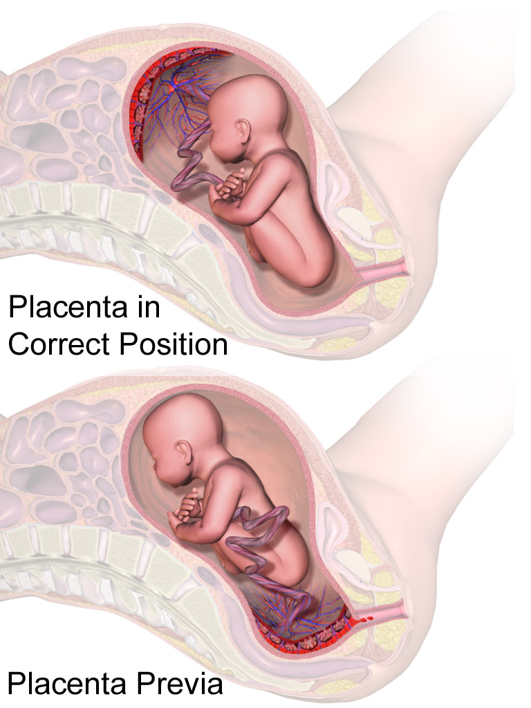
A diagram of the two positions, not an ultrasound. The upper drawing shows the placenta high on the wall, the way out clear beneath the baby. The lower shows a placenta praevia, the placenta covering the neck of the womb and blocking the baby’s path.
A placenta over the way out changes everything.
A placenta that reaches down over the neck of the womb is a placenta praevia, sitting square in the path the baby must take to be born. The neck is the door a placenta across it bars, a baby behind it unable to come out the usual road. This is the finding the scan looks hardest for, since a praevia missed is a praevia that bleeds. The bleeding is the danger. Toward term the womb softens and stretches, a placenta over the neck tearing at its edge, the blood the mother’s own and the loss fast. A praevia found early is watched, the birth planned by a cut long before the labour that would tear it. A low-lying placenta is one whose edge nears the neck of the womb, watched the same way for the bleeding it can bring. Many a placenta low at twenty weeks has risen clear by term, the lower womb having grown and drawn the placenta up, the early low edge no cause for alarm on its own. A reader who finds a low placenta early says so plainly and sets a scan for later, the question of the birth held open until the womb has grown. A placenta still low in the last weeks is the one that shapes the birth, the baby brought into the world by a planned cut to keep the mother from the bleed a labour would start. A praevia comes in degrees, a placenta whose edge just reaches the neck a lesser worry than one planted square across it, the scan measuring how far the cover runs, the degree it finds read into the plan for the birth. A praevia is no fault and no failure, only an accident of where the placenta chose to land, the scan there to find it before the birth. A placenta tearing off the wall before its time, an abruption, brings a bleed that comes with pain and a womb gone hard, a different trouble from the silent praevia, told apart by the pain it brings.
A praevia is the clearest reason a scan changes a birth. A baby cannot be born past a placenta in the way. A planned cut, set before the labour, lifts the baby out over the placenta with no bleed. The scan that names a praevia weeks ahead gives a team the time to plan the safest birth.
Bleeding from a praevia is a sudden gush of bright blood that comes without a pain to warn of it. A mother who has been told her placenta is low knows to come in at the first sign, the scan having armed her ahead of the bleed. A reader names the praevia so the warning can be given, the word the thing that keeps a mother safe. The bleed can come in the night with no cause at all, a reason a mother is warned to keep a way to help close by. The first bleed of a praevia is often a warning, a small loss that stops on its own, the herald of the heavier bleed to come, a mother who bleeds brought in and watched and the birth weighed against the loss. The bleed comes without pain, the silence itself the clue that sends a reader to look at the placenta over the neck. A mother bleeding from a praevia is laid up and watched, no examining hand put inside her where it might start the bleed afresh, the scan reading the placenta from outside the one safe look. A mother carrying a praevia is told to rest and to keep near help for the weeks before her planned birth.
The early low placenta is the commonest reason a scan is set for later. A placenta low at twenty weeks is read again at thirty-two or beyond, the later scan finding it risen clear in the great majority. The follow-up settles the question the early scan only raised, a low placenta read again later, the second look the one that calls the birth. The praevia caught on a routine scan, before any bleed, is the one a team handles best, the warning early and the plan made calm. A praevia that has not moved by term ends almost always in a planned cut, the day set ahead of the labour and the mother brought in before the womb can start to open.
Reading the grade
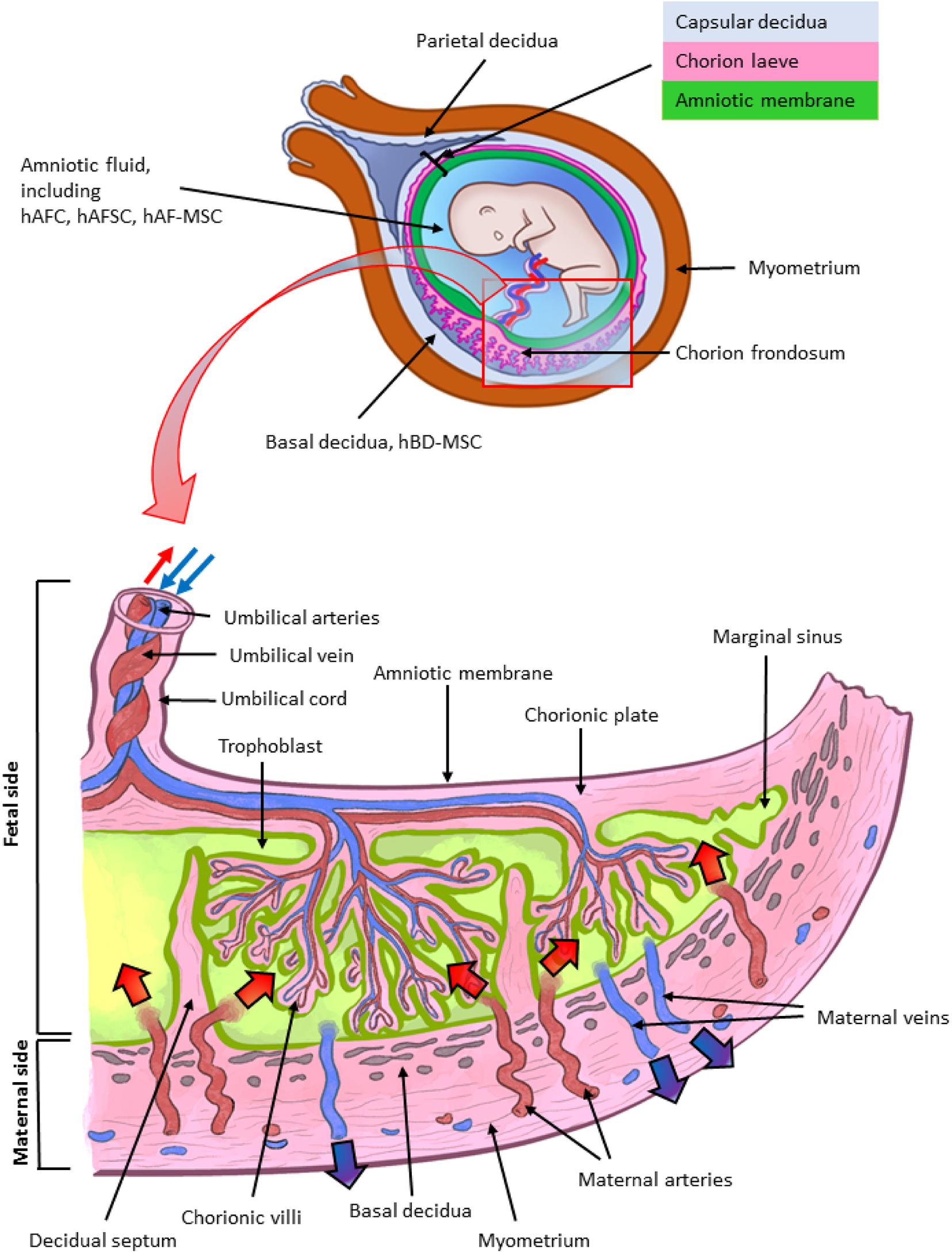
A diagram of the placenta’s structure, not an ultrasound. The cross-section shows the smooth plate facing the baby, the branching villi of its body, and the rough plate rooted in the wall of the womb, the layers the grade reads for the marks of age.
The placenta ages over the pregnancy, the scan reading that ageing in a grade. The grade runs from zero to three, the higher numbers marked by calcium streaking the placenta and breaking it into lobes. A higher grade marks a placenta further along, maturing toward the end of its work. The placenta keeps no clock a reader can read straight, the grade only a rough hand on the dial of its age. A reader judges a placenta by how it works far more than by its grade. A placenta of any grade can carry a healthy baby, the number no measure of the child inside. The grade reads three things, the smooth plate facing the baby, the body of the placenta, and the rough plate against the wall, calcium creeping in along these in order, the plate against the wall roughening first, the body breaking into lobes, the plate facing the baby last to indent. A grade zero placenta is a plain even sheet, its face smooth and its body without a mark, the look of a placenta early in its life.
A grade read against the age tells whether the placenta is keeping pace. A grade three at term is the expected mark of a placenta near the end of its work, its calcium the wear of a job nearly done. The grade is read for the age it shows up at, a grade three weeks early the one that worries. The calcium that grades a placenta is the same mineral that hardens a bone. The grade is logged at each scan, the record showing how far along a placenta has come. The placenta is born after the baby, the third stage of labour, peeling off the wall and passing out behind, a scan rarely watching that last act. A baby’s lung maturity is judged today by better tests than the grade, which serves as a marker of the placenta alone.
A placenta old before its time is the worry the grade can flag. A grade three found weeks before term is a placenta ageing early, its calcium a sign it may be failing the baby sooner than it should. A reader who finds an old placenta on a young pregnancy reads it beside the growth and the flow, the grade a flag to look harder, the answer found in those.
The grade is a soft sign, read with a light hand. Two readers can grade the same placenta a step apart, the calcium a matter of the eye. A grade three placenta reads coarse on the screen, its lobes ringed with bright calcium that throws small shadows, a look a reader knows at a glance. The grade has fallen out of the front rank of placental tests, the growth and the cord flow telling more about how a placenta works, a reader noting it and moving on with no baby delivered on a number from zero to three.
Growing too deep
A placenta that has grown too deep into the wall of the womb holds on after the birth, the accreta that cannot let go without tearing the womb and bleeding hard. The scan looks for the signs of it ahead of the birth, on the mothers at greatest risk. The accreta missed is the one that kills, the reason a scan hunts it on every mother with a scar. The accreta runs in a spectrum graded by how deep the placenta has burrowed, the deeper the invasion the worse the bleed when it is parted. The risk climbs steeply for a mother with past cuts, the scar tissue the ground the placenta roots into. The mother at risk is known long before the scan, her history of cuts and her low placenta marking her out, a reader bringing extra care to her scan where the signs of an accreta are easy to pass over.
The danger sits where a placenta praevia meets an old scar. A placenta lying low over the scar of a past cut can root itself into the scar, growing deep where the wall is weak. The scan reads the line between the placenta and the wall, the loss of that clear dark line the sign of a placenta that has grown in, pools of flowing blood within it adding to the worry. The join between them is the line that tells whether the placenta will let go after the birth. The scan reads three signs in all, the loss of the dark line, the dark pools of flowing blood within the placenta, and a bulge of placenta into the bladder above, any of the three on a low anterior placenta over an old scar sending a reader to look harder. Colour laid over the join lights up the tangle of vessels an accreta drives into the wall, a riot of flow filling it, the busy vessels one more sign of a placenta digging in. The worst of the spectrum grows clean through the wall and out, a percreta reaching the bladder beyond that turns a birth into major surgery. The accreta is read for whether it reaches the bladder, a colour scan of the line between the womb and the bladder showing vessels that cross where none should, the worst of these a bladder wall studded with flow on a low anterior placenta.
An accreta found ahead of the birth saves lives. An accreta caught early turns an emergency into a planned operation, the scan the thing that buys the warning. An accreta is among the gravest things a placental scan can find, the one that turns a routine birth into a fight for a mother’s blood. A team warned of it brings the mother to a centre ready for the heavy bleeding it can bring, the birth planned with blood and surgeons on hand. The scan that catches it early turns what would be a disaster into a managed risk. The accreta asks for a team, a place, and a plan, none a scan can summon on the day, so the warning has to come weeks ahead. A mother with an accreta is delivered at a centre with a team ready, the baby brought out high through the womb and the placenta often left in place or the womb removed to stop the bleed. The accreta is a rising danger of modern obstetrics, its numbers up where caesarean births are common, a reader trained to spot its signs heading off the shock it would once have sprung in the operating room. An accreta on an anterior placenta sits right under the cut a surgeon would make, a team warned of it planning a different way in to reach the baby around the placenta and spare the bleed.
What the look cannot settle
The scan has its limits with the placenta, a reader naming them plainly. A grade read by eye swings between readers, a low placenta early, an accreta deep in the wall, each a thing one look cannot fully settle. The placenta has a normal thickness too, a sheet a few centimetres deep against the wall, a placenta swollen thick or thinned to a strip read for what it means, a thick one sometimes the mark of a mother’s diabetes. The position read at one scan can read different at another, a baby shifting or the bladder full changing how the lower edge looks, a reader reading it with the bladder part-empty and the baby still for the truest view. The deepest questions a placenta raises, an accreta or a hidden lobe, are read best by a specialist with the time and the tools, a handheld reading the position and the gross signs and passing the rest up the line. The honest read names what it sees and flags what it cannot, a doubtful low edge written down as doubtful, the placenta one chapter of a longer story. The placenta read once is a snapshot, its low edge and its grade both things that move, the next scan the one that settles them. The screen tells the placenta’s position and its gross health, the readings a clinic leans on day to day.
On a handheld
The placenta is an easy target for a handheld, a broad bright band against the dark womb, plain to the eye from the first sweep. A clinician finds it, names its wall, and runs the probe down to the neck, the whole read, the wall, the edge, the grade, the signs of trouble, the work of a minute folded into the sweep that sizes the baby. The placenta filmed and saved travels to a specialist as well as any cart’s image, a reader who doubts an edge sending the film on for a second eye. A clinician trained on the placenta reads its seat and its edge with a confidence the skill of the eye grants, the eye that knows the normal reading the odd at a glance. The scan reads the placenta again after the birth, hunting a piece left behind in the womb that could go on to bleed.
The reach carries the placental check to where it counts. A bleeding mother far from a hospital is read where she is, a placenta over the neck found at once on the first sweep. The image saves to the phone, the placenta filmed, ready for a specialist to read from afar. The scan that finds a praevia early changes the whole of a pregnancy’s plan, the mother sent to keep near help and to book the birth at a centre, a flag a handheld can raise in a rural clinic months ahead. A placenta read on a handheld where no scanner stands can change where a mother gives birth, a praevia caught turning a home delivery into a hospital one, the probe reaching the mothers furthest from care. The placental scan rides in a coat pocket, carried to the bedside wherever a mother is met.
Common questions about placental position and grading
What is placenta praevia?
A placenta praevia is a placenta that reaches down over the neck of the womb, blocking the path the baby must take to be born. It is the placental finding a scan looks hardest for, since it bleeds toward term and means the baby must be born by a planned cut. A scan names it weeks ahead so the birth can be planned safely.
Will a low placenta found early stay low?
Usually not. Many a placenta low at twenty weeks has risen clear of the neck by term, the lower womb having grown and drawn the placenta up. A reader who finds a low placenta early sets a scan for around thirty-two weeks, the later scan settling whether the placenta still sits in the way.
What does placental grading mean?
The grade, from zero to three, reads how far the placenta has aged by the calcium laid down in it, the deposit streaking the placenta and breaking it into lobes. A high grade is normal near term. A high grade weeks before term can be a sign of a placenta ageing early.
What is placenta accreta?
An accreta is a placenta that has grown too deep into the wall of the womb and cannot peel away cleanly after the birth, bleeding heavily when it is forced. It is likeliest where a low placenta lies over the scar of a past cut. A scan reads the line between the placenta and the wall for the signs of it.
Can a handheld ultrasound check the placenta?
Yes, easily. The placenta is a broad organ against the wall of the womb, found in seconds on the first sweep. A clinician measures its lower edge against the neck of the womb and reads its grade at the bedside, the check reaching mothers far from a scanning centre.
About the Author
Julien Mercier
Senior R&D Engineer · Medical Ultrasound Transducer Development
Senior R&D Engineer with an M.S. in Applied Physics and over 15 years of experience in medical ultrasound transducer development, specializing in the design verification and performance testing of high-frequency imaging transducers. Currently leading the development and verification of the company’s next-generation high-frequency linear-array transducer, responsible for imaging performance evaluation and reliability analysis in preclinical testing. Brings extensive hands-on experience in piezoelectric element tuning, beamforming parameter optimization, and system-level performance testing.