Follicle monitoring is the series of transvaginal scans that tracks the growing follicles in the ovaries through a fertility cycle. Each scan counts the follicles and measures them one by one, building a record of how they grow from one visit to the next. The numbers time the two decisions a stimulated cycle turns on: when to trigger the eggs to mature, and when to collect them.
The cycle the scans follow
A stimulated fertility cycle sets out to grow a crop of follicles together. Daily hormone injections drive the ovaries to bring on a whole group of follicles at once, each a small fluid sac holding an egg. The aim across the cycle is a cohort of follicles maturing roughly in step, ready to give several eggs at a single collection. The scans are how that cohort is watched from the day stimulation begins to the day the eggs are taken. The crop is what makes the cycle work. A cohort of ten or fifteen eggs raises the odds that some will fertilize, develop and reach transfer. The injections push the ovaries to bring on that crop together. The scans confirm the ovaries are answering. The whole design of a stimulated cycle rests on growing a group of follicles at once.
The follicle is the structure the whole process turns on. It reads on the scan as a round dark pocket of fluid in the ovary, its egg too small to see within it, its size standing in for the maturity of the egg inside, a few millimeters early in the cycle and near twenty when the egg is ready for collection. The scan reads the egg’s readiness through the size of the sac around it.
Timing is the heart of the cycle. The scans supply it. The eggs mature on a schedule the injections set in motion, a schedule that runs over roughly eight to twelve days of stimulation for many cycles. The eggs ripen within a window the scans track. The trigger has to land inside it, when the cohort holds the largest number of mature eggs. The scans place the cycle on that schedule, visit by visit, so the trigger lands at the right point. The schedule cannot be rushed or held. Each follicle takes its own days to mature. The cohort moves together at a pace the injections set. The scans read where the cohort has reached on its own clock, never forcing it, only timing the trigger to the day it arrives. Reading that clock correctly is the central task of the monitoring.
The monitoring runs as a series of scans across the cycle. A scan every two or three days through the stimulation builds a picture of the follicles growing, each visit added to the one before. A nurse or a clinician reads the count and the sizes at each scan, adjusts the plan, and books the next. The cycle is steered by this run of scans across its length, each one a reading of where the follicles have reached.
The baseline scan
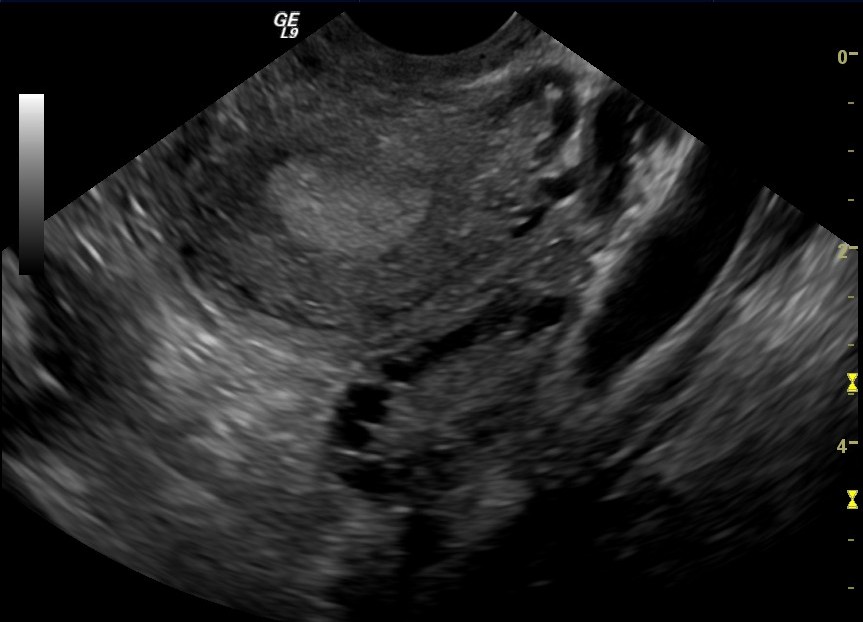
An ovary carrying multiple follicles on a transvaginal scan, each a small dark pocket of fluid. This is a polycystic ovary; a stimulated ovary in a fertility cycle shows the same crowd of follicles, which the scan counts and measures. The probe label and depth scale are the scanner’s own.
The monitoring opens with a baseline scan before stimulation begins. With the ovaries still at rest, the scan counts the antral follicles, the small resting sacs of two to nine millimeters that sit in each ovary at the start of a cycle. This antral follicle count gives a measure of how the ovaries are likely to respond to stimulation, read from the close view that resolves these small early follicles clearly. The antral count is a number with meaning. A count read across both ovaries gives a forecast of the crop the cycle is likely to yield, a guide the clinician sets the plan by. The small resting follicles are only a few millimeters across, sitting at the edge of what a scan can resolve, which is why the close transvaginal view is used to count them. The baseline number opens the record the whole cycle is tracked against.
The baseline reading shapes the plan for the cycle. A high antral count points toward a strong response and a lower drug dose, with an eye kept on the risk of over-response. A low count points toward a gentler outlook and a dose set to coax what follicles there are. The clinician reads the baseline count, along with hormone levels, to set the starting dose for the patient in front of them. The dose is set to fit the ovaries the scan reads. A clinician matches it to the antral count and the hormone levels, choosing a starting amount judged to bring on a good crop safely. The baseline scan turns a general protocol into a dose chosen for the patient.
The baseline scan also checks the ovaries and the pelvis are ready. It looks for a cyst left from a previous cycle, reads the lining of the uterus at its thin resting state, and confirms there is nothing to delay the start. A clean baseline clears the cycle to begin. The first dose of stimulation follows from there. The baseline rules out a false start. A cyst making its own hormones, a lining not yet shed, a follicle left active from before: any of these can throw a cycle off. The baseline scan finds them before a single injection is given. A clean baseline is the green light the cycle waits for.
Measuring the follicles
A follicle is measured by its diameter, taken as the average of its width across two or three directions. The sonographer freezes a clear view of the follicle, lays the calipers across it, and records the size in millimeters. Because a follicle is a rounded sac, averaging its diameters gives a truer figure than a single line across it. Each follicle large enough to count is measured this way and its size written down. The measurement follows a set method at every clinic. A follicle is read at its widest clear view, the calipers laid inside its bright rim, and two or three diameters averaged into one figure. Doing it the same way each time lets the sizes from one scan be compared with the last, the growth read as a true change in the follicle. The method is what makes a run of scans add up to a record.
The sizes climb across the cycle as the follicles grow. A follicle gains roughly one to two millimeters a day under stimulation, so a cohort that reads at ten millimeters on one scan reads nearer fourteen two days later. The run of scans tracks that climb, each visit recording the follicles a few millimeters larger than the last. The rate of growth itself tells the clinician whether the dose is driving the cohort as it should. A steady climb across the scans is the sign of a cohort responding well. Follicles that gain their millimeter or two a day, scan after scan, trace the smooth rise the cycle is looking for. A clinician reading that climb sees the stimulation working and holds the course. The shape of the growth, read across the run of scans, is as telling as any single size.
A cohort grows with a spread of sizes. The scan reads the whole range. A few leading follicles run ahead of the group, a band of middle follicles follows, and a tail of smaller ones sits behind. The scan measures the leaders that will set the timing and counts the cohort that will give the eggs. Reading the spread across the cohort is what lets the clinician judge when the largest number of follicles will be ready together.
The record built across the visits is the real product of the monitoring. Each scan adds a line to a chart of follicle sizes against the days of the cycle, a chart that shows the cohort rising toward maturity. A clinician reads that chart to see the cycle whole, the growth so far and the day the follicles will reach the size for a trigger. The monitoring is as much this running record as any single scan.
The measurements have to be exact. The close view is what makes them so. A follicle of fifteen or eighteen millimeters has to be told from one of twenty, because those few millimeters decide the timing of the whole cycle. The transvaginal probe, an inch or two from the ovary and running at a high frequency, draws a follicle edge a caliper can sit on cleanly. The precision the cycle runs on comes from reading the ovaries this close.
Counting across both ovaries
Each scan reads both ovaries in turn, counting and measuring the follicles in each and adding them to a total for the cycle. The ovaries often respond unevenly, one carrying more follicles than the other, so the scan reads each on its own and records the two counts together. The number that guides the cycle is the whole cohort across both ovaries, the leaders that set the timing and the count that gives the eggs. The two ovaries are read as one cohort for the timing. A follicle leading in the right ovary and one leading in the left both count toward the day the cohort is ready, their sizes read together. The scan keeps the two counts separate on the record, for the picture of how each ovary has answered, and adds them for the number that times the trigger. The whole crop, across both sides, is what the cycle is timed by.
The numbers that time the trigger
The trigger is the injection that ripens the eggs for collection. Its timing is read off the follicle sizes. The cohort is judged ready when its leading follicles reach a size in the high teens to around twenty millimeters, with a good number of the others following close behind. At that point the eggs in the larger follicles are mature enough to collect. The clinician calls the trigger. The size band is a guide read with judgment. A clinic looks for the leading follicles to sit in the high teens to around twenty millimeters, the range in which the eggs inside are mature enough to fertilize. The exact day is chosen by reading where the whole cohort sits against that band. A scan a day apart can move the call, which is why the scans come close together as the trigger nears.
The reading balances the whole cohort. Triggering on the day the greatest number of follicles sit in the mature range gives the largest crop of usable eggs. A scan that finds the leaders at twenty millimeters with a strong group close behind reads as a cohort ready to trigger. The judgment weighs the whole group, looking for the day that yields the largest crop of mature eggs together.
The collection follows the trigger on a fixed clock. The eggs are taken about thirty-six hours after the trigger injection, the window in which they have ripened fully and before the follicles release them on their own. The final monitoring scan, read against that clock, is what sets the hour of the trigger so the collection lands where it should. The whole run of scans points to this single timed decision. The thirty-six hour window is fixed by the biology. The trigger injection sets the final ripening in motion, a process that takes about a day and a half to finish. The collection is booked to land at its end. A trigger called too early or too late by hours shifts the eggs out of their best window, which is why the final scan times it to the hour.
The lining read alongside
The scan reads the lining of the uterus at the same time as the follicles. The endometrium thickens across the cycle under the rising hormones. The scan measures it and reads its pattern. A lining that has grown to a healthy thickness, with the layered appearance that marks it receptive, is a sign the uterus is preparing in step with the follicles. The lining reads as a number and a pattern. Its thickness, measured across the cavity, rises across the cycle toward a healthy depth. Its appearance takes on a layered look, three lines across it, that marks a lining readying to receive an embryo. The scan records both at each visit, watching the uterus come ready alongside the ovaries.
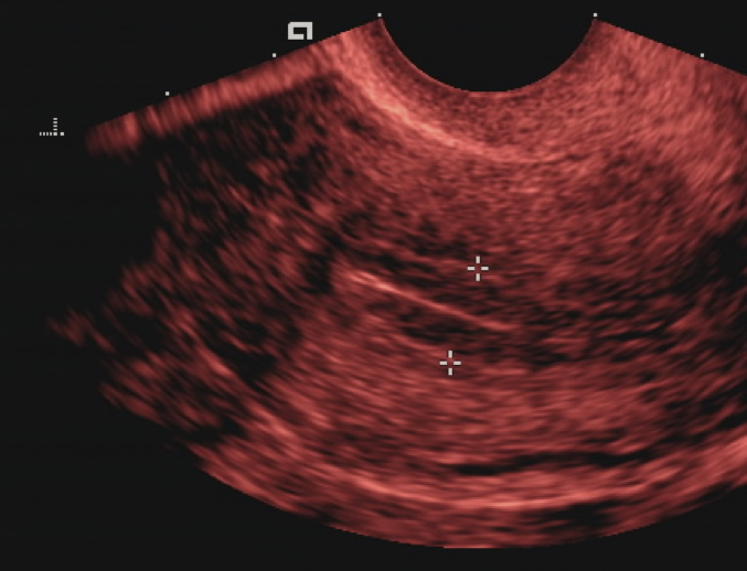
The endometrium, the lining of the uterus, measured on a transvaginal scan with calipers across it. The monitoring reads the lining alongside the follicles, watching it thicken toward a receptive depth. The markers are the scanner’s own.
The lining reading feeds into how the cycle is managed. A lining growing well alongside the follicles supports going ahead with a fresh transfer after collection. The embryos can also be frozen for a later cycle, giving the uterus time to prepare. The scan reads the follicles and the lining together, the two halves of a cycle that has to come ready at once.
What the count guides
The running count guides the dose through the cycle. A cohort growing slowly, with fewer follicles than the baseline promised, may call for the dose to be raised or the stimulation extended a few days. A cohort racing ahead may call for the dose to be eased. The clinician reads each scan and adjusts the injections to keep the cohort on a course toward a well-timed collection. The adjustments are small and read off each scan. A cohort growing a little slowly earns an extra day or two of stimulation before the next look, or a small lift in the dose. The clinician makes these calls scan by scan, nudging the cycle toward the day the largest crop of usable eggs will be ready.
The count is also the main warning of over-response. A scan that finds a large number of follicles, many of them growing fast, flags a raised risk of ovarian hyperstimulation, where the ovaries swell and fluid shifts in the body after the trigger. Reading that risk early lets the clinic lower the dose, change the trigger, or freeze all the embryos to let the ovaries settle. The monitoring catches the over-response with time still to act on it.
The handheld probe suits the rhythm of a monitored cycle. A patient comes in every few days for a quick scan, and a portable probe gives that scan at the bedside, run from a phone or a tablet, without a booking into a scanning suite each time. A fertility clinic running many patients through monitoring at once reaches for probes it can keep in every room. The scan that has to happen often and quickly fits a tool that travels. The portability changes how a monitoring clinic runs. A probe kept in each room lets a patient be scanned the moment she arrives, the picture read on a tablet at the chairside. A clinic moving dozens of patients through monitoring on a busy morning depends on scans that take minutes and need no scanning suite. The handheld probe carries the monitoring at the pace a fertility unit works.
Taken together, follicle monitoring is the thread that runs the length of a stimulated cycle. From the baseline count, through the serial measurements of the growing cohort, to the final scan that times the trigger, the run of transvaginal scans turns a course of injections into a timed harvest of eggs. On a handheld probe at the bedside, repeated every few days, it gives the cycle the numbers it is steered by from the first day to the last.
Common questions about follicle monitoring
What is follicle monitoring?
A series of transvaginal ultrasound scans through a fertility cycle that counts and measures the growing follicles in the ovaries. Each scan records how many follicles there are and how large each one is, building a picture of the cohort growing over the days of stimulation. The numbers time the trigger injection and the egg collection. The scan reads the lining of the uterus at the same time.
How often are the scans done?
Every two or three days through the stimulation, which runs over roughly eight to twelve days for many cycles. The cycle opens with a baseline scan before stimulation begins, then scans repeat across the stimulation as the follicles grow, with the visits often closer together toward the end as the timing of the trigger nears. A handheld probe makes these frequent quick scans easy to fit in.
How big should a follicle be before the trigger?
The leading follicles are usually in the high teens to around twenty millimeters when the trigger is called, with a good number of the others following close behind. The judgment weighs the whole cohort, looking for the day the largest number of follicles sit in the mature range together. The egg collection then follows about thirty-six hours after the trigger injection.
How is a follicle measured on the scan?
By its diameter, taken as the average across two or three directions because the follicle is a rounded sac. The sonographer freezes a clear view, lays the calipers across the follicle, and records the size in millimeters. The close transvaginal view draws a clean edge a caliper can sit on, which is what lets a fifteen-millimeter follicle be told from a twenty-millimeter one.
Why does the scan check the lining of the uterus?
Because the uterus has to come ready in step with the follicles. The endometrium thickens across the cycle. The scan measures it and reads its pattern as a sign of how the uterus is preparing. A lining that has grown to a healthy thickness with a layered appearance supports a fresh embryo transfer. The embryos can also be frozen for a later cycle if the lining needs more time.
Can follicle monitoring warn of overstimulation?
Yes. A scan that finds a large number of follicles growing fast flags a raised risk of ovarian hyperstimulation. Reading that risk early lets the clinic lower the dose, change the trigger, or freeze all the embryos to let the ovaries settle. Catching the over-response on the scans, with time still to act, is one of the safeguards the monitoring provides.
About the Author
Julien Mercier
Senior R&D Engineer · Medical Ultrasound Transducer Development
Senior R&D Engineer with an M.S. in Applied Physics and over 15 years of experience in medical ultrasound transducer development, specializing in the design verification and performance testing of high-frequency imaging transducers. Currently leading the development and verification of the company’s next-generation high-frequency linear-array transducer, responsible for imaging performance evaluation and reliability analysis in preclinical testing. Brings extensive hands-on experience in piezoelectric element tuning, beamforming parameter optimization, and system-level performance testing.