Early Pregnancy 5 to 8 Weeks Transvaginal Ultrasound Handheld
An early pregnancy ultrasound between five and eight weeks is a transvaginal scan that reads the first structures of a pregnancy from close range. In these weeks the pregnancy is only a few millimeters across, sitting deep in the pelvis where the close view reaches it clearly. The scan answers the questions that press earliest: whether the pregnancy is in the uterus, whether it is alive, and how far along it is.
What the early scan is for
The first question the scan answers is where the pregnancy sits. A pregnancy belongs inside the uterus, in the lining at the top of the cavity. The scan looks for the gestational sac in that place and confirms it is there. Settling the location early is the heart of the early scan, because a pregnancy growing outside the uterus needs to be found early, before it grows. The close view reads the uterus and the spaces beside it in the detail that question takes. The location reading rests on a simple sign. A sac that sits high in the lining, surrounded on all sides by the bright tissue of the uterus, is where a pregnancy belongs. The scan confirms that the sac lies within the muscle of the uterus, ringed by it on every side. Establishing that early, before the pregnancy grows, is the deepest reassurance the first scan can give.
The second question is whether the pregnancy is growing as it should. The scan reads the sac, then the yolk sac inside it, then the embryo, then the flicker of a heartbeat, each appearing in its turn over these weeks. Finding the expected structure for the dates gives a clear answer. The third question follows from the embryo itself: measured end to end, it dates the pregnancy more closely than any other method, fixing how far along it is to within a few days. The order of appearance is itself the reading. Each structure has a window in which it is expected, the sac first, the yolk sac next, the embryo and its heartbeat after. Finding the structure that fits the dates tells the sonographer the pregnancy is keeping pace. A scan that reads the expected stage for the weeks has found a pregnancy developing as it should.
A fourth question the scan settles is the number. One sac holds one pregnancy, and a second sac means twins. The early scan counts the sacs and, a little later, the embryos and their heartbeats. Knowing the number from the start shapes the whole of the care that follows. These four readings, location, growth, dates and number, are the work of the early pregnancy scan. The count is clearest in these early weeks. Two separate sacs, each with its own developing pregnancy, mark a twin pregnancy plainly before the embryos crowd together later on. The scan also reads how the sacs sit, whether they share a lining or keep their own, a detail that shapes how a twin pregnancy is looked after. Counting early gives the care that follows its starting point.
Week by week, what appears
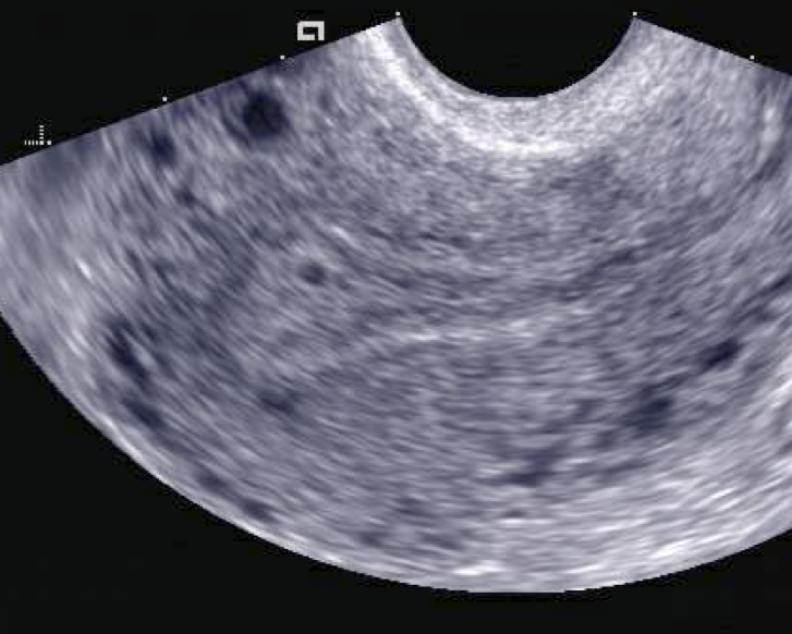
A gestational sac of about two millimeters on a transvaginal scan, the first sign of a pregnancy, set in the lining at the top of the uterus. A sac this small reads clearly only from the close transvaginal view, around five weeks. The markers are the scanner’s own.
Around five weeks from the last period, the gestational sac comes into view. It reads as a small dark pocket, two to three millimeters across, set into the bright lining at the top of the uterus. At this stage the sac is the whole of what the scan finds, a round fluid space with a bright rim. Its position high in the lining, off to one side of the cavity line, is the first sign that the pregnancy has settled where it should. At this earliest stage the reading asks for care. A small fluid collection in the lining can resemble a sac before a true pregnancy is confirmed within it. The sonographer looks for the bright, even rim and the position high in the lining that mark a real gestational sac. A sac with no yolk sac yet, on a single early scan, is read as a finding to follow, with a repeat scan to confirm.
By about five and a half weeks the yolk sac appears inside the gestational sac, when the sac has grown to roughly five or six millimeters across. The yolk sac reads as a fine bright ring within the dark pocket, the first structure that confirms a true pregnancy taking shape inside the sac. Its presence is a reassuring early marker, a step on the way to finding the embryo itself. The yolk sac has a size and a shape the scan notes. A normal yolk sac is round, thin-walled and a few millimeters across, growing slowly over the early weeks. The structure feeds the embryo before the placenta takes over. Its clean appearance is one of the markers of an early pregnancy on track. The scan records it as the confirmation that the sac holds a developing pregnancy.
Near six weeks the embryo shows as a small thickening at the edge of the yolk sac, the fetal pole, a few millimeters long. Within this pole, at about the same time, the scan catches the first flicker of a heartbeat, a tiny pulsing that color or motion settings can confirm. Seeing cardiac activity is the clearest sign of a living pregnancy the early scan can give. A pole of a certain length with a beating heart marks a pregnancy reaching its early milestones on time. The heartbeat is read with motion mode as well as the eye. A tracing taken across the flickering pole draws the beat as a wave, giving a rate the scan can record. Catching the heartbeat is often the moment an early scan turns on, the sign a woman has come hoping to see. The scan holds on it long enough to be sure of a steady, regular beat before moving on.
Through seven and eight weeks the embryo grows quickly, lengthening from a few millimeters toward a centimeter and a half. The heartbeat strengthens and quickens, climbing from around a hundred and ten beats a minute toward a hundred and seventy over these weeks. The shape of the embryo begins to form, a head end and a body taking rough shape on the screen. Each scan in this window reads an embryo visibly larger than the week before. Detail begins to emerge alongside the growth. By eight weeks the limb buds appear as small swellings, the head takes a clearer shape, and the early movements of the embryo can flicker across the screen. The pregnancy that began as a dark pocket a few millimeters wide now reads as a recognizable embryo with a strong heartbeat. These weeks carry the pregnancy from a first sign to a clearly formed early fetus.
Why the scan goes inside
The structures of these weeks are too small for any route except the close one. A sac of two or three millimeters, a yolk sac finer than a grain of rice, an embryo a few millimeters long: these live below what a scan from the lower belly can resolve across the depth of the pelvis. The transvaginal probe sits an inch or two away and runs at a high frequency, the combination that brings a structure this small into clear view. An early pregnancy scan is a transvaginal scan because the things it reads exist at the edge of what the close route alone can show. The frequency is the reason. A probe close to the uterus runs at six to twelve megahertz, the band that draws a structure of one or two millimeters as a clear shape. From this range the sac, the yolk sac and the early pole each read as distinct forms the sonographer can measure. The whole early pregnancy scan depends on the detail that closeness and a high frequency bring together.
Reading viability
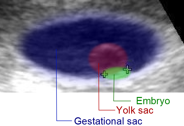
An early pregnancy with its three structures colored and labeled: the gestational sac, the yolk sac within it, and the embryo, where the heartbeat is read. The color and the labels are added to the scan to show the parts.
Viability turns on the heartbeat, and the heartbeat appears on a timetable the scan reads against the dates. A living embryo of a few millimeters shows a beating heart. The cleanest sign of a healthy early pregnancy is a fetal pole of adequate length with clear cardiac activity, found at the week the dates predict. When that sign is present, the scan gives the reassurance an early pregnancy often comes for. Cardiac activity carries a weight no other early sign matches. A pregnancy with a clear heartbeat at the expected stage has a strong chance of continuing. Every further week the heartbeat holds raises that chance. The scan records the presence of the beat, its rate, and the length of the embryo it belongs to, building the picture that a pregnancy is established and growing. This is the reading the early scan is built to deliver.
An early scan sometimes reads a stage earlier than expected for the dates, a sac with no yolk sac yet, or a pole with no heartbeat seen. A reading like this, on its own, settles little, because the dates carry their own uncertainty and a pregnancy a few days behind looks much like one that has stopped. The careful answer in these cases is to wait and scan again, giving the pregnancy the days it needs to declare itself. A repeat scan a week or more later reads growth where the first scan left a question.
The thresholds that call a pregnancy lost are set deliberately high, to keep a viable pregnancy from ever being called otherwise. An empty sac is called a failed pregnancy only once it has grown past about twenty-five millimeters with no embryo within it. A visible embryo is called a loss only once it reaches about seven millimeters in length with no heartbeat seen. These cut-offs leave a wide margin, and a reading short of them calls for a repeat scan and a wait. The reason for the wide margin is the cost of a wrong call. A pregnancy wrongly called lost, then acted on, ends a pregnancy that might have continued. The guidelines that set the cut-offs at twenty-five millimeters and seven millimeters were written to make that error all but impossible, accepting a repeat scan and a week’s wait as the price of certainty. A sonographer holds to those numbers exactly, never calling a loss on a borderline reading.
The heart rate itself carries information once a beat is found. A rate climbing week by week, from around a hundred and ten near six weeks toward a hundred and seventy by nine or ten weeks, tracks a pregnancy developing on course. A slow rate early on is read with caution and followed up on a later scan. The number is recorded at each visit as one more marker of how the pregnancy is progressing.
Reading viability in these weeks is as much about timing as about any single image. A structure missing today may be a structure only a few days early. The scan reads each finding against the dates, holds back from a hard conclusion where the margin is narrow, and uses the repeat scan to turn an uncertain first look into a clear answer. Patience built into the reading is what keeps the early scan safe. The repeat scan does the work a single look cannot. A pregnancy that read as uncertain on the first scan shows its growth, or its lack of it, over the days that pass. A sac that has enlarged and gained a yolk sac, an embryo that has lengthened and kept its beat, settles the question the first scan left open. The interval is part of the diagnosis, as much as any single image is.
Dating from the scan
The embryo dates the pregnancy more precisely than anything else available. Measured from the top of its head to the base of its rump, the crown-rump length maps onto the weeks of pregnancy along a curve that holds tightly in the first trimester. A measurement in these weeks fixes the dates to within about five days, the firmest estimate the whole pregnancy will receive. A dating done early carries through every scan and decision that follows.
Accurate dates matter for the rest of the pregnancy. They set the timing of later scans, the reading of screening tests, and the judgment of whether a baby later is growing on track or arriving early. An early pregnancy scan, by measuring the embryo when the dating curve is at its steepest and surest, gives the pregnancy a backbone of dates that everything afterward is read against. This is one of the quiet, lasting jobs the early scan does. The measurement is taken with care. The sonographer freezes a clear long view of the embryo, lays the calipers from the crown to the rump along its length, and reads the figure off the curve built into the machine. Taken this way in the early weeks, the crown-rump length gives a due date that holds within a few days. A dating set here is rarely revised by a later scan.
The scan in the early pregnancy unit
Early pregnancy scans happen wherever a woman first raises the question, often in an early pregnancy unit attached to a clinic or an emergency department. A handheld transvaginal probe suits this setting closely. It runs from a phone or a tablet, travels to the room the patient is in, and reads the pelvis at the bedside without a trip to a scanning suite. A clinic seeing women through the uncertain first weeks reaches for a probe it can keep close at hand. The handheld form fits the rhythm of early pregnancy care. A woman may need several scans across the early weeks, each a quick look that a portable probe gives without a booking and a wait. The same probe serves the clinic room, the bedside and the early pregnancy unit, the picture running to a screen the clinic already owns. The scan goes to the woman, in the place she is already being seen.
The early scan carries weight for the woman beyond its images. A first look that finds a heartbeat is a moment of reassurance after weeks of waiting and wondering. A scan that raises a question, or finds a loss, is a hard moment that the sonographer handles with care, explaining what the screen shows and what comes next. What the probe reads, the sonographer carries to the woman, the news handled with care either way.
The repeat scan is built into how the early pregnancy unit works. A first scan that lands in the uncertain window, too early to confirm or too close to the threshold to call, sets up a return in a week or two. The interval gives a developing pregnancy the time to show its growth and a clearer answer to emerge. Working in pairs of scans across an interval is how the unit reads the weeks that a single look cannot settle. The interval is chosen to match the question. A sac that should grow a millimeter or so a day is given a week before the next look, enough time for a clear change to show. A pregnancy waiting on a heartbeat is given the days that carry it from the stage it reached to the stage a beat is expected. The length of the wait is part of the reading, set to let the pregnancy answer for itself.
Taken together, the early pregnancy scan from five to eight weeks does a defined and important set of jobs. It places the pregnancy in the uterus, confirms a heartbeat, fixes the dates, and counts the number, each from a close transvaginal view of structures only millimeters across. On a handheld probe at the bedside, repeated where a single scan leaves a question, it carries a woman through the uncertain early weeks of a pregnancy with the answers ultrasound can give.
Common questions about the early pregnancy scan
When can a transvaginal scan first see a pregnancy?
Around five weeks from the last period. The gestational sac comes into view as a small dark pocket two to three millimeters across, set in the lining at the top of the uterus. The yolk sac follows near five and a half weeks, and a fetal pole with a heartbeat appears near six weeks. These structures are only millimeters across, which is why the close transvaginal view reads them weeks before a scan from the belly could.
When does the heartbeat appear on the scan?
Near six weeks, once a fetal pole of a few millimeters has formed. The heartbeat shows as a fine flicker within the pole, confirmed with motion or color settings. A pole of adequate length with clear cardiac activity is the cleanest early sign of a living pregnancy. If a short pole is seen with no heartbeat yet, the scan is repeated after an interval before anything is concluded.
How accurate is dating from an early scan?
Reliably accurate in these weeks. The crown-rump length, measured from the head to the rump of the embryo, maps onto the weeks of pregnancy along a curve that is steep and reliable in the first trimester, fixing the dates to within about five days. This early measurement gives the firmest dating the pregnancy will receive. It is carried through every later scan and test.
What does an empty gestational sac mean?
It depends on the size of the sac. A small sac with no yolk sac or embryo yet may be too early to read. The scan is repeated after a week or more to allow growth. A sac is called a failed pregnancy only once it has grown past about twenty-five millimeters with nothing within it. That threshold is set high on purpose, to leave a wide margin and avoid calling a viable pregnancy lost.
Why is the scan done transvaginally this early?
Because the structures are tiny. A sac of two to three millimeters, a yolk sac, and an early embryo sit below what a scan from the lower belly resolves across the depth of the pelvis. The transvaginal probe, close to the uterus and running at a high frequency, brings these millimeter structures into clear view. The route is chosen for the detail the early weeks need.
Is the scan safe in early pregnancy?
Yes. Diagnostic ultrasound has a long record of safe use in pregnancy, including the early weeks. The transvaginal route adds only a covered probe a short way inside the vagina. The scan is done when it answers a question that matters, such as the location, the viability or the dates. It is a routine and well-established part of early pregnancy care.
About the Author
Julien Mercier
Senior R&D Engineer · Medical Ultrasound Transducer Development
Senior R&D Engineer with an M.S. in Applied Physics and over 15 years of experience in medical ultrasound transducer development, specializing in the design verification and performance testing of high-frequency imaging transducers. Currently leading the development and verification of the company’s next-generation high-frequency linear-array transducer, responsible for imaging performance evaluation and reliability analysis in preclinical testing. Brings extensive hands-on experience in piezoelectric element tuning, beamforming parameter optimization, and system-level performance testing.