





































Equine Reproductive Ultrasound Scan Sites Stallion And Mare
An equine reproductive ultrasound reads a mare’s or a stallion’s breeding tract through a probe passed into the rectum, run over a fixed set of sites in the same order every time. In the mare the probe reads the ovaries, the uterus, and the early pregnancy, finding the follicle that times a breeding and the small dark vesicle of a conception. In the stallion it reads the testes and the glands behind them. The scan is the eye on a breeding animal’s tract, the picture showing the vet what a hand inside can only partly feel.
The scan goes in through the rectum
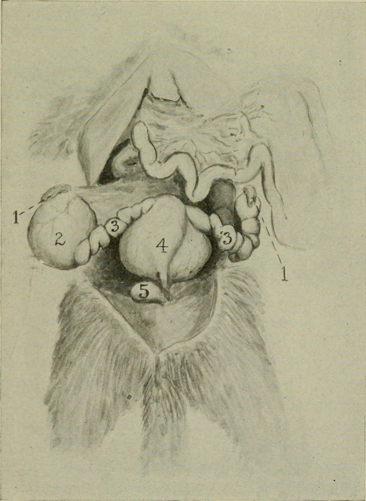
The mare’s tract sits deep in the pelvis, out of reach from the outside. The vet passes an arm into the rectum with a long straight probe in the hand, and reads the tract through the thin wall of the gut. The probe lies right above the uterus and the ovaries, close enough to draw them in fine detail. A rectal scan asks for care, the gut wall thin and the arm worked gently to keep from tearing it, the mare held still in stocks for the safety of the vet and the animal alike.
The scan runs over the tract in a set order. The vet starts at the uterine body where the horns meet, follows one horn out to its ovary, comes back to the body, and follows the other horn to the other ovary. The whole tract is covered the same way on every mare, nothing left unseen. A scan run to a routine misses nothing, the order itself the guard against an unread horn or an unchecked ovary.
The hand reads along with the probe. The arm feels the size and the tone of the uterus, the firmness of an ovary, the grip of the cervix. The probe turns each of those into a picture. The two senses build one read of the tract, the feel and the image checked against each other. A vet who has felt a thousand tracts reads the scan faster for it, the hand finding the ovary and steadying it under the probe. The two together miss less than either alone.
The mare’s ovaries
The ovaries are the first thing the scan is run to read. Each ovary carries the follicles and the corpus luteum that mark where the mare stands in her cycle, the structures that tell the vet when she will breed. A follicle reads as a round black pocket of fluid on the ovary, growing larger through the days before ovulation until it reaches the size of a large grape. The vet measures it across, follows it from one scan to the next, and reads its growth to call the day of ovulation. A breeding or an insemination dropped onto that day catches the mare at the one window she will hold to a covering. The corpus luteum forms on the spot a follicle leaves after it ovulates, a denser grey structure that tells the vet the mare has already ovulated. The ovaries read together give the whole of the cycle, the follicle that is growing toward a heat and the corpus luteum left from the last one.
A follicle close to ovulation changes its look. It softens, loses its round shape, and thins at the wall when the egg is ready to leave. The vet reads those changes to put the ovulation within a day, the timing that decides a breeding’s success. A mare in a strong heat may carry two large follicles at once, the vet reading both to know whether she will drop one egg or two. A double ovulation raises the chance of twins, the reason the follicle count feeds straight into the twin check weeks later.
The ovaries also tell of trouble. An ovary grown large around a single thick-walled structure points to a hormone-producing tumour, the kind that turns a mare’s behaviour and stops her cycling. The scan reads the shape and the make-up of the growth, the picture that shows the vet what the ovary has become. The granulosa cell tumour is the growth a breeder dreads, a large lumpy ovary that floods the mare with hormones and shuts her cycle down. The scan reads it early, the firm honeycomb of the growth a pattern the vet learns to spot at a glance.
The follicle count reads the mare’s value as a breeder. A pair of ovaries carrying a good store of follicles is a mare with seasons of breeding ahead, the scan reading the reserve a hand cannot count. An old mare whose ovaries have gone small is read for the breeding life she has left. A mare scanned before she is bought is read for her ovaries as much as her legs, a barren or failing tract a fault that no soundness in the limbs can make up for. The scan reads a breeding prospect a buyer cannot judge from the outside.
The two ovaries are read one after the other. The vet follows the horn out to reach each one, lifts it into view against the probe, and reads it from end to end before crossing to the other. A mare is never called open or in heat on one ovary alone, the pair read together for the full story. A double ovulation shows as two ripe follicles, one on each ovary or both on one, the finding noted for the twin check to come.
The uterus and the heat
The uterus reads the mare’s readiness to breed. Through the wall of the gut the probe shows the uterine body and the two horns, the tissue changing its look across the cycle. In heat the uterus fills with fluid in its folds, the cross-section of a horn showing a pattern of dark spokes a vet learns to read at a glance. The pattern has a name a vet uses, the cartwheel or the orange-slice, the spokes of fluid standing out in the cut face of the horn. The depth of the edema grades the heat, the spokes filling in toward a full wheel by the time the follicle is ripe. A vet reads the heat off the uterus and the follicle together, the two lining up to call the day a mare should be covered or bred.
That spoked pattern is the sign of a mare coming into heat. The folds of the uterus swell with fluid through the heat, read alongside the growing follicle to call the right day for breeding. The pattern fades after ovulation, the uterus turning quiet once the mare has left her heat. A uterus that stays full of fluid after the heat has passed reads as a problem, the fluid a sign of infection or of poor clearing that keeps a mare from holding to a breeding. The scan finds that retained fluid early, the cause of a mare failing to catch read on the screen.
The early pregnancy and the twin
The scan finds a pregnancy days after the breeding. A conception reads as a small round black vesicle of fluid in the uterus, seen from as early as twelve to fourteen days. The vet runs the probe along both horns and the body to find it, the early vesicle small enough to miss on a quick look. The vesicle grows a few millimetres a day, reaching the size of a small grape about a week after it first shows. The vet reads its size to age the pregnancy to within a day or two.
The first scan is run to find a twin. A mare cannot carry two foals to term. A twin pregnancy left alone loses both and risks the mare. The vet checks every part of both horns for a second vesicle, the early scan timed before the vesicles settle in place.
A twin found early is dealt with on the spot. Before about day sixteen the vesicle still moves freely, open to the vet pressing one against the probe and pinching it out, the other left to grow alone. The mare carries a single foal from a pregnancy that would have lost both. The reduction works best in the days the vesicles still roll free of each other, one held against the gut wall and pressed out with the other sitting clear of it. Done in time, it saves a breeding that twins would have ended.
The timing of that first scan is fixed by the twin check. A scan run too late finds the vesicles settled and hard to separate, the window for pinching one out already closed. The vet books the mare for around fourteen to fifteen days after breeding, early enough to catch a twin while it can still be reached. The mare comes back at around twenty-five days for the heartbeat, and again later for the sexing and the growth, a string of scans spaced across the early pregnancy. Each visit reads the foal at a fixed stage the vet knows to expect.
The order never changes
The vet scans the same sites in the same order every time. Body, one horn, one ovary, back, the other horn and ovary.
Following the horns
The horns are the road to the ovaries. From the uterine body the vet follows one horn out along its length, reading the tissue along the way, until the horn ends at the ovary. The ovary is lifted into the window and read, then the vet traces back to the body to start the other side.
The sweep finds what a single view would miss. A vesicle tucked at the tip of a horn, a follicle on the far face of an ovary, a small twin hidden against the body: each turns up only on a full sweep of the whole tract. The vet covers all of it before calling the mare.
The same road serves every kind of scan. The follicle check, the heat read, the pregnancy test, the twin hunt all run along the horns from the body to the ovaries. A vet learns the road once and runs it for every question the breeding season brings. The road is a short one, the landmarks fixed, the cervix at the start, the fork of the horns, the bend out to each ovary. A vet new to the work learns the turns in a season and runs them by feel ever after.
The growing foal
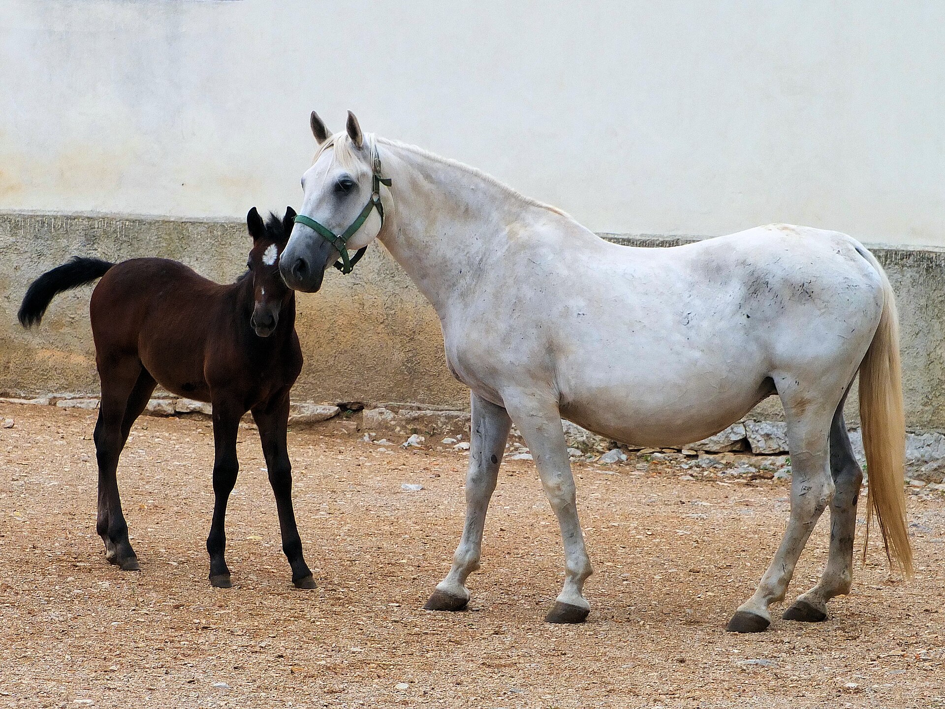
A mare back in foal is scanned again to follow the pregnancy. The heartbeat of the embryo flickers on the screen from around day twenty-five, the first sign of a live foal. The embryo proper shows by then as a small mass within the vesicle, the heartbeat a flicker at its edge. The vet reads it to confirm the pregnancy is holding, a check run a few weeks after the first.
The sex of the foal reads on the scan from around two months. A trained eye finds the genital tubercle, a small bright spot whose place along the body marks a colt from a filly, the answer read in a narrow window around two months. A breeder learns the sex of a foal months before it stands, planning the sale or the keeping from the early answer. The growth of the foal is followed through the months, its size and its parts measured against the stage it should be at. The growth is checked against a chart of what a foal should measure at each month, a small or quiet foal read for trouble in the pregnancy. A breeder follows a valuable foal through the year with a scan every few weeks.
The stallion
The stallion is scanned for the soundness of his breeding. The testes are read from the outside, a probe laid on the scrotum reading each one for its size, its make-up, and any lump or change inside. A testis that has grown small or uneven is read for the trouble behind a stallion’s poor fertility. The scrotal scan asks for a quiet stallion and a gentle hand, the probe laid on the skin of the scrotum with gel, each testis read along its length and across its width.
The size of the testes reads the stallion’s output. A bigger testis makes more sperm. The scan measures each one to weigh a stallion’s likely fertility. The two testes are measured in length, width, and height, the figures put into a sum that reads the daily output of sperm a stallion can give. A breeder reads the testes of a young stallion to judge what he will give before a season’s bookings are taken.
The make-up of the testis reads for disease. A dark patch, a bright fleck, or a fluid pocket in the even grey of a testis marks an injury, an infection, or a tumour, the scan telling a treatable trouble from one that ends a stallion’s career. The picture sends one stallion to treatment and reads another as finished at stud.
The glands behind the testes come into the scan as well. The accessory glands, the ones that make the fluid of the semen, are read through the rectum the same as a mare’s tract. The glands sit deep against the floor of the pelvis, reached only by the probe. A swollen or fluid-filled gland on the scan points to the seat of an infection that no outside check would find. A blocked or infected gland that fouls the semen is found on the scan, the cause of a fertility problem read where a hand cannot reach.
The stallion scan rounds out the breeding picture. A run of open mares from a single stallion sends the vet to scan the male, the picture reading whether the trouble lies in him. A breeding operation reads both sides of its stock, the mares and the stallion each scanned for the part they play. A stud that scans its stallions each season catches a fading testis or a new growth before it costs a year of foals.
The probe for the job
The work runs on a long straight probe. A rectal linear probe reaches the length of the arm and lays a broad window against the uterus and the ovaries. The frequency is set high enough to draw the fine detail of a follicle wall or an early vesicle, the depth shallow because the probe sits right on the tract. A frequency of around five to seven and a half megahertz suits the work, high enough for the fine detail of an early vesicle and matched to the shallow depth the rectal window asks for.
The stallion’s testes ask for a different probe. A linear probe laid on the scrotum reads the testis from the outside, the same kind of probe a clinic uses for any shallow scan. The one handheld carries both probes, the rectal wand for the inside work, the surface probe for the scrotum. A vet on a stud carries the one machine from the mare’s stocks to the stallion’s box, the two probes covering every reproductive scan a breeding yard asks for in a day.
When the picture is hard to read
The scan has its blind spots. A rectum full of gas or dung throws shadows across the tract, a poorly prepared mare giving a poorer picture. The vet clears what can be cleared and works the probe to find a clean window onto the organs.
The early vesicle tests the eye. A conception of twelve days reads as a black spot a few millimetres across, easy to take for a pocket of fluid or to miss in a fold. The vet runs the probe slowly over every part of the tract, the early scan asking for a careful eye and a steady hand. A vesicle missed on the first scan turns up larger a week later, the reason a vet who finds nothing early books the mare back for a second look before calling her open either way.
The read comes with the season’s work. A vet who scans mares through a breeding season reads the cycle, the heat, and the early pregnancy at a glance, the eye trained on hundreds of tracts. The sites stay the same on every mare, the skill built up over the hundreds passed under the probe. The first season is the slow one, the eye learning to tell a follicle from a coil of bowel and a vesicle from a pocket of fluid. By the second the read comes fast, the sites and their signs known by heart.
What the sites add up to
The equine reproductive scan is a fixed tour of a breeding tract. The probe goes in through the rectum and reads the ovaries, the uterus, the horns, and the early pregnancy of a mare, the testes and the glands of a stallion. Every site is covered in the same order, the whole tract read on every animal. The tour takes a few minutes on a quiet mare, the probe reaching every site from the one entry. Nothing of the tract is left to a guess.
The sites read together time the whole of the breeding. The follicle calls the day to breed, the spoked uterus confirms the heat, the vesicle marks the conception, the twin check guards the mare, the heartbeat and the growth follow the foal. Each answer comes from a site the vet knows to reach. The scan answers in one visit what once took weeks of waiting and guessing, the breeding season run off a string of pictures the vet draws beside the mare. A stud books its coverings around the scan, the vet’s read setting the day for every mare on the list.
The scan reads what a hand alone once only guessed at. The ovary and the early vesicle are seen now, the breeding timed off the picture the vet draws each visit. A season planned around the scan runs on certainty a hand inside could never give.
The mare and the stallion each get their scan in the breeding year. The vet reaches the same sites, reads the same structures, and answers the same questions for every animal the season brings. The probe on the tract is the tool a modern breeding operation runs on. The mare’s scan and the stallion’s scan together read the whole of a breeding, the female tract and the male soundness covered by the one probe across the season. A breeder who scans both ends of the cross leaves nothing of the year to chance.
Common questions about equine reproductive scanning
Where does the vet scan a mare for reproduction?
Through the rectum. The vet passes an arm into the rectum with a long probe and reads the uterus and both ovaries through the gut wall, following each horn out from the uterine body to its ovary. The scan covers the ovaries for follicles, the uterus for the signs of heat, and the horns for an early pregnancy.
How early can ultrasound find a pregnancy in a mare?
From around twelve to fourteen days after breeding. The conception reads as a small round black vesicle of fluid in the uterus. The first scan is timed early on purpose, to catch a twin while one vesicle can still be pinched out, before about day sixteen.
Why is the twin check so important in mares?
A mare cannot carry two foals to term. A twin pregnancy left alone usually loses both and can harm the mare. The early scan finds a second vesicle in time for the vet to pinch one out, leaving a single healthy foal to grow.
What is scanned on a stallion?
The testes and the glands behind them. A probe on the scrotum reads each testis for its size and its make-up, finding a lump, an injury, or a change that hurts fertility. A rectal scan reads the accessory glands that make the fluid of the semen. The scan weighs a stallion’s breeding soundness.
What probe is used for equine reproductive scanning?
A long rectal linear probe for the mare’s tract and the stallion’s glands, reaching the length of the arm to lay a window against the organs. A surface linear probe reads the stallion’s testes from the scrotum. The frequency is set high for the fine detail of a follicle or an early vesicle.

About the Author
Julien Mercier
Senior R&D Engineer · Medical Ultrasound Transducer Development
Senior R&D Engineer with an M.S. in Applied Physics and over 15 years of experience in medical ultrasound transducer development, specializing in the design verification and performance testing of high-frequency imaging transducers. Currently leading the development and verification of the company’s next-generation high-frequency linear-array transducer, responsible for imaging performance evaluation and reliability analysis in preclinical testing. Brings extensive hands-on experience in piezoelectric element tuning, beamforming parameter optimization, and system-level performance testing.