





































Exotic Pet Rabbit Bird Reptile Ultrasound Handheld Veterinary
An exotic-pet ultrasound reads the insides of a rabbit, a bird, or a reptile through a small high-frequency probe, finding the few clear windows that the animal’s air, scales, or shell allow. The patient is tiny, its anatomy unlike anything in a cat or dog, the scan working around an anatomy the sound can barely cross. It is run on a fragile animal that tolerates handling poorly, the whole scan over in a minute or two, the probe finding what it can in the time the patient will give.
A different patient on the table
An exotic patient breaks every habit a vet learns on cats and dogs. It is a fraction of the size, its organs packed into a body that may weigh as little as a sparrow. Its insides sit where no dog’s ever would, in places and at depths a cat-and-dog scan never has to reach. A vet trained on the steady anatomy of the dog meets in the exotics a dozen body plans at once, the bird, the lizard, the snake, the tortoise, the rabbit each built to its own design, each read by its own map.
The scan bends to the patient. A smaller probe, a higher frequency, a lighter touch, a shorter session: each is set to a body that a cat-and-dog routine would crush. The vet who scans exotics learns a new map of the insides and a new gentleness in the hand. The settings are set fresh for the species on the table, the depth down to a centimetre or two, the gain trimmed for a thin body, the focus set close under a skin that sits a hair’s breadth from the probe.
The air that fights the sound
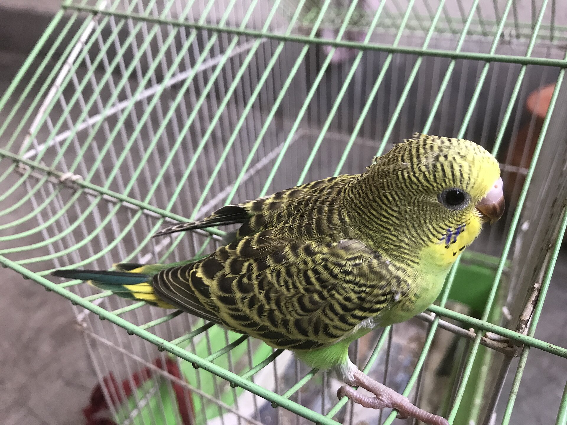
The exotic patient is full of the one thing ultrasound cannot cross, which is air. Sound travels through tissue and fluid and turns back at air, so a pocket of gas throws a wall across the picture and hides whatever lies behind it. A bird carries great air sacs that reach right through its body, light bladders of air that lift it in flight and turn every beam of sound back, so a scan of a bird works in the narrow lanes between them. A reptile of some kinds, the snake and the chameleon among them, carries air sacs of its own that block the beam the same way. A rabbit packs a gut that never stops working and is never empty of gas, the bowel loops throwing their shadows across the belly the probe is trying to read. The exotic scan is, more than anything, a hunt for the few windows where tissue meets the probe with no air in the way, the vet learning where on each kind of animal those clear lanes lie.
The air also turns the scan into a finding. Free fluid in a space the air usually fills, a coelomic cavity gone dark with effusion, reads loud and clear on a patient whose air usually hides the view. A bird that scans easily, its organs laid open where the air sacs once blocked them, is often a sick bird, the fluid of its illness the one thing that opened the window the air kept shut.
The gas is read for itself as well. A rabbit’s gut blown tight by gas and gone still is a rabbit in stasis, an emergency the scan names from the shadows it usually fights. On the right patient, that gas is the diagnosis the scan came to find. A gut that has stopped moving, flat under the probe, tells the same tale, the stillness as loud a sign as the gas itself.
The scan reads the air sacs for their own troubles too. A bird’s air sac thickened, flecked, or flooded with fluid speaks of an infection caught in the space that usually defeats the probe outright, the hunt to reach it paying off in the answer. An air sac so thickened or so clouded that the probe reads it at all is a finding in itself, the clear space turned opaque the mark of the disease.
Reading around the air is a skill more than a setting. A vet works the probe into the angle that slips past a gas pocket, presses just enough to push a loop of bowel aside, and waits for the moment a moving gut clears the view. The patience to find the window is half the exotic scan. The skill comes with the cases, a vet learning over a year of birds and reptiles where the lung ends and the liver begins, which way to tip a tortoise to bring an egg to the glass.
Scanning around a shell
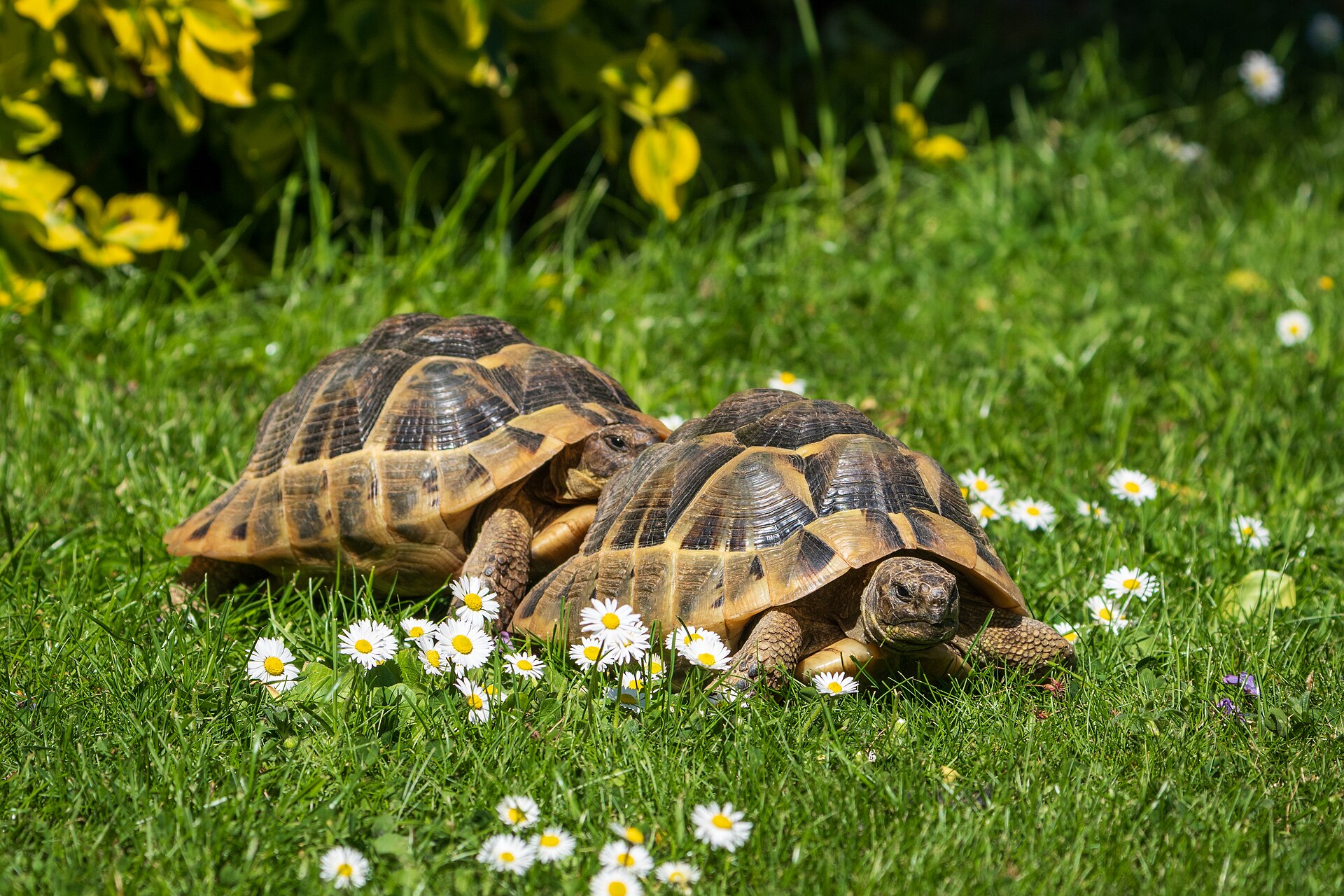
A tortoise carries its troubles behind a wall the sound cannot cross. The shell that guards it turns back every beam, so the scan reaches the insides only through the soft gaps the shell leaves open. The probe never crosses the bone and horn of the shell, the scan built entirely around the openings nature left for the legs and the neck.
Those gaps have names a vet learns. The prefemoral fossa, the soft hollow in front of each hind leg, is the main door into a tortoise, the probe slipped in there to read the bladder, the eggs, and the organs of the coelom. A second window opens at the neck, in front of a foreleg, for a look at the heart and the liver. The animal is held upright and rocked gently, the insides shifting toward the window to meet the probe. The fossa is a small one, the angle tight, the probe worked in past the skin of the leg pocket to fan across the coelom from the one opening the shell allows on each side.
The lizard and the snake give easier ground. Their skin, soft between the scales, takes the probe across much of the body, the scan run along a snake’s length or across a lizard’s flank with far less of the shell’s resistance. A little water or gel works the probe past the scales to the tissue beneath. A snake is read along its length in sections, the heart a third of the way down, the organs strung out one behind the next in a line the probe follows from head to tail. The vet learns the order of a snake’s insides by heart, the same string of organs in the same line on every snake brought in.
The probe for a small patient
A tiny patient asks for the smallest, quickest probe a clinic owns, its high frequencies drawing the fine detail an exotic scan lives on. The probe a clinic buys for cats’ superficial work does double duty on the whole of a budgerigar. The shallow reach of a high frequency reads a finch from end to end, the depth and the detail the exotic scan asks for.
The footprint matters as much as the frequency. A small flat head, or a tightly curved one, fits the shallow curve of a little body and the narrow gap of a shell window. A pad of gel or a bath of warm water floats the probe over a surface it could not press against directly. The standoff brings a curved little body into focus the probe could never reach laid flat against it.
A minute is all the patient gives
An exotic spends its strength fast under a hand. A scan done in that first minute takes the picture before the patient pays.
The egg and the follicle
The reason the probe comes out more than for anything else is the reproductive tract. A bird, a reptile, a rabbit can carry a clutch of eggs or a string of follicles no other test can read, an egg gone wrong among the commonest emergencies the exotic vet meets.
The follicles read as a row of round, even pockets along the ovary, growing through the breeding season. The scan counts them, measures them, and watches them ripen, a window onto a breeding state the body’s outside keeps wholly hidden. A reptile’s follicles regressing, shrinking back unspent, tell a different story than a clutch building toward eggs. A row of follicles holding steady through the season is a bird in lay, the count and the size read off the screen to judge whether breeding runs to plan or has begun to fail. The count read early sets the breeder’s expectation, a clutch of four seen on the screen before a single egg is laid.
The egg itself is the thing the scan is run to find. A bird or a reptile that strains, sits fluffed up, or stops passing droppings may be bound by an egg it cannot push out, a killer the exotic vet races to name. The scan finds the egg, reads its shell and its size, and shows whether it sits where it should or has slipped where it should not. It tells the soft, shell-less egg from the hard, the egg a little help will pass from the one lodged fast against a deformed shell or a narrowed passage. It reads the others stacked behind, the clutch the vet must reckon with before reaching for a needle or a knife. A scan that maps the eggs turns a guess into a plan, the line between an animal that lays its clutch and one lost in the straining.
The female rabbit asks a question of her own. An intact doe carries a high risk of a cancer in her womb, the scan reading the uterus for the thickening, the cysts, or the mass that marks it, a finding that sends her to surgery before it spreads. The womb a vet cannot feel is the one the probe brings into view. A doe scanned before she is spayed shows the vet what the surgery faces, a thickened horn or a mass read on the screen turning a routine neuter into a cancer caught in time.
Reading the coelom
A bird and a reptile keep their organs in one shared cavity, the coelom, with no diaphragm to split the chest from the belly. The scan reads that whole space through its few windows, the liver, the heart, the gut, the kidneys, and the gonads all met from the same handful of approaches. The vet learns the coelom as one room, the heart high under the keel, the liver below it, the gut and the kidneys and the gonads ranged behind, each found by aiming the probe from the spot the air leaves clear. The handful of approaches stays the same from one bird to the next, a routine the vet runs the same way each time.
The liver is the organ the scan reads best. Swollen, bright, or shrunken, it tells of the diseases that fell exotic pets in numbers, the fatty liver of an overfed parrot, the infection that spreads through a coelom. A liver read against the size it should be is half of many an exotic diagnosis. A parrot fed too long on seed lays down fat through its liver until the organ swells, its grey gone the pale of stored fat, a change the scan reads early enough to turn the diet before the liver fails.
Fluid in the coelom is the loud find. A coelom gone dark with free fluid speaks of heart failure, a tumour, an infection, or a liver giving way, the scan drawing a sample under the probe to name which. The coelom filled is an exotic in trouble, read in a moment once the fluid has cleared the air from the view. A tap drawn under the probe sorts the blood from the clear seep from the cloudy fluid of an infection, the colour and the cells naming a cause the picture had only pointed toward.
The rabbit’s belly
The rabbit is the exotic a clinic scans more than any other, its belly holding troubles of its own. A rabbit’s thin body wall lets a high-frequency probe read the whole of its belly with ease, the belly open to a careful sweep.
A rabbit’s bladder runs thick with chalky sludge, the calcium it cannot clear settling into a sand that shows bright on the scan and packs into stones. The probe reads how much has gathered, whether a stone has formed, and whether the bladder behind it is straining, the picture that tells a flush from a surgery. The sludge a rabbit cannot pass thickens over weeks into a paste, the bladder straining against a load the scan measures before it hardens into a stone no flush will shift.
The gut tells the rabbit’s gravest tale. A bowel blown tight by gas, its loops swollen and no longer stirring, is a gut in stasis, the slow shutdown that kills more pet rabbits than any single disease, named on the scan by the still, swollen loops.
The scan reads the rest of the belly in the same pass. A kidney scarred, a liver swollen, an abscess walled somewhere it cannot be felt: each turns up on the sweep that started with the bladder or the gut, the one probe reading the whole of a small patient at a sitting. A rabbit hides its illness until it is far along, a prey animal built to show nothing, so the scan that reads the belly early catches what the rabbit’s own stillness conceals.
When the patient cannot wait
An exotic in a crisis gives the scan its hardest test and its highest value. A bird gasping, a tortoise straining, a rabbit gone off its food and hunched: each is an emergency the scan can sort in minutes, an egg, an effusion, a gut shut down, a bleed. The owner brings in an animal hunched and fading, the one thing inside that will kill it found on the scan in minutes.
The speed is the mercy. An exotic that cannot be handled for long gives a vet a narrow window, a scan that reads the trouble in it sparing the animal the longer workup it might not survive. The picture found fast is the one that saves the small patient.
What the scan cannot reach
The exotic scan leaves much unseen, its blind spots known to an honest reader. The air defeats it where the air sits, a lung deep behind an air sac, an organ tucked behind a loop of gas, beyond the reach of any window. A bird’s lung sits locked against the body wall behind the air it breathes, a place the probe is turned away from on the healthiest patient, read only when disease has changed the ground.
The shell keeps its own secrets. A tortoise organ that sits away from a prefemoral window stays hidden, the shell allowing the probe no other way in, a limit no skill can fully beat.
The size sets a floor under what the scan resolves. The smallest patients, a finch, a tiny gecko, carry organs near the limit of what any probe can separate, the picture grainy where the structures are finer than the sound can split. A heart the size of a lentil asks more of a machine than a clinic handheld can give. The finest probe still meets a floor, the structures of the smallest patients packed closer than the sound can tell apart, the read on a creature that size as much a matter of knowing the anatomy as seeing it.
The temperature shifts the picture in a reptile. A cold reptile slows its body until its organs shrink and shift, the scan read on a chilled patient telling a different tale than the same animal warmed to its proper heat. A reptile is brought up to temperature before the read is believed.
The scan names the trouble it can reach and refers the rest. The deep look, the named disease, the surgery to follow belong to the exotic specialist and the workup beyond the handheld, the first scan pointing the way. The handheld finds what it can reach and refers the rest, the line between the two drawn by an honest reader.
Where the handheld fits
The handheld suits the exotic patient better than the cart-bound machine ever did. It comes to the animal in its own carrier or on the consult table, the scan run where the patient is calmest, the small probe and the quiet box adding nothing to the fear that already runs high. A quiet machine that neither roars nor towers keeps a stress-prone exotic calmer than a clinic full of equipment ever could.
The portability is the point with a patient this fragile. A scanner carried to the table, run in a minute, and put away spares an exotic a journey and a wait it might not have the strength for. The scan goes to the patient that cannot easily be brought to the scan. A bird carried in a cloth, a tortoise lifted from its box, a snake uncoiled on the table is scanned where it sits, the machine carried over to it.
The handheld settles the first questions and points the way on. Reading a tiny heart in full, naming a coelomic mass, planning the surgery on an egg-bound reptile belongs to the specialist and the referral, with the handheld settling only how hard and how soon to chase the trouble. It is the eye that opens the hidden patient in the first minutes. From that first look the vet knows whether the trouble can wait for the morning or must be met within the hour.
A window onto a hidden patient
An exotic-pet ultrasound opens a body built to keep its secrets. It works around the air, the shell, and the size that hide a rabbit, a bird, or a reptile from every other look, finding the windows where the probe can read the insides of the smallest patients.
What it finds in those few clear lanes, an egg, a flooded coelom, a gut shut down, a swollen liver, can decide whether a small life is saved from a trouble no hand could feel. No other test in the clinic reaches so far inside a budgerigar or a gecko, the whole of it the probe laid on and the picture read with barely a hand raised against the animal. That is what the small probe earns its place doing.
Common questions about exotic pet ultrasound
Can ultrasound be used on rabbits, birds, and reptiles?
Yes, with a small high-frequency probe and a gentle hand. The scan works around the air sacs of a bird, the shell of a tortoise, and the gas of a rabbit’s gut, finding the clear windows where it can read the insides. It reads the liver, the gut, the kidneys, the reproductive tract, and any fluid in the body cavity.
Why is ultrasound harder on exotic pets than on cats and dogs?
Their bodies fight the sound. Birds and some reptiles carry air sacs, tortoises a shell, rabbits a gas-filled gut, none of which the sound can pass through. The patients are tiny, their organs near the limit of resolution, their tolerance for handling short, so the scan is quick and worked through a few narrow windows.
What is the commonest reason to scan an exotic pet?
The reproductive tract. A bird or reptile bound by an egg it cannot pass is one of the commonest exotic emergencies, the scan finding the egg, reading its size and position, and showing the clutch behind it. The scan also reads follicles, a rabbit’s uterus, and the diseases of the liver and gut.
How are tortoises and turtles scanned through their shell?
Through the soft gaps the shell leaves. The main window is the prefemoral fossa, the hollow in front of each hind leg, where the probe slips in to read the bladder, the eggs, and the coelomic organs. A second window at the neck reads the heart and liver. The animal is held upright and rocked gently to bring its insides toward the probe.
Can a handheld ultrasound be used on exotic pets?
Yes, and it suits them well. A small handheld with a high-frequency probe comes to the patient, runs in a minute, and adds little to the stress a fragile exotic feels. The full study and the surgery stay with an exotic specialist, the handheld the fast first look that points the way.

About the Author
Julien Mercier
Senior R&D Engineer · Medical Ultrasound Transducer Development
Senior R&D Engineer with an M.S. in Applied Physics and over 15 years of experience in medical ultrasound transducer development, specializing in the design verification and performance testing of high-frequency imaging transducers. Currently leading the development and verification of the company’s next-generation high-frequency linear-array transducer, responsible for imaging performance evaluation and reliability analysis in preclinical testing. Brings extensive hands-on experience in piezoelectric element tuning, beamforming parameter optimization, and system-level performance testing.