





































Pet Hospital Handheld Ultrasound What To Consider Before Buying
Choosing a handheld ultrasound for a pet hospital comes down to matching the machine to the patients on the table and the vets who will hold it. A few things decide the right buy: the probe, the image, the build, the ease of use, and the price set against what the scan brings in. Each one is weighed against the work the clinic does day to day, ahead of the longest spec sheet in the brochure.
Start with the patients
The buy starts with a question the spec sheet cannot answer: what walks through the door. A clinic of cats and small dogs needs fine detail held close under the skin above all, the shallow reach of a small-animal machine the thing all its patients ask for. The patients set the depth the sound must reach and the detail it must show. A pocket machine sized for kittens and toy breeds leaves a clinic stuck the day a mastiff comes in bleeding, the depth it lacks exactly what the big patient needs.
The exams the clinic runs shape it further. A practice doing pregnancy scans and bladder checks asks less of a machine than one reading hearts and chasing bleeds. The work the clinic means to do, written down before any demo, is the truest guide to what it should buy. A practice planning to grow into cardiac or exotic work buys for that plan now, sparing itself a second machine in a year.
The honest list is short. A clinic names the scans it runs today, the ones it means to grow into, the species it sees, and the vets who will hold the probe, then reads the spec sheets against that list, the list leading and the numbers following. That list outlives the demo and the sales talk, the clinic that wrote it down holding to its own work through every pitch.
The probe is the choice that decides it
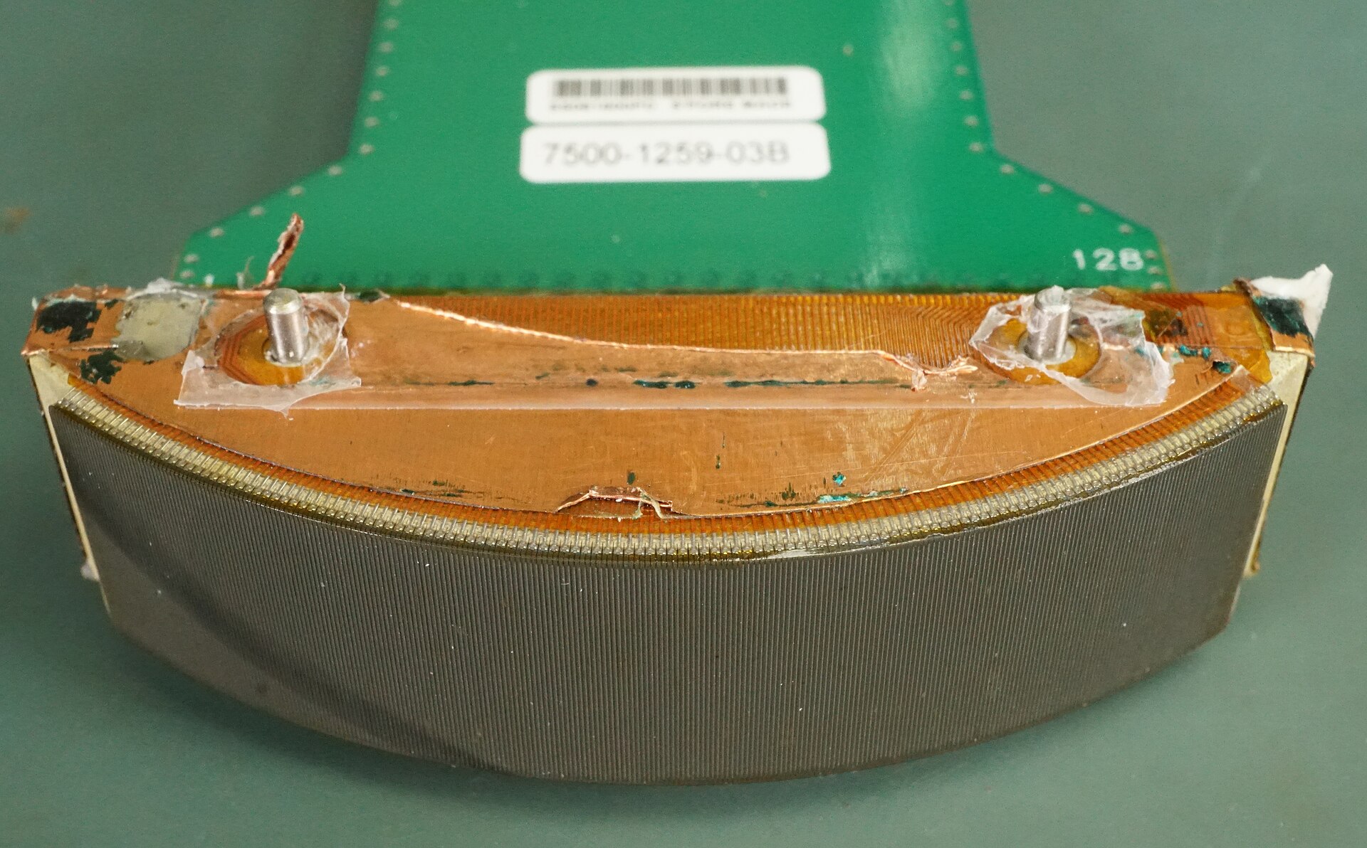
The probe decides more about a handheld than the screen or the software ever will. It is the part that touches the patient and shapes the sound, and the wrong one turns a costly machine into a poor scanner. A clinic chooses the probe first and the rest around it. The screen, the battery, the software all serve the picture the probe draws, a screen only ever as good as what the probe hands it.
A micro-convex probe is the workhorse of small-animal practice. Its small curved head fits between the ribs and under the costal arch, into the tight windows a dog or cat gives, and fans a wide beam from a narrow point of contact, so a whole organ falls onto the screen from a press the size of a fingertip. It runs a range of frequencies that reads the fine grain of a cat’s kidney close under the skin and still reaches the deep abdomen of a barrel-chested dog. That reach is what makes it the one probe a clinic buys if it buys only one, the scan it covers running from a pregnancy count to a bladder stone to a swollen spleen to a focused look at the heart. A practice can work for years on this probe alone, adding the others only as the caseload asks for them. The curved head and the mid-range frequency together cover more of the small-animal day than any other single choice on the shelf.
A linear probe earns its place on the superficial work. Its high frequency draws fine detail from the structures close under the skin, the tendons, the lumps, the thyroid, the eye, at a resolution the curved probe cannot match. A practice that reads many lumps and small parts wants this probe beside the first. Its flat face sits on a clipped patch of skin and draws the surface in a sharpness the curved probe was never built for, the tendons, the eye, and the small mass given an edge the deeper probe leaves soft.
The heart asks for a probe of its own. A phased-array head, small of footprint and built to fan between the ribs, lays a clean picture of the beating chambers and carries the Doppler that times the flow. A clinic moving into cardiac work reaches for this one, or for a micro-convex that does a fair job of the focused heart scan when the budget holds to a single probe. The full cardiac study, with its M-mode and its spectral Doppler, asks for the dedicated head and the software to drive it, the step a clinic takes once the heart work earns its own machine.
The large-animal work changes the list again. A clinic that scans cattle or horses needs a long rectal probe and the depth to reach a deep abdomen, a different machine entirely, the territory of its own buying guide. The small-animal hospital is spared that reach and chooses among the probes the patients on its table need. A clinic working both worlds finds the barn machine and the consult-room machine rarely turn out to be the same one, a reason many keep one of each.
One probe or several
A single-probe machine keeps things simple. It does one thing well, suits a clinic with a narrow caseload, costs little, and asks nothing of the vet but to pick it up and scan. The price stays low, the choices few, a fair buy for a practice that knows exactly the one job it needs done.
A machine that takes more than one probe, swapped on a connector or carried as separate wireless heads, grows with the practice. The engineering of how a probe connects and changes is a study of its own, set out elsewhere in this library. The choice a clinic makes here is simpler: how many kinds of patient and exam the one machine must serve. A practice that buys a second probe later wants a machine that will take it, the connector and the software ready for the head the caseload has not yet called for.
An image good enough to trust
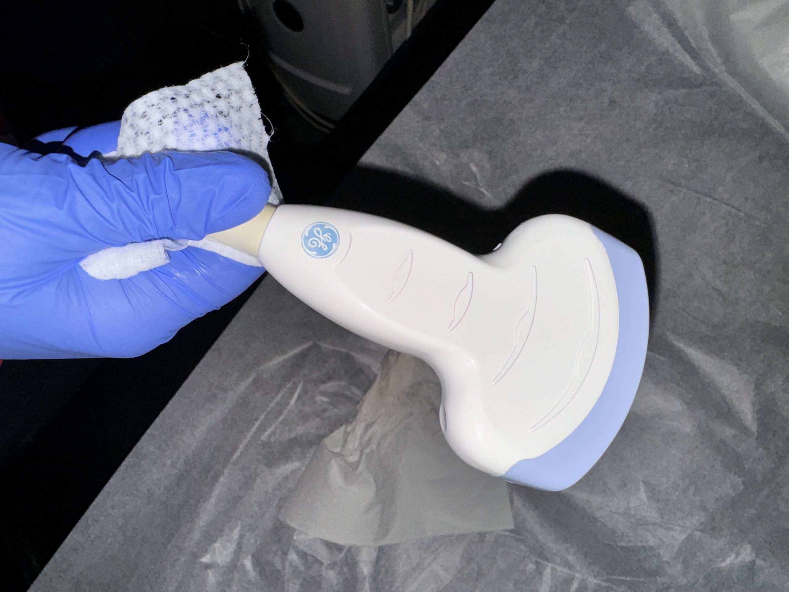
An image is bought to be trusted, a picture so rough it cannot be read earning nothing whatever its price. The resolution decides how fine a detail the scan resolves, the wall of a thin vessel, the early change in a kidney, the small mass under the skin. The penetration decides how deep that detail still holds, the reach down into a deep-chested dog the test of it. A picture that turns to grey soup past a few centimetres fails the patients a clinic needs it for above all, the large and the deep. The clinic reads the demo on its hardest patient, judging the picture where the real work will test it.
The two pull against each other, the right balance following the patients. A clinic of small animals leans toward the higher frequencies that draw fine detail close in, the same range pulled lower the deeper the patient runs. The machine that serves a mixed caseload carries the whole span, dialled to the patient on the table. A probe that spans a broad band of frequencies frees the one machine to read the kitten and the mastiff alike, the range set to whoever is in the room.
A demo on the clinic’s own patients tells more than any brochure. A vet scans a familiar case, a cat’s kidney, a dog’s heart, a known lump, and reads whether the picture is one a diagnosis can rest on. The image that convinces in the room is the one to buy. The clinic runs the demo on its own hard cases, the overweight cat and the gassy gut of a real waiting room that a polished sales clip never has to show.
The numbers on the page guide the eye, never standing in for it. A high frequency printed in a spec means little if the picture it draws is noisy, so the read in the hand outranks the figure on the sheet every time.
The unit that gets used is the one that earns out
A machine returns nothing from a cupboard. Used daily, it pays for itself many times over.
Easy enough for the whole team
A handheld earns its keep only when the whole team can use it. An intuitive screen and a few clear controls put the probe in the hand of every vet on the rota, the new graduate and the locum scanning as readily as the specialist. The fewer the dials between the vet and the picture, the more the machine is used, the using of it the whole of where its value lies.
The clever machine does some of the work itself. Presets tuned for a cat’s abdomen or a dog’s heart set the depth, the gain, and the focus at a tap, automatic optimisation cleaning the picture without a hand on a dozen dials. A new graduate reads a useful scan on the first day, the machine carrying the settings the experience has not yet taught. The same presets steady the hand of a seasoned vet in a hurry, the right depth and gain laid on at a tap, ready before the emergency needs them.
The training that comes with the box decides how fast that ease arrives. A short course and a run of guided cases turn a nervous first user into a confident one, the scan folded into the clinic’s day far sooner than a manual alone would manage. A maker that sends a trainer to the clinic, or runs a webinar a nervous vet can follow, turns a boxed machine into a used one inside a week, a kind of help to weigh in the buy.
Built for a busy clinic
A handheld lives a hard life in a working practice. It is carried room to room, set down on wet tables, splashed with gel and worse, and dropped now and then by a tired hand at the end of a long shift. The build has to take it.
A sealed body, wiped down a dozen times a day, survives the cleaning a clinic gives it between patients, the probe and the screen cleaned without water creeping into the seams. A unit that cannot be cleaned properly is a unit that spreads what it touches.
The weight and the shape matter in a busy room. A light machine moves from the consult table to the kennel to the prep room without a second thought, the scan run wherever the patient happens to be. The grams add up over a day of scans, a heavy probe tiring the hand that holds it steady before the morning is out.
A handheld that drops and keeps working is a handheld a clinic can trust. The toughest of them shrug off the knocks of a busy day, the screen and the probe built to survive a fall from a table that a long shift makes likely.
The cleaning a clinic gives a machine is harder on it than any drop. Disinfectant wiped on a dozen times a day, gel worked into every seam, the odd splash of blood or urine: a body that takes all of it without clouding its screen or loosening its seals is the one still earning its keep in five years.
Cordless and how long it lasts
A wireless handheld frees the vet from the cord and the cart. The probe links to a tablet or a phone, the picture carried over the air, the whole scanner small enough to slip in a coat pocket between calls. A clinic already running on tablets folds the scanner into hardware it owns, the picture landing on a screen the team already reads from. The link itself is read for how steady it holds, a picture that freezes or drops mid-scan a worse failing than a short battery.
The freedom lasts only as long as the battery. A unit that holds a full morning of scans on one charge keeps the clinic moving through its list, the charge that lasts the first thing to ask after, with a spare battery and a fast charger close behind.
Colour, and the extras that earn their place
Colour Doppler lifts a handheld from a grey picture to a reading of the blood. It paints the flow through a kidney, the vessels feeding a lump, the leak across a heart valve, a layer of meaning the grey image alone cannot give. A clinic doing any heart or vascular work wants colour on the machine. The same colour reads the twist in a torsion, the flow into a tumour, and the leak of a worn valve, a tool that earns its place across the whole of the caseload, the heart only one part of it.
The measuring tools earn their place too. A set of calipers and a few built-in calculations turn a picture into a number a record can hold, the size of a kidney, the count of a litter, the thickness of a wall. The scan that measures cleanly saves the vet the sums. A clinic that keeps its scans on file builds a record it can read back, this year’s kidney set beside last year’s, the picture as useful in the watching as in the first look.
The small conveniences add up over a year. A machine that saves an image to the patient file in a tap, that shares a clip with a specialist over the air, that logs a measurement without a written note, folds the scan into the day and saves a step at every turn.
What it costs and what it returns
The price is read against the return, never on its own. A handheld brings scans in-house that once went out to a referral or a travelling sonographer, each one a fee kept and a diagnosis made the same day. A machine used daily pays back its price inside a year in many a busy clinic. The scan also wins work a clinic would once have turned away, the pregnancy check and the quick belly look done on the spot and kept in the clinic. Each scan held in-house is a small return that adds up across a year into the price of the machine and more.
The cheapest machine is rarely the best value. A unit bought on price alone, with a probe that does not fit the work or an image so coarse the eye cannot lean on it, costs more in missed diagnoses and idle days than it ever saved at the till. The value sits in the scanning the machine does well, a thing the invoice figure never shows.
The running costs hide behind the sticker. A probe that cracks and costs a fortune to replace, a subscription locked behind the software, a battery that fades in a year: each adds to the true price, the questions to ask before the money moves. A probe replacement quoted at half the price of the machine, a yearly software fee, a battery sold as a wear part: the true cost is the sum of all of it across the years the machine serves.
A clinic buys for the years ahead as much as the day. A machine that grows with the practice, taking a new probe or a new tool when the caseload widens, outlasts one bought to the edge of today’s need and outgrown in a season. A machine bought a notch above today’s need carries the practice through its next few years of growth, the cost of the headroom paid back the first time the caseload widens into it.
The support behind the box
A handheld is only as good as the help behind it. A maker who answers the phone, sends a loaner when a unit goes down, and updates the software over the years keeps a clinic scanning through the life of the machine. A maker that has been in the market for years, with a clinic or two willing to vouch for the service, is a safer bet than a name new to the trade. A cheap unit from a seller gone quiet after the sale is a machine alone the day it breaks, the saving at the till handed back with interest. The questions come before the money, the maker’s record on repairs and updates and loaners read as carefully as the spec sheet.
The warranty sets the floor under the buy. A long cover on the probe and the body, the parts likeliest to take a knock, counts for more than a short one whatever the headline price, the clinic spared the bill for the first hard fall. A warranty read closely tells a clinic what the maker expects of its own machine, the cover a quiet measure of the years it means the box to last in a working clinic.
Matching the buy to the practice
The right handheld is the one that fits the patients, the vets, and the purse of the clinic that buys it. There is no single best machine, only the best match for the work a given practice does. The clinic that knows its own work picks the machine that fits that work, the cleverest features on a mismatched one counting for nothing the day-to-day work never calls on.
The small clinic of cats and dogs lands on a light micro-convex handheld with a clean image and a simple screen, a linear probe added when the lump work grows. Scale the practice up into cardiac and exotic cases and the buy grows with it, the multi-probe machine, the colour, and the depth to match, often more than one unit, a tough handheld for the wards beside a finer machine for the imaging room.
The buy is made on a demo in the clinic, never on a brochure alone. A clinic scans its own patients on the machine it is weighing, asks the questions of cost and support and durability that the spec sheet leaves out, and chooses the one that earns its place on the table. No two clinics land on the same machine for the same reasons, the buy fitted to the work, the team, and the budget of the one practice making it.
A handheld bought to fit the practice is a tool the whole team reaches for, the scan run on the patient in the room while the owner waits, the answer found in minutes that once took a referral and a week. That is the buy to make.
Common questions about buying a handheld ultrasound
What is the first thing to weigh when buying a handheld ultrasound for a vet clinic?
The probe, matched to the patients and exams the clinic runs. A micro-convex probe covers the run of small-animal abdominal and pregnancy work, a linear probe adds the fine detail for lumps and small parts, and a phased-array probe suits cardiac scanning. The buy starts from the work the clinic does, ahead of any spec sheet.
Which ultrasound probe does a small-animal clinic need first?
A micro-convex probe, with a frequency around 3 to 9 MHz. Its small curved head fits between the ribs and reads a cat’s kidney or a dog’s full abdomen, and it does a fair job of a focused heart scan. A clinic buying one probe for general work buys this one.
How do I know if the image quality is good enough?
Run a demo on the clinic’s own patients. Scan a familiar case, a kidney, a heart, a known lump, and read whether the picture is one a diagnosis can rest on. The image that convinces in the room outranks any frequency printed in a brochure.
Is a wireless handheld ultrasound a good buy for a busy clinic?
For a busy clinic, usually yes. A wireless probe links to a tablet or phone and moves room to room without a cart, the scan run wherever the patient is. The thing to check is the battery, how many patients it scans on one charge, and whether a spare and a fast charge are offered.
Does a cheap veterinary ultrasound save money?
Rarely. A machine bought on price alone, with a probe that does not fit the work or an image so coarse no eye can trust it, costs more in missed diagnoses and idle days than it saved. The value is read against the scanning the machine does well and the years it lasts, far from the figure on the invoice.

About the Author
Julien Mercier
Senior R&D Engineer · Medical Ultrasound Transducer Development
Senior R&D Engineer with an M.S. in Applied Physics and over 15 years of experience in medical ultrasound transducer development, specializing in the design verification and performance testing of high-frequency imaging transducers. Currently leading the development and verification of the company’s next-generation high-frequency linear-array transducer, responsible for imaging performance evaluation and reliability analysis in preclinical testing. Brings extensive hands-on experience in piezoelectric element tuning, beamforming parameter optimization, and system-level performance testing.