Subcutaneous Mass Ultrasound Differentiation Handheld Probe
A subcutaneous mass ultrasound reads a lump under the skin to tell the everyday from the dangerous. A high-frequency linear probe shows what a lump is made of, how deep it sits, and how much blood runs through it. Nearly every lump proves to be a fatty lipoma or a fluid-filled cyst, named on sight and left alone. The scan’s real work is to catch the rare lump that breaks the friendly pattern and send it on.
The common lumps
A lump under the skin is the bread-and-butter of soft-tissue ultrasound. The great majority are harmless: a soft fatty lipoma, a cyst of trapped skin, a swollen vessel, a reactive node. The scan reads the lump in seconds and, in the common cases, answers the patient’s one question on the spot. What is this lump, and does it need anything done? Many patients come with a lump they have felt and turned over and feared, a lump reading to a frightened mind as a cancer until told otherwise. The scan’s first gift is often the plain reassurance that a soft, classic lipoma is nothing to fear. The body grows lumps as it ages, a fair back or arm carrying a few by middle life. The scan meets them every day, the question always the same: is this one of the harmless many, or the rare one that is not? The commonest worry a clinic hears is a new lump. The commonest answer the scan gives back is reassurance, a soft, shallow, classic lump named and the patient sent home easy. A lump’s story matters as much as its look, how long it has sat, whether it hurts, whether it has changed, all read beside the picture to settle the call.
The first question is whether a lump holds fluid or solid tissue. A press of the probe answers it, a fluid lump squashing and shifting under the touch. A fluid lump reads dark inside, the echoes even all through, the sound passing cleanly to a bright back wall behind it. A solid lump holds its shape and carries echoes through its body, a texture the probe reads for the clues to what it is. The press is the oldest trick the probe owns here, telling fluid from solid faster than the eye alone, the hand feeling a fluid lump answer the push before the screen even confirms it.
Depth comes next. It weighs more than size. The probe reads where a lump sits in the layers, in the fat just under the skin or down below the tough sheet of fascia that wraps the muscle. A lump held in the fat, above that sheet, is the company the common benign lumps keep. The fascia is the line a reader watches every lump against. The deep fascia is a bright, taut line the probe finds with ease, the floor of the fatty layer. A lump that sits wholly above it keeps the company of the benign.
The fatty lump
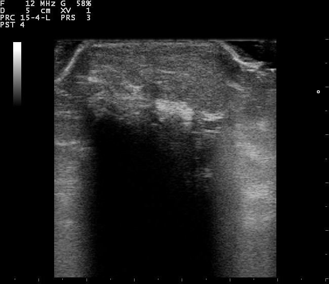
A lipoma on a 12 MHz linear probe, an oval mass lying in the fatty layer with the faint parallel streaks of its fibrous septa. It reads close to the shade of the fat around it, the soft, quiet look a reader names on sight.
A lipoma is the commonest lump of all, a soft ball of fat that sits in the fatty layer where it belongs. It reads close to the shade of the fat around it, oval, its long axis lying parallel to the skin. Thin bright lines run through it, fine streaks of fibrous tissue laid along its length, the feature that names a lipoma on sight. It squashes under the probe and springs back, soft all through. A lipoma can grow large and stay soft all the same, a hand-sized lump in the back that has sat there for years. Size in a soft, streaked, fatty lump is no cause for alarm on its own. The streaks are the signature, finer and more even than the jumble inside a tumour. A reader who finds those parallel lines in a soft oval has the diagnosis in hand, the rest of the scan a confirmation. A lipoma changes little over years, the same soft oval scan after scan, and a reader reads that steadiness as one more mark of the benign. The fat of a lipoma is the patient’s own fat gathered into a ball, doing the body no harm where it sits. A lipoma asks nothing of the patient once it is named, a lump to live with, left where it lies.
A lipoma carries little or no flow. A flick of colour Doppler finds a vessel or two threading the septa, with nothing else to see. A soft, oval, fatty lump with its parallel streaks and its quiet Doppler is a lipoma a reader names with confidence and leaves to the patient. The lump that grows fast, hardens, or fills with flow is the one that earns a second look. A few fatty lumps wear a different coat. An angiolipoma carries more vessels and can ache. A lump with thick, uneven septa, or a touch of flow, is watched more closely than a plain one. The plain lipoma, soft and streaked and quiet, stays the rule.
The lump full of fluid
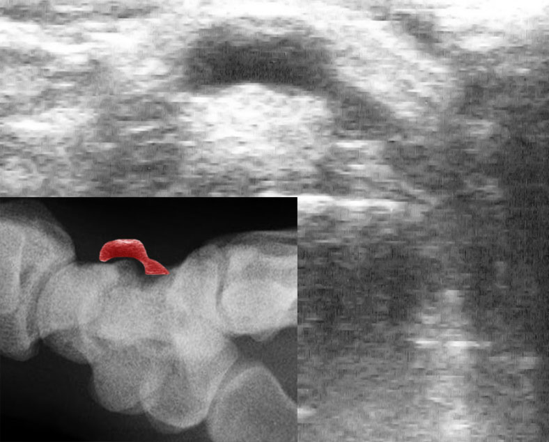
A ganglion cyst on ultrasound, the dark, well-defined pocket of jelly that swells beside a joint. The inset, an X-ray of the wrist with the ganglion marked in red, shows where it sits.
A cyst is a bag of fluid the skin has trapped. The commonest is the epidermoid. It sits round and well-walled in the fat, its dark inside filled with the soft debris of trapped skin that throws a faint, swirling echo. The sound passes through it to brighten the tissue behind. A fine tract may run from the cyst to a little pit on the skin, the punctum that gives its origin away. The debris inside a cyst can pack so tight it mimics a solid lump, a trap for the eye until the press gives it away. A firm push sets the contents swirling inside the wall, the move no solid tumour makes. Pressing is the surest of the tests, the fluid giving way under the probe, the give felt in the hand and seen on the screen at once. An epidermoid that has burst spills its keratin into the fat and flares into a red, painful swelling that mimics an infection. The history of a long-standing lump gone sore points the reader back to the cyst behind it.
A cyst presses differently from a solid lump, giving a little and shifting its contents. Left alone, it sits quiet for years. An inflamed cyst swells, reddens, and lights up with flow around its angry wall, a state that can mimic an abscess. The reader reads the wall and the flow to tell a quiet cyst from an inflamed one.
A ganglion is a cyst of another kind, a pocket of jelly that swells beside a joint or a tendon, commonest at the wrist. It reads as a clean dark pocket, well-walled, often with a thin stalk running back to the joint it came from. The probe traces that stalk and names the lump, sparing a patient the worry of a solid tumour. The jelly inside a ganglion reads a shade brighter than water, a near-solid darkness that can fool a quick look. The thin stalk to the joint is the giveaway the probe follows to settle the call. A sebaceous lump and an epidermoid read much alike on the screen, both walled pockets of trapped skin and oil, and the scan need not split the two to call them benign. A thick, irregular wall on a cyst, or a knot of solid tissue growing from it, is the rare sign that lifts it out of the routine.
An abscess is fluid of a worse kind, a pocket of pus in an angry, painful patch. It reads as a messy dark collection, its contents shifting and swirling under a press, ringed by tissue lit up with the flow of inflammation. The history of pain and heat, read beside the picture, points to an abscess a needle can drain. An abscess answers a history the picture cannot give on its own: days of pain, heat over the skin, a fever. The scan reads the messy collection and the inflamed rim, the story naming it and pointing to the drain.
The signs that warn
A few signs lift a lump out of the harmless crowd.
Depth is the first and the heaviest warning. A lump that sits below the fascia, down among the muscle and deep to the fat, has left the company of the common benign lumps. The deep position alone marks a lump for a closer look. A reader who finds a mass below that sheet treats it with care whatever else it shows. Size adds its weight next. A lump grown past five centimetres has earned a referral, its bulk a warning even when the rest of the picture looks bland. A small lump is no guarantee of safety, a fair share of sarcomas sitting under that size. A large one tips the scale toward concern on its own. The look inside tells the rest. A lump that reads uneven, its body a jumble of light and dark with no clean pattern to it, has none of the order a lipoma or a cyst shows. Flow seals it. Colour Doppler that lights up a mass through and through, vessels running in a chaotic tangle deep in its body, is the mark of a tumour growing its own blood supply. The combination outweighs any one sign. The picture rarely flies every flag at once, a deep lump sometimes reading bland and a shallow one sometimes reading angry. The reader counts the flags a lump shows and lets the count set the level of concern, no one mark deciding it alone. A dark, structureless pool inside an otherwise solid mass is necrosis, the dead centre of a tumour outrunning its own blood, a sign that leans hard toward the malignant. The edges deceive as well: a mass that pushes a clean, smooth margin can still be a sarcoma, the tidy border no promise of safety here the way it reads in other organs. The worst lumps grow. A mass that has swelled noticeably over weeks, by the patient’s own account, carries that growth as a flag in its own right, the speed of it a warning the still picture cannot show. A lump that comes back where one was cut out before is read with double care, a recurrence at an old scar the way some sarcomas announce themselves. A sarcoma favours the thigh and the limbs, a deep mass in a big muscle the classic seat of one. A deep, large, uneven lump packed with disordered flow carries every red flag at once. The reader who sees them sends the patient on without delay. The flags are read together, never one at a time, the whole set the measure of a lump’s risk.
The fascia is the line a reader fixes every lump against. A solid mass below it, down among the muscle, is a mass to image and to biopsy until it is proven harmless. The position settles much of the question before the texture is even read. A reader spends real care pinning a lump’s place against that bright sheet, since the answer to where so often leads to the answer to what. A lump that straddles the fascia, part in the fat and part in the muscle, is read with the same care as one wholly below, a mass that has breached the sheet earning its referral on that alone. A mass fixed to the tissue around it, one that will not slide under the probe, adds its own quiet warning. A lump anchored in place earns a closer read.
Ultrasound points the way; it rarely gives the final name. A lump with every red flag is a lump under suspicion until a biopsy reads its cells. The scan’s job is to sort the lumps that can be left from the ones that must be sent, and to move the worrying one on before it grows. A confident benign call is earned only on the classic looks, never forced onto a mass that does not fit them. The grey picture has a ceiling it cannot pass, two solid lumps reading alike, one holding a benign tumour and the other a sarcoma. No tilt of the probe parts them, the needle the test that reads what the picture only ranks. The referral is no failure of the scan. The scan has done its work the moment it sorts the safe from the doubtful, and handing the doubtful on is the right end of a good read.
The safe path is to refer the doubtful. A mass that fails to read as a clear lipoma or a clear cyst, that sits deep, that has grown, or that carries flow, is handed to a specialist for imaging and a needle. The cost of sending a benign lump for a scan it did not need is small. The cost of calling a sarcoma a lipoma is the one that cannot be undone. Everything that falls short of a clear lipoma or cyst is sent for an MRI and a core biopsy, the two tests that settle a soft-tissue mass. The handheld feeds that pathway, catching the lump early and starting it on its way.
The lumps in between
Not every lump reads as a clean lipoma or a clean cyst. A solid lump that is neither plainly fatty nor plainly fluid sits in the middle ground, neither named nor dismissed. The honest answer there is that the scan cannot say for certain, and an uncertain lump is a lump to send on, never to guess at.
Some lumps in that middle have benign names of their own. A nerve sheath tumour reads as a fusiform mass strung on a nerve, a tail of nerve running from each end. A pilomatrixoma, a hard lump under a child’s skin, carries bright specks of calcium. A reader who knows these names spares a patient a needless fright, an alarming unknown turned into a named, watched lump. The unfamiliar middle-ground lump is read as a reason to refer. A foreign body lodged under the skin reads as a bright fleck with a dark tail, a splinter of wood or glass the probe finds where an X-ray sees nothing, the story of a wound pointing the reader to look for it.
The middle ground is where caution pays. A reader leans toward care there, naming only the lumps that fit a benign pattern cleanly and sending the rest. The scan that errs toward referral in doubt is the scan that never misses the sarcoma hiding as an ordinary lump. The reader who refers a benign lump now and then is the safe one. Trusting the grey too far is how a sarcoma passes as a lipoma. The honest report names what it can and flags what it cannot, a reader writing that a lump reads as a likely lipoma, or that it cannot be called and needs more. The clear lipoma and the clear cyst make up the bulk of a clinic’s day, the handful that fit neither the few that earn the care. The few that fall in the middle are the reason a reader scans carefully, never in haste.
Reading the depth
The layers of the skin read as a stack on the screen: the bright skin on top, the darker fat below it, the bright sheet of fascia, then the muscle beneath. A reader places a lump in that stack at a glance, the layer it sits in half the diagnosis. The probe is held light for this, since a heavy hand presses a soft lump flat and hides the depth itself the reader is trying to read. The lightest touch that keeps the lump in view is the one that keeps its true place. A lump read in two planes is a lump pinned down, its length along the skin and its depth into the layers both taken. The cross view catches a mass dipping toward the fascia that the long view alone would miss. A reader reads a lump’s depth against the layers the body offers at that spot, thin over a shin, thick across a back. A reader sweeps the whole lump in both planes before fixing its depth, the deepest edge of a mass the one that decides its layer, a lump that looks shallow at its centre dipping below the fascia at its margin. The depth is measured from the skin down, the number logged for a later scan to read the lump against its old place in the layers. Depth read right is half the work of a soft-tissue scan done.
On a handheld
The skin and the fat suit a pocket probe as well as any tissue. A lump sits a finger’s breadth under the surface, square in the sharp range of a small linear head. A clinician who feels a lump reads it on the spot, its make-up, its depth, and its flow all there in a two-minute scan, the patient’s question answered in the same visit. The scan asks nothing of the patient but to bare the skin over the lump. No fasting, no dye, no wait, the answer given in the room where the lump is felt. The probe goes to the patient in the clinic chair, no trip to a scanning room asked of someone who came in over a lump. Wherever the lump sits, the skin over it is bared and read where the patient is.
The reach carries the scan to the lump wherever it is found. A worrying mass met in a clinic or a rural ward is marked at once for a referral. The image saves to the phone, the lump measured and its flow filmed, ready for a specialist to read from afar. A lump watched over months is scanned again with the old images in hand, its growth plain across the series. A lump filmed today and again in three months tells its story in the comparison, the old clip beside the new on the one screen, a lump that has changed a lump to send on. A clip of a classic lipoma saved on the phone teaches the next learner the look of the harmless, the soft oval and its parallel streaks there to study away from the patient. The cost of the tool has fallen far enough that a GP surgery or a skin clinic can own the probe that reads its own lumps, the work that once meant a referral to imaging done in the room where the lump is felt. The whole read of a skin lump, from the first feel to the clear answer, rides in a coat pocket the clinician already carries.
Common questions about subcutaneous mass ultrasound
What does a lipoma look like on ultrasound?
A soft, oval lump in the fatty layer, close to the shade of the fat around it, its long axis lying parallel to the skin. Thin bright lines run through it, the fibrous streaks that name it. It squashes under the probe and carries little or no flow on colour Doppler. These signs together let a reader name a lipoma with confidence.
How does ultrasound tell a cyst from a solid lump?
A fluid cyst reads dark inside, the echoes even all through, the sound passing cleanly to brighten the tissue behind. A press sets its contents shifting. A solid lump holds its shape under the press and carries echoes through its body. The press and the look together separate the two in seconds.
What signs make a subcutaneous lump worrying?
A lump that sits deep, below the fascia among the muscle, that has grown past five centimetres, that reads uneven inside, or that lights up with disordered flow. A lump that has grown fast or come back after removal adds to the concern. Any of these sends the lump on for imaging and a biopsy.
Can a small lump still be cancer?
Yes. A fair share of sarcomas measure under five centimetres, so size alone is no guarantee of safety. The depth, the texture, and the flow weigh as much as the size. A small lump that sits deep or reads uneven is sent on the same as a large one.
Can a handheld ultrasound assess a lump under the skin?
Yes. The skin suits it well. A lump sits shallow, within easy reach of a small linear probe. Its make-up, depth, and flow read in a couple of minutes. A clinician reads a lump at the bedside, names the clear lipoma or cyst, and marks the worrying one for a referral in the same visit. A lump answered at the bedside is a worry lifted in the same hour, the patient spared the wait for a scan at a distant department. The skin lump is the kind of question a pocket probe was made to answer.
About the Author
Julien Mercier
Senior R&D Engineer · Medical Ultrasound Transducer Development
Senior R&D Engineer with an M.S. in Applied Physics and over 15 years of experience in medical ultrasound transducer development, specializing in the design verification and performance testing of high-frequency imaging transducers. Currently leading the development and verification of the company’s next-generation high-frequency linear-array transducer, responsible for imaging performance evaluation and reliability analysis in preclinical testing. Brings extensive hands-on experience in piezoelectric element tuning, beamforming parameter optimization, and system-level performance testing.