





































Canine Feline Abdominal FAST AFAST Handheld Veterinary Ultrasound
An abdominal FAST scan checks a hurt animal for free fluid in the belly, the blood a torn organ spills after a trauma. An ultrasound probe runs four quick points across the abdomen, reading each for the dark pocket of fluid that does not belong there. The whole scan takes a couple of minutes at the table, and it can be repeated as often as the animal needs watching.
The one question first
A FAST scan asks one thing before all others. Is there free blood in the belly. An animal hit by a car, kicked, or fallen from a height can tear its spleen or its liver and bleed into the abdomen, the bleed hidden under an unbroken skin. The scan finds that hidden blood in minutes.
The scan is run after a trauma above all, and after any sudden collapse a hidden bleed might explain. A ruptured tumour on the spleen drops a dog weak and pale with no blow at all, the scan reading free fluid whatever opened the vessel.
Free blood pools where the belly is lowest. It gathers in dark pockets between the organs, pushed there by gravity, held back by no wall. The probe goes looking for those pockets at the points they collect.
The answer steers the next hour. Blood found in the belly sends an animal toward a transfusion and a surgeon, the scan sorting the bleeding patient out of a crowd of battered ones long before the bloodwork is back. The speed is the point, an answer won at the table while the animal is still being settled. A team can move on a FAST read in the first minute, the drip started and the blood ordered before a single test has run.
The four points
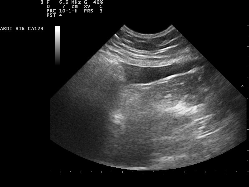
The scan runs a fixed circuit of four spots, the same four every time so none is forgotten. The first sits just behind the breastbone, the probe aimed up under the ribs at the spot where the liver meets the diaphragm. The dark wedge of fluid shows here between the liver lobes, gathering in the angle where the soft liver presses against the muscular dome above it. This one window does more than any of the others. Through the same arch of the diaphragm the probe reads the sac around the heart, a dark rim there meaning a pericardial bleed that can squeeze the heart still. It reads the space around the lungs in the chest beyond, a band of black there meaning fluid pressing the lung. A pericardial bleed, a chest filling with fluid, a belly filling with blood: the three gravest things a blunt trauma does can all be caught from this one spot under the breastbone, which is why the reader lingers here longest and reads it with the closest care of the four.
The second spot sits on the left flank, over the seat of the spleen and the left kidney. Blood gathers in the gutter between the spleen and the body wall, an easy place to read on an animal lying on its right side. The spleen is a common source of the bleed, so this view earns close attention. A small bleed often shows here as a fine dark line hugging the tip of the spleen, easy to miss on a hurried sweep.
The third spot sits low on the midline, over the bladder. Fluid pools behind and around the bladder at the lowest point of the belly, the place a small bleed shows first. A torn bladder leaks its urine here too, a flood that reads much the same as blood until a needle tells them apart. The view is taken with the bladder in sight as a marker, the corners around it read for the dark slivers that pool against the body wall.
The fourth spot sits on the right flank, over the liver and the right kidney. It mirrors the left, catching fluid in the gutter on the other side. Read in turn, the four points sweep the whole lower belly where free fluid runs, the circuit built so a single pass misses nothing.
Each point takes only seconds. The probe is laid, the screen is read for a dark pocket, the finding is noted, and the probe moves on. A practised hand runs all four in under two minutes.
Where the fluid hides
Free fluid obeys gravity. It runs downhill to the lowest part of the belly and pools there, so the spots the scan checks are chosen as the low points an animal makes when it lies on its side. The bladder view sits lowest of all on a small animal, the first place a free pocket gathers.
A full bladder helps the search, its dark pool of urine a landmark the eye finds fast, a thin rim of free fluid showing sharply against it. The reader uses that pool to orient the probe before reading the corners around it.
The position of the animal sets where to look. Gravity fills the down flank on an animal lying on its side, the low spot behind the bladder on one that stands, and the probe is worked into whichever gutter has filled. The reader thinks in terms of where water would run before laying the probe down. An animal that cannot be moved is scanned where it lies, the reader finding the low point of whatever position it has settled into.
What free fluid looks like
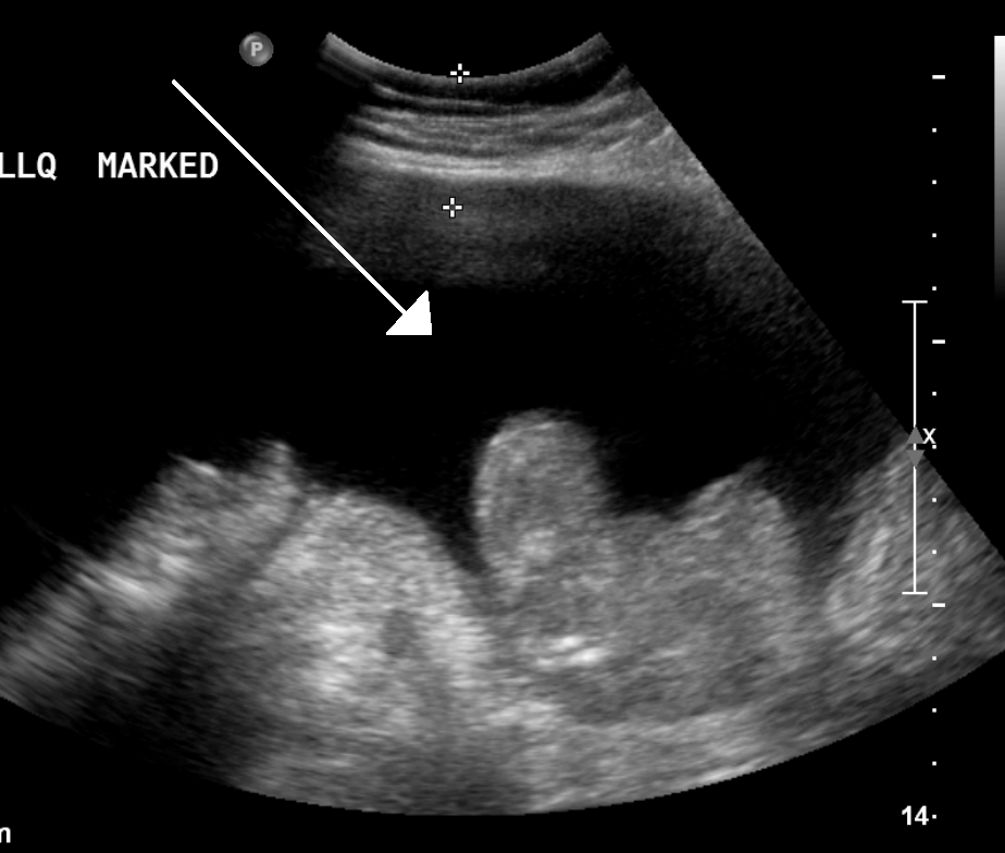
Free fluid reads black on the screen. Blood, urine, and the watery effusions all return little sound, showing as dark pockets against the grey of the organs around them. The darkness alone is the first clue, a patch of black where solid tissue should sit. The screen is turned up bright for the search, the gain pushed so a thin pocket of black stands out against the noise of the tissue around it.
The shape tells it apart from the organs. Free fluid pushes into the angles between organs and takes their edges, drawn out into sharp-cornered triangles and slivers, a shape the rounded bulk of a cyst or a full bladder never takes. A triangle of black wedged into the corner where two organs meet is the classic mark the reader hunts for.
The amount shows in how the fluid spreads. A bleed starts as a thin black line tracing an organ’s edge, grows into a pool that floats the bowel loops, and at its worst rounds out the whole belly. The reader reads the spread at each point and carries the picture from one to the next.
A trained eye learns the traps. A pocket of fat can read dark and mimic fluid, a vessel in cross-section can show as a black circle, a fluid-filled stretch of bowel can pass for free fluid until it is watched moving. The reader rocks the probe and watches a moment to tell a true free pocket from a look-alike that belongs.
The depth of the pocket guides the needle that may follow. A broad pocket of free fluid is a simple target for the tap that names it, the needle dropped under the probe straight into the black, the safest pocket and the surest angle picked before the needle goes near.
Fluid first, the source later
The scan finds the blood before it finds what is bleeding. Stopping the bleed is the surgeon’s work, in its own time.
Putting a number on it
The four points carry a score, one point for each spot that holds free fluid. The total runs from zero, a dry belly, to four, a belly awash with fluid at every point, the abdominal fluid score.
The number stands in for the volume, one more point added for each site the blood reaches. A high score, nearing four, is a large bleed that drops the blood pressure and calls for a transfusion. A torn spleen often stops its own small bleed under watching and support, the low score holding steady or fading on the repeat scan. The reader scores the belly the same way every time, so one animal’s number means the same as the next. The thresholds are read with judgment, a score of three on a small cat carrying more weight than the same three on a large dog, the body size set against the pooled blood.
The score earns its keep in the watching that follows. A number written down at the first scan is the mark every later scan is read against. The score is quick to take, a few seconds added to a scan already run, and it turns a vague sense of how much blood into a figure a team can act on and track.
The number is read with the animal, never alone. A small score on an animal crashing toward shock counts for more than the number suggests, the bleed perhaps sitting where the scan reads it poorly. The gums, the heart rate, and the blood pressure are weighed alongside the score on the screen.
Scanning it again
The scan is built to be repeated. A first FAST on a hurt animal is a single reading of a moving situation, the bleed still running or already clotting as the probe is laid down. One scan cannot say which way it is going.
A second scan an hour later tells the direction. A score that has climbed since the first marks a bleed still running, the belly filling as the team watches, the cue to move toward surgery or a transfusion. A steady number lets the team breathe, the bleed settling on its own and the animal earning more time under watching.
This tracking is the scan’s quiet strength. A bleed small at midnight can be large by two, caught by the second look, the change read off the rising score. An animal stable on paper can be bleeding slowly inside, the serial scan the thing that shows it before the blood pressure falls. The serial scan turns a single uncertain picture into a trend a team can read, the direction of the bleed counting for more than any one frame.
The timing of the next scan is set by how the animal is doing. The shakier the patient, the sooner the next look, the probe picking up a bleed before the bloodwork can show it. The scan costs the animal nothing, run as often as the doubt demands.
What the fluid turns out to be
The scan finds the fluid; naming it takes a needle. A sample drawn from the pocket the scan points to, read by eye and under the microscope, tells blood from urine from bile from the cloudy fluid of an infection. The scan and the sample work as a pair.
Blood in the belly after a trauma is the common find, a torn spleen or liver bleeding into the abdomen. Urine points to a torn bladder or a torn ureter, a different emergency with its own repair. Bile or gut content points to a rupture that turns septic fast. The colour and the cell count of the tapped fluid sort one from the next. The look of the fluid on the screen gives a first guess before the tap, a pocket that swirls and settles hinting at fresh blood, the needle settling the matter for certain.
A fluid that clots in the syringe, or one that matches the blood drawn from a vein, reads as fresh bleeding. The tap closes the question the scan opened, the black pocket on the screen given a name the treatment can follow.
Past the belly
The first FAST point looks through the diaphragm as well. Aimed up under the breastbone, it catches fluid in the sac around the heart and the space around the lungs, a pericardial or pleural effusion read on the same probe that swept the belly. A breathless animal with a quiet heart earns this look at once.
The same approach scans the chest in its own right. A thoracic FAST reads the lung surface for the sliding that a healthy lung shows, the loss of that slide marking air trapped between the lung and the chest wall after a trauma. The probe that finds blood in the belly finds the collapsed lung and the squeezed heart with a few more touches. The chest scan reads in seconds, the lung either sliding clean or held still under trapped air, the answer as quick as the belly’s.
A trauma rarely stays in one cavity. An animal hit hard enough to tear its spleen can crack a rib into a lung or bruise the heart, so the scan that clears the belly carries on to the chest in the same sitting. One probe, one pass, covers the cavities a blow can fill.
When the number climbs
A high score is a call to act. Fluid at every point, a score of four, on an animal gone pale with a racing heart is a large hemorrhage that will not wait, the scan sending the team toward blood products and an open belly without delay. The score of four is the one number that ends the watching and starts the cutting, read off the screen in the time it takes to name it.
The climbing score decides between watching and cutting. A bleed that fills the belly faster than support can hold the animal up is one a surgeon has to reach, the rising number the clearest sign the bleeding has outrun the watching.
What the scan cannot see
The scan has real blind spots an honest reader works around. It finds the fluid, leaving the organ that bled it unnamed. A bellyful of blood tells a vet to act, the torn spleen or liver found later by the surgeon or the full scan, a job the FAST leaves undone.
Small volumes slip past early. A bleed so slight it pools no readable pocket can read as a dry belly on the first scan, which is why a hurt animal earns a second scan before it is ever cleared. The first dry scan rules little out for good.
Some bleeds hide where the scan reads poorly. Blood behind the lining of the back wall, in the retroperitoneal space, pools out of the path the four points sweep. A fat or deep-chested animal, or a belly full of gas, gives a picture harder to read, a small pocket lost in the dimness.
The scan does not weigh the blood. It shows the dark pockets and counts the points, the reader judging the volume by eye and the score. The true measure of the blood lost is read off the animal, the gums and the pulse and the pressure, with the scan as one piece of the call. A scan read in isolation can mislead, a dry belly on a crashing animal sending a team looking the wrong way, which is why the picture is always weighed against the patient.
Where the handheld fits
The handheld scanner brings FAST to the table where the animal lands. A pocket probe laid on a clipped or wetted belly runs the four points in the triage room, the cage, or the back of a van on the way in. The scan travels to the patient, saving a crashing animal the trip to a scanner.
The scan answers the triage question and hands the rest on. Naming the bled organ, grading the damage, and planning the repair is the work of a full abdominal study or a surgeon, the FAST the fast first read that points the way. It is built for the first minutes and hands the full workup on.
A vet learns to run a FAST with far less training than a full scan asks. The four points are fixed, the question is single, and a steady reader picks up the skill in a short course and a run of cases. That low bar is what puts the scan in every emergency room. A nurse or a junior vet can run the circuit and flag a positive belly for the senior to act on, the scan spreading the first read across a whole team.
The animal behind the fluid
A FAST is read on the patient in front of it. A dark pocket on the screen is read differently on a stable animal and on one sliding into shock, the same picture weighed against the pulse and the gums and the story of the injury. A vet who knows what a car does to a dog reads the belly already half-expecting the torn spleen, the scan confirming or clearing the fear in a glance.
The scan guides the hands without replacing them. A vet still feels the belly, reads the heart, and watches the breathing, the FAST adding the one thing the hands cannot reach, the sight of the blood pooling inside.
The minutes that decide
A FAST scan turns the first uncertain minutes after a trauma into a decision a team can act on. It reads the belly for the hidden blood, scores how much, and points the way toward the transfusion, the surgery, or the watchful wait.
The scan is run again and again as the hours pass. Each reading sets the last in motion, the rising or steady score telling whether the bleed has grown or settled, the animal tracked through the night on a probe that costs it nothing.
What the scan buys is time and a direction. It catches the bleed that an intact skin hides, before the blood pressure falls, while there is still room to act. The animal that would once have been watched and lost is the one the scan now sends to the table in time.
It asks little of the animal, a clipped patch and a few minutes on its side. The difference it makes is between a bleed caught early and one found too late, read off four dark corners of a belly in the space of a breath.
The skill spreads wider every year, the hardware shrinking and the training with it, until the first scan on a hurt animal is as routine as a stethoscope on its chest. A FAST is the eye that sees the bleed an intact belly hides, and it has become the first thing a good emergency room reaches for.
Common questions about a FAST scan
What is an abdominal FAST scan in dogs and cats?
It is a quick ultrasound that checks an injured animal for free fluid in the belly, usually blood from a torn organ. The probe reads four set points across the abdomen for the dark pockets where fluid collects. The whole scan takes a couple of minutes and can be repeated as the animal is watched.
What is the abdominal fluid score?
Each of the four points that holds free fluid scores one, adding to a number from zero to four. The total runs from zero, a dry belly, to four, fluid at every point. The score stands in for how much blood has pooled, and a number rising across repeat scans marks a bleed still running.
Can a FAST scan tell where the bleeding is coming from?
Not usually. The scan finds the free blood and grades how much, the torn organ found afterward by a surgeon or a full abdominal study. Naming the source and planning the repair is a separate step that the FAST points toward.
Why is the FAST scan repeated?
One scan is a single look at a moving situation. A bleed small at the first scan can be large an hour later, so the scan is run again to read the direction. A score climbing across repeat scans is a bleed still running, the cue to act before the blood pressure falls.
Can a handheld scanner run a FAST exam?
Yes, and it is built for it. A pocket probe runs the four points at the table where the animal lands, in the triage room or the back of a van. The skill takes far less training than a full scan, which is why the FAST sits in nearly every emergency room.

About the Author
Julien Mercier
Senior R&D Engineer · Medical Ultrasound Transducer Development
Senior R&D Engineer with an M.S. in Applied Physics and over 15 years of experience in medical ultrasound transducer development, specializing in the design verification and performance testing of high-frequency imaging transducers. Currently leading the development and verification of the company’s next-generation high-frequency linear-array transducer, responsible for imaging performance evaluation and reliability analysis in preclinical testing. Brings extensive hands-on experience in piezoelectric element tuning, beamforming parameter optimization, and system-level performance testing.