





































Feline Hypertrophic Cardiomyopathy HCM Ultrasound Handheld Diagnosis
Hypertrophic cardiomyopathy thickens the muscle of a cat’s left ventricle until the wall crowds the pumping chamber smaller. An ultrasound is how the disease is found and measured. The scan reads the thickened wall straight off the screen, puts a number to it, and reads the swollen atrium that warns of what the disease can do next.
What the thickening does
Hypertrophic cardiomyopathy is a disease of the heart muscle itself. The wall of the left ventricle grows abnormally thick, muscle laid down in excess until it crowds the chamber it surrounds. The thickened muscle turns stiff. A stiff chamber fills poorly between beats, the heart that should relax to take in blood no longer able to let go.
The thickening grows from a fault in the muscle itself, often one a cat is born with, the heart laying down too much muscle over the cat’s early years. The fibres of a thickened heart sit in disarray under the microscope, a tangle that helps explain the stiff filling and the stray beats the disease throws.
The disease is the heart trouble cats meet more than any other. Its risks are the gravest a feline heart carries: a clot thrown to the legs, a lung filling with fluid, a heart that stops with no warning at all. The scan exists to find the thickened muscle before any of that arrives. A cat found early can live out a full life with the disease watched and held in check.
Measuring the wall
The diagnosis rests on a number. The scan measures the thickness of the left ventricle wall at the end of filling, the moment the chamber sits at its fullest. A wall thicker than about six millimetres at that moment marks the disease, give or take the fraction that puts a cat in a grey zone. The number sounds small, only a few millimetres of muscle, a single millimetre enough to carry a cat from a healthy wall to a diseased one.
The measure is taken at more than one place. Thickening sits unevenly in many cats, heavy in places on the wall between the ventricles, spread thin in places around the outer free wall. A reader who measures a single spot can miss the disease hiding a centimetre away, so the wall is checked along its length and across the chamber. A handheld probe sweeps the whole wall in one pass, the reader pausing on the thickest stretch to drop the calipers.
A reading is weighed against the cat in front of it. A big-framed Maine Coon carries a thicker wall in health than a small domestic cat, so the number is read with the breed and the body in mind. The scan is frozen on a clean frame, the calipers laid wall to wall, the figure noted for this cat and set against the next scan a year on. One borderline millimetre rarely settles the matter on its own.
The reading is taken at the same point in every heartbeat, the instant the chamber has filled and the wall has thinned to its least. A measurement caught at the wrong moment, mid-squeeze, reads too thick, a false alarm off a healthy heart. The handheld freezes the loop and steps back to the right frame before the calipers go down, a small discipline that keeps a healthy cat from being called sick.
The thickening that is not the disease
A thick wall on the screen does not always mean hypertrophic cardiomyopathy. Several other troubles thicken a cat’s heart muscle, each to be ruled out before the diagnosis is made.
An overactive thyroid drives the muscle to grow. High blood pressure does the same, the heart thickening under the load. Each leaves a wall that reads thick on the scan for a reason that has nothing to do with the primary disease.
A dehydrated cat fools the scan in the other direction. An under-filled chamber draws its walls together until they read falsely thick, a pseudo-thickening that vanishes once the cat is given fluid. A wall measured on a dry cat is read again on a watered one before it is believed. The same caution covers a cat scanned in a panic, a racing heart and a tight chest skewing the picture until the cat has settled.
The order of work follows from this. A cat with a thick wall has its thyroid checked and its blood pressure read, the name of the primary disease earned only by a wall left thick once those causes are excluded. The scan finds the thickening and hands the question of its cause to the rest of the workup.
A treated thyroid or a controlled blood pressure can let a thickened wall settle back toward normal, the kind of thickening that lifts once its cause is removed. The reading that called a thick wall is taken again weeks later, the muscle measured once the cause behind it has been dealt with.
A stiff chamber
The harm of the disease lies upstream of the pump. The thickened chamber can no longer relax to draw blood in between beats. Pressure backs up behind a heart that will not loosen, the build-up driving the disease toward its dangers.
The scan reads this stiffness through its effects more than the wall alone. A chamber that fills under high pressure pushes that pressure back into the atrium behind it. The atrium answers by swelling, the swelling the mark the reader hunts for, the sign that the stiff heart has begun to cost the cat.
The bulge that blocks the flow
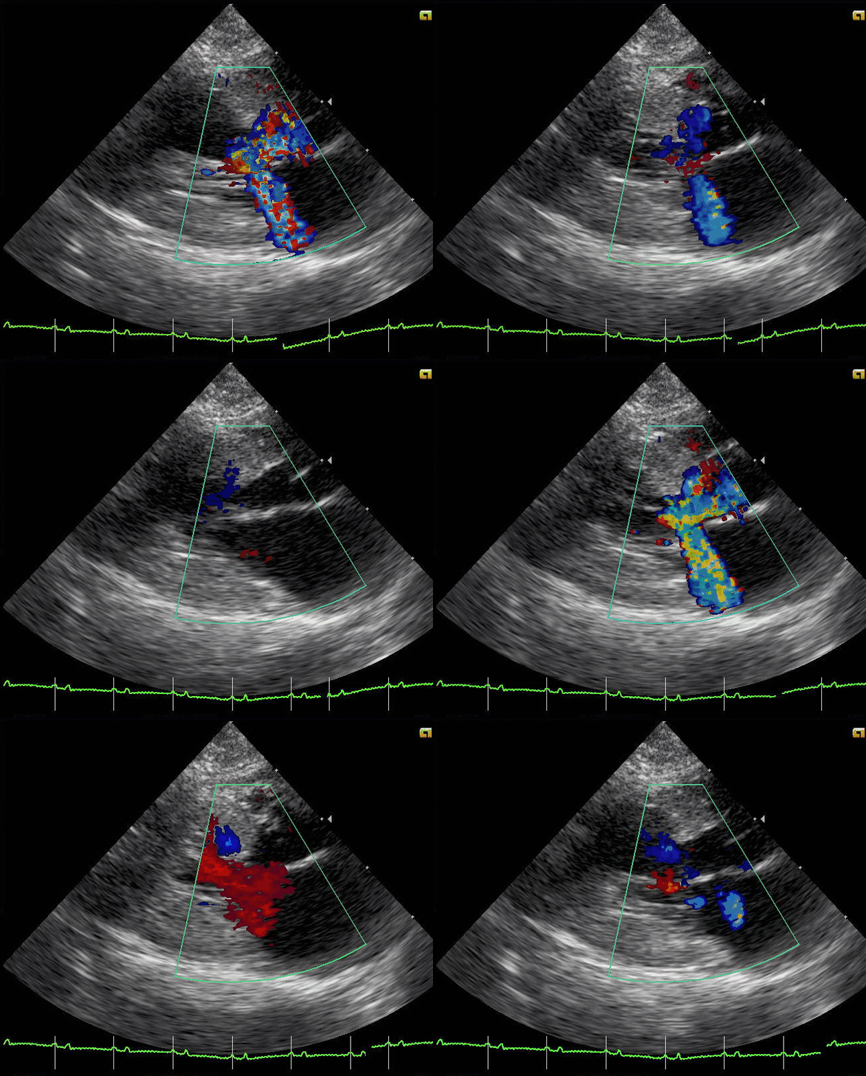
In some cats the thickened wall bulges into the path the blood takes on its way out. The leaflet of the mitral valve gets dragged into that narrowed path with each beat, swinging forward to choke the outflow further. This is the obstructive form of the disease, the choked flow reaching the vet’s stethoscope as a murmur.
The scan shows the bulge and the swinging valve directly. A colour study lays the disturbed flow speeding through the narrowed channel, the moving picture catching the valve leaflet pulled forward in real time. The murmur that sent the cat in is read on the screen for what drives it, a finding the ear could only guess at. The strength of a murmur is a poor guide to the danger behind it, so the scan reads the chambers under the sound to weigh what it means.
The atrium behind it
The left atrium is the chamber the reader watches hardest of all. It sits just behind the stiff ventricle and takes the full brunt of the pressure that backs up out of a heart no longer able to relax, swelling slowly across the months and years the disease runs its course. Its size is read against the aorta lying beside it on the same frozen frame, the ratio of atrium to aorta the single number a heart scan gives that weighs heaviest in what comes next. An atrium swollen well past the aortic root has crossed into the range where the real dangers gather, the range where fluid begins to threaten the lungs and a clot begins to threaten the legs. The reader takes that measurement on every scan and sets it against the one before, since an atrium climbing year on year is the surest sign the disease has started its move toward a crisis.
The blood inside a swollen atrium moves slowly. On the scan that sluggish blood can show as a faint swirling haze, a smoke-like drift that means it is close to standing still. Standing blood is clotting blood, the haze a warning that a clot is forming in the chamber before it has fully formed.
A cat showing that haze sits on the edge of the disease’s cruelest turn. The slow blood in the stretched chamber is the seed of the clot that can break loose and ride out of the heart. The reader who sees the haze flags the cat for the drug that fights the clot at once.
The atrium carries the prognosis. The further it has swelled past the aorta, the higher the cat’s risk, the cat with a greatly swollen atrium steered onto a clot-preventing drug and watched the closest of all. The number that drives that decision is read off the screen in seconds.
The atrium is read on every visit for one more reason. The pull of the disease shows in the trend across scans, the chamber widening from one year to the next, with time still left to act on it.
Found before the crisis
The scan finds the thick wall in a cat that looks perfectly well. That is the only good time to find it.
The clot it can throw
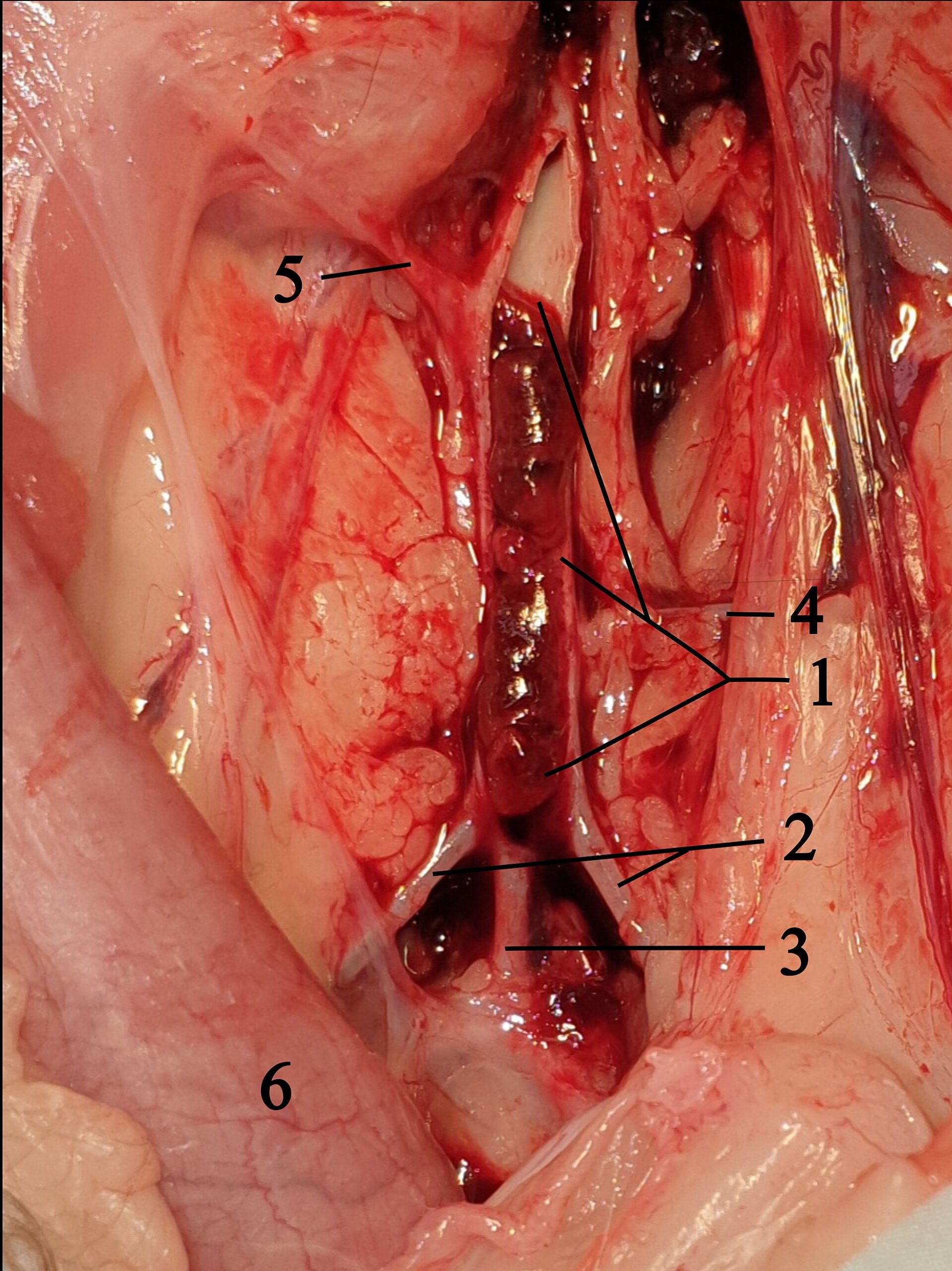
The worst thing this disease does begins in the swollen atrium. Blood pooling in that stretched chamber clots, a piece of it breaking free to ride the bloodstream out of the heart. It travels down the main artery to where the vessel splits to the hind legs and lodges there, a saddle of clot blocking the blood to both legs at once.
The cat goes down in sudden agony. The hind legs turn cold, the pads pale, the pulse in the legs gone, the cat crying out at a pain that comes on in minutes. A vet who knows the disease recognises it on sight, the back half of the cat struck useless in minutes. The sight is unmistakable once seen, sending an owner who knows the breed’s risk racing the cat in, the speed of treatment counting for everything.
The scan ties the catastrophe back to its source. A heart scan on a struck cat reads the thick wall and the swollen atrium that bred the clot, sometimes the clot itself still sitting in the chamber. The diagnosis that took weeks of guessing in another age is made at the table in minutes. A clot lodged at the split of the artery is read on the scan as a still grey plug where blood should be flowing, the cause of the cat’s agony shown plainly on the screen.
Finding the disease before this day is the whole reason the at-risk cat is scanned. A clot that has already lodged is a hard thing to undo, the outcome often grim even with every effort. A swollen atrium caught on a screening scan, long before any clot, is a cat put on a drug that thins the odds of the clot ever forming. The scan turns a sudden catastrophe into a risk that can be managed ahead of time. The drug pushes the clot further off, the difference often deciding how long a cat keeps its legs under it. The earlier it is started, the better it holds the danger back.
When the lungs flood
The other crisis fills the lungs. Pressure backing up from the stiff heart pushes fluid out of the blood into the air spaces, bringing the cat in panting, its mouth open, drowning slowly on its own fluid. A cat in that state is an emergency that counts its time in hours. The cat hides the early breathlessness well, keeping still and quiet, until the lungs are wet enough to force the open-mouthed breathing that brings it in at last.
The scan reads the flooded lung and the failing heart in one sitting. Bright lines rising from the lung surface show the fluid, the swollen atrium behind them naming the heart as its source. The picture points the treatment in minutes. A drug to pull the fluid off buys the cat its breath back, the heart brought under control alongside. A scan repeated once the fluid clears reads the heart on its own terms, the wall and the atrium measured cleanly for the long plan.
A flooded cat is scanned where it sits, the probe laid on the side of a cat that cannot lie down to breathe. The scan is kept short, the few measurements that matter taken fast, the full study left for once the cat can breathe again. A scan that confirms the heart as the source of the fluid in a minute or two spares a struggling cat the longer handling it could not survive.
The silent disease
Hypertrophic cardiomyopathy hides better than almost any feline illness. A cat can carry a badly thickened heart through years of eating, purring, and sleeping in the sun as though nothing is wrong. The disease asks nothing of the cat until the day it asks everything.
Many cats give no warning a careful owner could catch. The first sign is often the crisis itself, the clot already in the legs or the chest already flooded, sometimes a cat found dead with no sign at all the day before. A heart that hid its disease for years declares it all at once. The cruelty of the disease is that the first warning and the crisis so often arrive on the same day, which is the case the screening scan is built to prevent.
A vet sometimes catches a clue before that day. A soft murmur, an odd gallop sound, a skipped beat under the stethoscope on a routine visit: any of these sends a cat for the scan that reads the heart behind the sound. The clue is easy to miss, which is why the scan is what settles the question the stethoscope can only raise. Many cats with the disease have no murmur at all, the heart hiding behind a normal-sounding chest, which leaves the scan as the one sure way to find them.
Screening the breeds at risk
A few breeds carry this disease far more than the general run of cats. The Maine Coon and the Ragdoll head the list, each with a known fault in a heart-muscle gene passed down their lines. A scan run on a well cat of these breeds is screening, the search for a thick wall in a heart that has shown nothing yet. The risk runs in the bloodline, handed down through a line, so a pedigree carries as much weight here as anything on the screen.
The screening starts with a swab. A genetic test reads whether a Maine Coon or a Ragdoll carries the known mutation, telling a breeder and an owner the risk a cat was born with. A cat that carries the fault is scanned from young adulthood and rescanned each year, the wall and the atrium measured and tracked through the years the cat stays well. A clean scan at one age says nothing certain about the next, so the screening is repeated for the life of an at-risk cat.
A blood test runs alongside in many clinics. A marker the strained heart releases rises when the muscle is under strain, a high reading on a screened cat pointing toward a heart that needs the full scan. The test is a quick way to sort the cats that need a closer look from the many that do not, a first filter ahead of the probe. A normal result on the blood test holds only for the day it is taken, the screening still due again on its yearly round.
What the stage changes
The findings set a cat’s stage, the stage steering the care. The atrium carries the heaviest of it. As long as it stays close to normal, the cat is watched and rescanned, with little else done. The swelling that carries it past the safe mark lifts the cat into the band where a clot-preventing drug earns its place. The stage is not fixed for life. A cat moves up a band when its scans change, the care kept in step with the heart.
That drug is the heart of the matter for the higher-risk cat. A medicine that makes the blood slower to clot, started once the atrium has swelled, lowers the odds of the clot that paralyses the legs. The evidence behind it is why the atrium is measured with such care, since the measurement is what opens the door to the drug. A trial in cats found the drug cut the odds of a second clot in those that had already thrown one, the reason it is reached for the moment the atrium crosses the mark. No drug yet stops the wall from thickening in the first place, so the work falls on catching the disease early and guarding against its dangers.
A cat that has already reached a crisis is managed past it and held there. Fluid is pulled off a flooded chest, the heart rate is steadied, the clot risk is treated hard. The scan tracks the heart through all of it, reading whether the atrium settles or swells further over the months that follow.
The stage is written into the cat’s record at every visit, a line others can read and build on. A vet picking the cat up later sees at a glance where the disease has reached and what the last plan was. The record turns a single scan into a story the care can follow over the cat’s life.
How the handheld fits
The handheld scanner brings this work into the consulting room. A vet lays a pocket probe on a cat’s chest, measures the wall against the six-millimetre mark, reads the atrium against the aorta, and sorts the cat that needs a specialist from the cat that can be watched. The whole check takes a few quiet minutes on the table. A cat tolerates a pocket probe laid gently on a clipped chest far better than a long restraint, so the focused scan suits the feline patient that resents being held.
The pocket scan settles the urgent questions and refers the fine ones. Grading the disease in full, timing the drugs to the stage, and weighing the murmur with every measurement is the work of a complete study in a cardiologist’s hands. The pocket unit goes where the cat is, the consult room, the shelter, the cattery, well ahead of any trip to the referral clinic. The handheld finds the thick wall and the swollen atrium that send a cat toward that study, the findings hardest to come by in a cat that still seems well.
The cat behind the wall
The breed and the picture carry weight in the end. A young Maine Coon with a thick wall and nothing else to explain it points hard at the inherited disease. The breed and the story shift where the same thick wall points. A cat is read whole, the heart against the body it beats in, the wall against the cat’s own size and story.
The window the scan opens
Hypertrophic cardiomyopathy gives a cat years of quiet before it strikes. The years of silence the disease grants are exactly what a screening scan turns into time to act.
It reads the thick wall and the swollen atrium on a cat that still seems well, and it hands the vet the chance to guard against the clot and the flood before either arrives. Those are years a cat keeps, bought by a scan that looked while there was still nothing to feel.
The whole of it costs the cat a few minutes on its side and a clipped patch of fur. The difference it makes is between a heart found in time and a cat lost without warning.
Common questions about feline HCM
How is hypertrophic cardiomyopathy diagnosed in a cat?
An ultrasound of the heart is the test that confirms it. The scan measures the left ventricle wall at the end of filling, a wall thicker than about six millimetres marking the disease once other causes are ruled out. The scan also measures the left atrium, since a swollen atrium is the sign the disease has begun to threaten the cat.
Why does the scan measure the left atrium?
The atrium is measured because its size tells how dangerous the disease has become. A swollen atrium is where the clot forms that paralyses the legs and the sign that fluid may soon back into the lungs. Measuring it on every scan is how the risk is tracked over time.
Can a cat have hypertrophic cardiomyopathy with no symptoms?
Yes, and many do for a long time. A cat can carry a badly thickened heart for years and look and act entirely well the whole time. The first outward sign is often a crisis, a clot in the legs or a flooded chest, which is why at-risk cats are scanned before they show anything.
Which cats should be screened for the disease?
The breeds known to carry it, the Maine Coon and the Ragdoll above all, are screened with a genetic test and a yearly scan from young adulthood. A cat of any breed with a heart murmur, a gallop sound, or a clot is scanned to read the heart behind the finding.
Can a handheld scanner diagnose feline hypertrophic cardiomyopathy?
Yes, for the core questions. A vet measures the wall thickness and the atrium size on a pocket unit and picks out the heart that has thickened. Grading the disease in full and timing the treatment is the work of a complete echo with a cardiologist, the step the handheld points the cat toward.

About the Author
Julien Mercier
Senior R&D Engineer · Medical Ultrasound Transducer Development
Senior R&D Engineer with an M.S. in Applied Physics and over 15 years of experience in medical ultrasound transducer development, specializing in the design verification and performance testing of high-frequency imaging transducers. Currently leading the development and verification of the company’s next-generation high-frequency linear-array transducer, responsible for imaging performance evaluation and reliability analysis in preclinical testing. Brings extensive hands-on experience in piezoelectric element tuning, beamforming parameter optimization, and system-level performance testing.