





































Superficial imaging
What You Can See with High Frequency Linear Probe in Superficial Scan
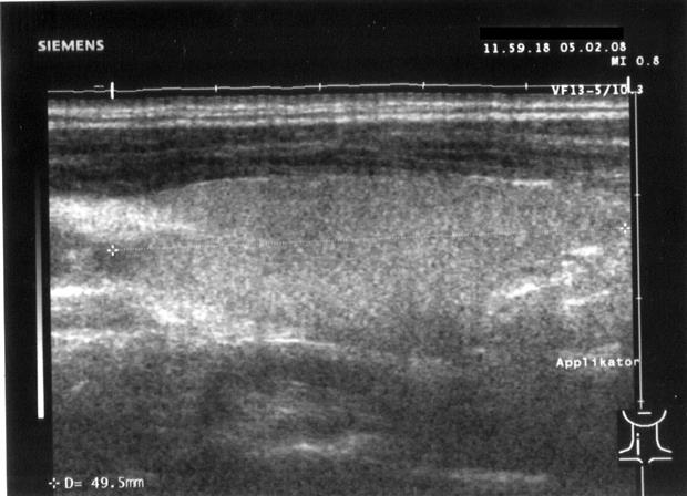
A high-frequency linear probe reads the body’s surface in fine detail. It runs at 7.5 to 18 megahertz across a flat wide face, a band that draws crisp structure in the first few centimetres and fades beyond them. The high frequency buys fine resolution and reaches only shallow, which is why the probe is built for what lies near the surface. Inside that range sit the vessels and nerves, the tendons and glands, the small structures a clinician reads at the surface, each shown with a clarity a lower-frequency probe cannot reach.
Why the high frequency stays shallow
Sound at a high frequency carries fine detail and fades fast. A short wavelength resolves two close structures that a long one blurs into one, which is why a 15-megahertz beam separates a nerve from the vessel beside it where a 3-megahertz beam would merge them. The same short wavelength is absorbed quickly by tissue, so the beam runs out of energy within four or five centimetres. The probe reaches no deeper, and it does not need to, since the structures it is built for sit in that shallow band.
Resolution is the payoff for staying shallow. A high-frequency linear probe resolves detail down toward a tenth of a millimetre near the surface, fine enough to read the layers of a vessel wall or the fascicles inside a nerve. That figure is the reason the probe is the tool of choice in vascular, musculoskeletal, and small-parts work, where the question turns on detail a millimetre across or less. The fineness is not even in every direction. The detail runs sharpest in the beam’s own direction and a little softer to the sides, which is why a reader turns the probe to lay a fine feature along that sharper line.
The vessels just under the skin
The vessels of the neck, the limbs, and the surface are the linear probe’s daily work. The carotid artery shows its wall in layers, the intima-media thickness read in fractions of a millimetre as an early mark of vascular disease. Any plaque is seen for how far it narrows the channel and whether its surface is smooth or ragged. The probe maps a vein before a cannula goes in, the needle watched into the vessel in real time, and it runs the two-point compression that reads a deep vein thrombosis in the leg.
The same face reads flow as well as structure. Colour Doppler laid over the grey image shows blood moving toward the probe in one colour and away in another, so a vessel lights up and a leaking or narrowed segment shows its disturbed flow. A pulsed-wave trace at one chosen depth reads the speed of that flow, the figure a vascular study turns on.
The smaller surface vessels read just as clearly. A forearm vein is mapped before a difficult cannula. An arteriovenous fistula in a dialysis patient is checked for its flow and for any narrowing along it. A temporal artery reads on the same shallow setting, its wall thickened in an arteritis, the probe pointing a biopsy to the worst segment. A small vessel in a finger or a skin flap shows its flow where a coarser probe finds nothing. Each sits in the shallow band the high frequency owns, held in the detail a deeper probe would lose.
The wall itself tells more than its thickness. A dissection waves a flap inside the lumen, and an aneurysm balloons the wall past its normal calibre. A clot fills the channel with material that reads soft and dark when fresh and brightens as it ages, the probe watching whether flow has cut a path back through it. Each of these reads in the fine grain the high frequency lays on a vessel a centimetre under the skin.
Nerves, tendons, and muscle
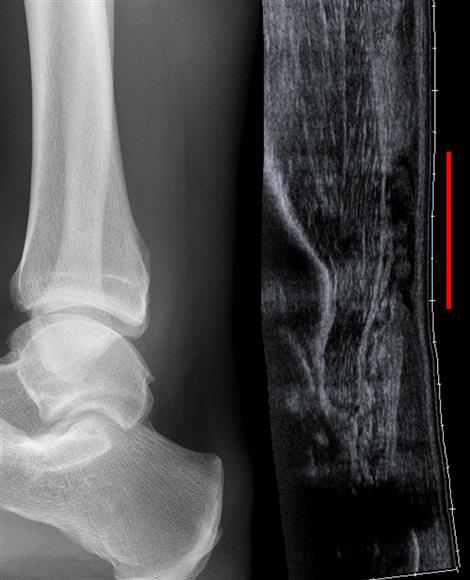
The musculoskeletal structures of the limbs are where the high-frequency face shows its full reach. A peripheral nerve reads as a bundle of dark fascicles wrapped in a brighter sheath, the honeycomb pattern a clinician learns to recognise, and the probe follows that nerve along its course to find where it is trapped or swollen. A regional anaesthesia block leans on this picture, the needle guided to the nerve under live view so the drug lands where it should and not in the vessel alongside. A tendon shows its tight parallel fibres, the fibrillar pattern that brightens when the beam sits square to it, and a tear reads as a gap or a dark swelling in that orderly grain. The probe watches the tendon move as the joint flexes, a dynamic look no static image gives, so a tear that hides at rest opens up under motion. Muscle reads as darker tissue veined with bright connective lines, and a strain or a bleed shows as a disruption of that architecture. The bursa over a joint, normally a thin film, shows its swelling when inflamed. A foreign body lodged in soft tissue, a splinter of wood or glass that an X-ray misses, throws a bright reflection the linear probe catches and a needle can then be guided to. In all of this work the structures sit a millimetre or two apart and move as the limb moves, so only a high-frequency face read in real time tells one from the next. The same scan that finds a swollen nerve maps the vessel beside it, so the needle that follows reaches the nerve and stays off the vessel. The picture holds the fine grain a fixed low-frequency probe would smear into a single grey band. The depth at which all this reads is itself a clue. A structure’s distance under the skin, measured off the same image, tells a swollen node by where it sits and fixes the depth of a foreign body before a needle goes for it. The high frequency that draws the fine grain holds the depth scale finely too, so the reading carries not only what a structure is but how far down it lies, the figure a guided needle is set by.
The thyroid and the lymph nodes
The thyroid sits a centimetre or two under the skin of the neck, squarely in the linear probe’s range. The gland reads as an even mid-grey tissue, and a nodule stands out against it. The probe measures the nodule, reads whether it is solid or fluid-filled, and looks for the fine specks of calcium and the irregular margins that mark a nodule worth sampling. A lymph node nearby shows its shape and its internal structure, a healthy node carrying a bright fatty centre at its core.
That detail feeds a structured read. A clinician scores a thyroid nodule on its features, the composition, the shape, the margin, the bright specks, each read off the high-frequency image, and the score steers whether a needle goes in. The linear probe gives the picture those features are read from, in the seconds a bedside scan allows.
The gland’s blood flow reads on the same scan. Colour Doppler over the thyroid shows a nodule’s vascularity and lights up the raised flow of an inflamed or overactive gland. A diffuse disease, a thyroiditis or a Graves gland, reads in the texture and the flow across the whole gland, its change spread everywhere at once, the high-frequency picture holding the fine detail a coarser probe would smear over.
The salivary glands sit at the surface too. The parotid and the submandibular gland read as even tissue on the high-frequency face, a stone in a duct showing as a bright speck with a shadow behind it, a mass standing out against the gland around it.
An enlarged parathyroid gland behind the thyroid comes into view on the same scan, the small structure the high frequency resolves where a lower-frequency probe would pass over it.
All within a few centimetres of the skin
All of these structures sit within a few centimetres of the surface, the shallow band where the high frequency reads at its finest.
Skin, fluid, and the small structures
The layers of the skin itself read on the highest-frequency probes, the dermis and the tissue under it separated cleanly enough to measure a lesion’s depth before it is removed. A collection of fluid under the skin, an abscess or a cyst, shows as a dark pocket the probe sizes and a needle drains under guidance. A swollen joint at the surface, a wrist or a finger, shows its fluid and its lining. The probe reads the small structures a deeper scan would pass over, each sitting in the shallow band the high frequency was built to serve.
The lymphatics and the surface’s small structures read here as well. A swollen lymph node in the groin or the axilla, sitting shallow, shows its shape and whether its fatty centre survives. A ganglion at a wrist shows as a clear fluid pocket a needle can be guided into. A haematoma or a seroma under a surgical wound shows its size and whether it is settling, checked at the bedside without opening the dressing. Each sits in the first centimetre or two, too shallow and too small for a deeper probe to read well.
Reading the surface well
The high-frequency picture carries a quirk the reader learns to handle. A tendon or a nerve reflects the beam brightly only when the beam sits square to its fibres, an effect called anisotropy. At an angle the same structure darkens, so a healthy tendon read off-square can mimic the dark gap of a tear. The reader rocks the probe to hold the beam square, reading the structure at the angle that fills it with signal. The rocking doubles as a test of what a structure is, since a fibred tendon or nerve shifts in brightness as the angle changes, a swing that names it as fibred.
The machine settings are tuned to the shallow field. The focal zone, the depth where the beam is narrowest and the detail sharpest, is dropped onto the structure of interest a centimetre or two down. The overall depth is set shallow, so the structure fills the screen rather than floating in a deep black field. The frequency is lifted toward the top of the probe’s band for the finest grain on a shallow target, then eased a step lower when a little more reach is needed. The gain is set so the tissue reads mid-grey and the structures stand against it. Each control is matched to the structure in front of the probe, the picture only as good as the setup behind it.
A few quirks of the picture read as information once known. A fluid pocket brightens the tissue behind it, the posterior enhancement that marks a cyst out from a solid lump. A strong reflector throws repeating lines beneath it, the reverberation a needle tip or a bright interface gives. The reader takes these as clues, each one pointing to what sits under the probe.
The high frequency does its work at the surface
A vascular service lives on this probe. It reads the carotid wall and the leg veins, maps a vessel before a line goes in, and follows the flow with Doppler, all inside the shallow band the high frequency owns.
A musculoskeletal clinic reads tendons, nerves, and small joints with it. It watches each structure move as the limb flexes, the dynamic look a still image cannot give.
A regional anaesthesia team guides every block under its picture, the needle steered to the nerve and clear of the vessel. A surgeon checks a lump or drains a collection with it at the bedside.
The handheld form puts that surface detail in a pocket. A clinician carries the high-frequency face to the bedside, the clinic room, the field, reading the surface structures where a cart could never follow.
The one thing the face does not do is reach deep. A question sitting below its few centimetres, a deep organ or a great vessel, passes to a lower-frequency probe, so the linear face travels alongside a convex one in a kit that reads the whole body.
Common questions about high-frequency superficial scanning
What frequency does a superficial linear probe use?
Roughly 7.5 to 18 megahertz, with some skin and small-parts probes reaching higher. The high frequency draws fine detail in the first few centimetres and fades beyond them.
How deep can a high-frequency linear probe see?
About four or five centimetres. The short wavelength that gives the fine detail is absorbed quickly by tissue, so the beam runs out of energy at shallow depth, which is enough for the structures the probe is built for.
What structures does it read best?
Vessels just under the skin, peripheral nerves, tendons and muscle, the thyroid and lymph nodes, skin layers, superficial fluid collections, and foreign bodies in soft tissue, all sitting in the shallow band.
Why is it used for nerve blocks?
It shows a peripheral nerve as a honeycomb of fascicles and the vessel beside it, so the needle is guided to the nerve under live view and kept out of the vessel, the drug landing where it should.
How does it help with the thyroid?
The gland sits a centimetre or two under the skin, in range. The probe reads a nodule’s composition, shape, margin, and any calcium specks, the features a structured score uses to steer whether a needle goes in.
Can it find a foreign body an X-ray missed?
Often, yes. A splinter of wood or glass in soft tissue throws a bright reflection the high-frequency probe catches even when it does not show on an X-ray, and a needle can then be guided to it.

About the Author
Julien Mercier
Senior R&D Engineer · Medical Ultrasound Transducer Development
Senior R&D Engineer with an M.S. in Applied Physics and over 15 years of experience in medical ultrasound transducer development, specializing in the design verification and performance testing of high-frequency imaging transducers. Currently leading the development and verification of the company’s next-generation high-frequency linear-array transducer, responsible for imaging performance evaluation and reliability analysis in preclinical testing. Brings extensive hands-on experience in piezoelectric element tuning, beamforming parameter optimization, and system-level performance testing.