Sonography Portable USG Equipment Evolution From Cart to Handheld Wireless
For its first four decades, a sonography machine was furniture. It stood on wheels. It drew power from a wall socket. It lived in a room a patient had to be brought to. The image it made was good, often excellent. The price of that image was a heavy console, a rack of electronics, a probe tethered by a thick cable. The story of portable ultrasound is the story of how that furniture shrank, step by deliberate step, until the part that mattered fit in a hand. The screen became a phone someone already owned.
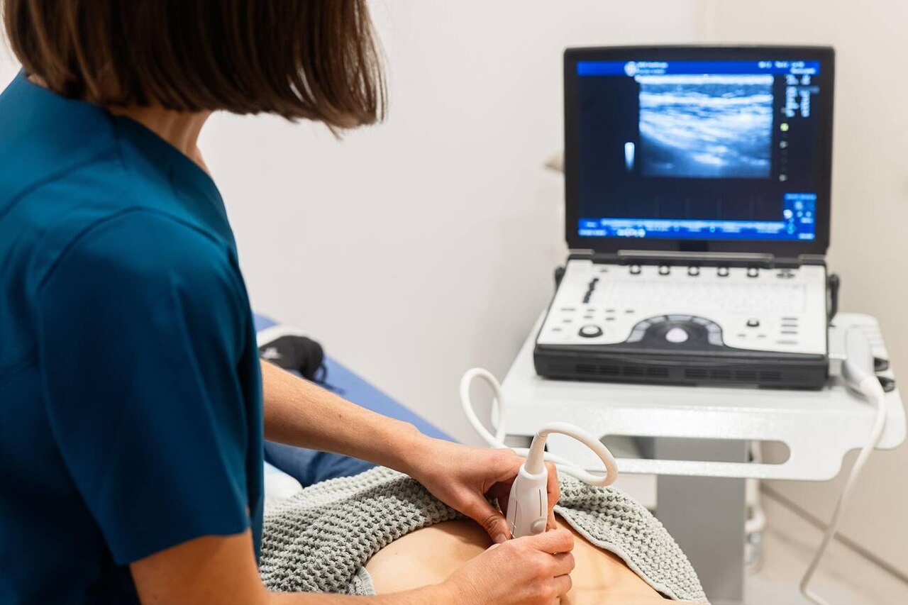
A portable ultrasound machine wheeled to the bedside, its picture on the unit beside the clinician. (Goleisureintl, CC BY 4.0, via Wikimedia Commons)
The change was neither sudden nor accidental.
Each stage solved a specific limit of the stage before it. Each carried a cost the next stage had to answer. Read the sequence in order, and the handheld wireless probe makes sense. Its strengths come from somewhere. Its compromises were inherited from the cart it replaced.
The console era and what held it in place
Early diagnostic systems were built around a large beamformer. That bank of electronics times the firing of each transducer element. It assembles the returning echoes into a picture. It ran hot. It drew heavy current. It needed space. The transducer in the clinician’s hand was almost a passive antenna. The intelligence sat in the cabinet across the room. A cable of dozens of fragile coaxial lines joined the two. Moving the machine meant moving the whole cabinet. The cable length set a hard limit on how far the probe could roam.
This arrangement had a logic. Image quality depended on processing power. Processing power meant transistors. Transistors meant heat, bulk. A bigger box made a better picture. The clinic accepted the trade. The alternative was no bedside imaging at all. A shared machine wheeled between rooms was a reasonable compromise for a hospital that could schedule around it.
The limit it imposed was access. A scanner that lives in a room serves the patients who reach that room, on the schedule the room allows. The sick patient in a corridor waited. The casualty in a field clinic waited. The animal in a barn, the newborn in an incubator down the hall: each waited, or went unscanned. The equipment could not follow the question to where it arose.
The laptop turn
The first real loosening came when the beamformer shrank into a laptop-sized chassis. Makers moved much of the timing and image assembly onto integrated circuits built for the task. The console collapsed into a clamshell a clinician could carry in one arm. The probe still plugged in by cable. The device still wanted a flat surface, a power supply for long sessions. The machine had stopped being a room fixture. It had become a piece of luggage.
That mattered for emergency work in a way numbers do not capture. A laptop scanner could ride in an ambulance. It could sit on a crash cart. It could travel between a clinic’s three rooms without a booking. The picture sat a step below the flagship console. For a focused question it was already good enough to change a decision. The portable had found its purpose. It would never rival the cabinet on fine detail. It put a usable image where the cabinet could never go.
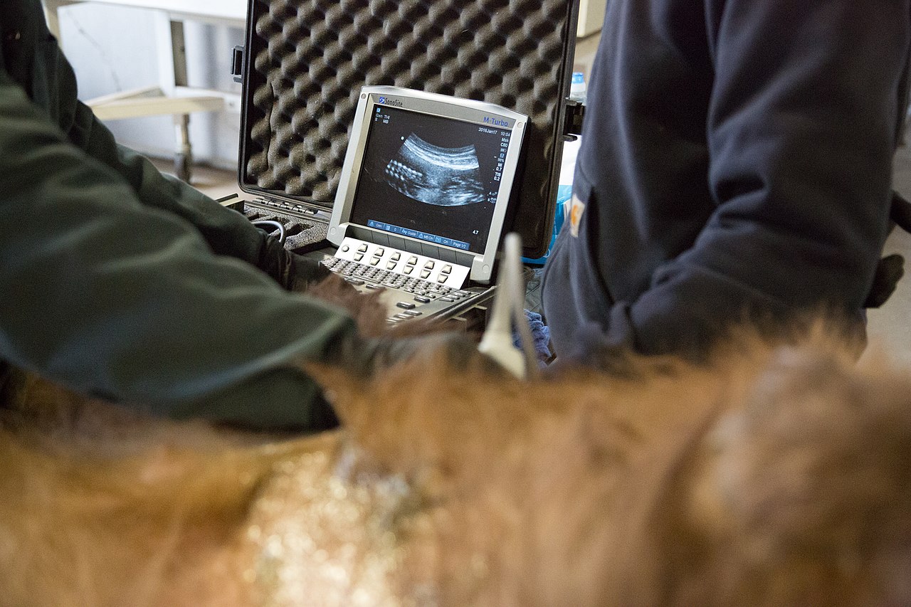
A portable laptop-class scanner carried into the field, far from any imaging suite. (California Department of Fish and Wildlife from Sacrame, CC BY 2.0, via Wikimedia Commons)
The laptop form taught the field a lesson it would lean on later. A clinician did not need every feature of a radiology suite to answer a bedside question. Strip the interface to the controls a focused exam uses, and the device grows faster to learn, faster to wield. That simplification set the direction for everything that followed.
The probe swallows the machine
The decisive leap miniaturized the beamformer until it fit inside the transducer housing itself. The electronics that form an image could now sit in the head a clinician already held. The separate box disappeared. The probe became the machine. A passive antenna on a cable turned into a complete scanner. It needed only power, a screen. Both came from a handset the clinician carried for other reasons.
Here the equipment crossed a threshold. A device that fits in a coat pocket, runs on a small battery, shows its picture on a phone, is no longer luggage. It is part of the clinician’s everyday kit, as ready to hand as a stethoscope. The exam could finally follow the patient anywhere. The scanner went wherever the pocket went. The question no longer waited for the room.
Shrinking the beamformer into the probe forced engineers to confront a problem the cabinet had solved with brute space. The way they answered it shapes how a handheld behaves in the hand today. Heat is the enemy of dense electronics. A beamformer packed into a sealed housing has nowhere obvious to shed the warmth it makes. The cart vented heat through fans, through open volume. The handheld has neither. So the design splits its effort between the picture and its own survival. It runs a power budget that trades continuous scanning against thermal headroom. It routes warmth into the casing through conductive paths. Some designs add a phase-change coating that soaks up a burst of heat, then releases it slowly. The device limits how long the highest-output modes run before it steps itself down to protect the array. Safety standards cap how warm the part of the probe that touches skin may grow. That ceiling, set for the patient, often decides the limit on a long scan. To stay beneath it the wand watches its own surface. It pulls back quietly: a lower frame rate, a narrower aperture, less transmit power. Each is a small subtraction from the picture, bought for another minute of safe scanning. A clinician who scans in short bursts rarely meets these limits. A clinician who runs a long continuous study does. The device’s behavior at that edge is the truest tell. How gracefully it derates. How warm the housing grows. How fast it recovers. That edge separates a well-built handheld from a cheap one far better than any headline number on the box. The cart never had to make these choices. A handheld is not a small cart. It is a different machine, answering a different set of constraints.
The wireless link
Cutting the cable was the last tether to fall. A wired handheld still bound the probe to its screen by a physical line. A wireless one sends its image over a radio link to a phone, a tablet, a laptop. The scanning hand is free of any leash. The convenience is obvious. The engineering behind it is less so. It explains the few quirks a wireless probe still carries.
A live ultrasound feed is a demanding stream. The probe compresses each frame. It pushes the frame across a wireless channel without a stutter the eye would catch. It keeps the lag between moving the probe and seeing the picture short, so hand and image feel joined. A dropped frame at the wrong moment hides the finding the clinician is hunting. So a wireless probe leans on a dedicated dual-band link, away from the crowded household channel. It manages its battery against the drain of scanning plus transmitting. It accepts a small, deliberate delay in exchange for a steady picture. These compromises buy the freedom of no cable. A buyer who understands them reads a spec sheet differently.
Freedom from the cable changed the ergonomics of the exam as much as its reach.
A clinician guiding a needle holds the probe in one hand, the needle in the other, the image floating on a screen propped where both can see it. A sonographer scanning a large animal stands clear of a kick, still watching the picture. The cable was a small thing in a clinic room. It had quietly shaped how every scan was performed. Removing it opened postures the tethered machine ruled out.
What “portable” and “USG” came to mean
The vocabulary trailed the hardware. “Portable” once described a console small enough to wheel without two people. Later it meant a laptop a clinician carried. Now it points to a probe in a pocket. “USG,” the shorthand for ultrasonography common in many regions, stayed constant. The equipment under the word changed past recognition. A buyer reading older listings has to translate. A device sold as portable in one decade outweighs a clinic’s whole handheld kit in the next.
The drift matters for procurement.
A search for portable sonography equipment returns machines from every layer of this history, priced for different work. Sorting them means asking what the word meant when the listing was written. Match that to the question the clinic needs answered. A label that has slid under its own feet for forty years cannot be trusted on its own.
What the picture shows
Shrinking the machine put real pressure on image quality. Pretending otherwise sets a buyer up to be let down. A handheld trades some penetration, some dynamic range, some of the deepest specialist modes, for the freedom of its size. The question a clinic should ask is whether the picture answers its question. Whether it equals a flagship console on a chart matters less.
For a focused exam the answer is steadily yes. A narrow question forgives a great deal. Finding free fluid. Judging whether a heart squeezes. Reading the artifacts a lung throws back. Guiding a needle into a vessel. Each reads from features a pocket scanner renders cleanly. The handheld struggles where the question turns subtle, on fine tissue texture deep in a large body, or a measurement that demands the steadiest possible image. A clinic that knows the edge plans around it. Matching the device to the depth a study needs is the single judgment that decides whether a portable purchase serves the work. That judgment rests on understanding what the small form gave up to become small.
Storage, sharing, the record
An image that lives on a phone has to leave it. The exam still belongs in the patient’s record. It still needs to reach a colleague for a second read. It still has to satisfy the rules that govern medical images. The handheld generation answered this by leaning on the networks the clinic already used. Studies travel to the central archive over the standard medical-imaging protocol. They sit where the institution keeps the rest of its records.
The shift is quiet but real.
A scanner that captures, stores, transmits from a pocket folds the once-separate steps of imaging and filing into one motion. A clinic adopting handhelds inherits both the convenience of that motion and the duty of getting it right.
Where the equipment stands now
The handheld wireless probe of today is the sum of these answered limits. It carries a beamformer that once filled a cabinet. It sheds heat through clever materials. It talks to a consumer handset over a private radio link. It slips into a pocket between patients. It does not match a flagship console on the finest detail, nor on the deepest specialist modes. For the focused questions that fill emergency, bedside, field work, the gap rarely decides the answer. The equipment found the job the cart could not do. It built itself around that job.
What the evolution did not do is abolish the cart.
A comprehensive study that sweeps an organ system, documents every structure, feeds a specialist’s report, still rewards the processing power a console provides. It rewards the large screen too. The handheld did not replace that work. It added a second tier beneath it, fast and mobile and always present. The pocket tier handles the urgent narrow question. The cabinet handles the thorough one. A clinic that grasps the two tiers buys for both. The patient gains from the pairing. The image that has to travel reaches the bedside in a pocket. The image that has to be exhaustive waits for the room that can produce it.
The line from cart to handheld is a single idea pursued without pause: bring the picture to the patient. Each generation moved the machine a little closer to the bedside. The wireless probe is the point where the distance finally closed.
About the Author
Julien Mercier
Senior R&D Engineer · Medical Ultrasound Transducer Development
Senior R&D Engineer with an M.S. in Applied Physics and over 15 years of experience in medical ultrasound transducer development, specializing in the design verification and performance testing of high-frequency imaging transducers. Currently leading the development and verification of the company’s next-generation high-frequency linear-array transducer, responsible for imaging performance evaluation and reliability analysis in preclinical testing. Brings extensive hands-on experience in piezoelectric element tuning, beamforming parameter optimization, and system-level performance testing.