How to Choose a Portable Ultrasound Machine for Clinic
A portable ultrasound machine is a clinical tool first. The right one answers the questions a clinic asks every day. The wrong one sits in a drawer. Choosing well starts before any spec sheet. It starts with the work the device has to do.
A clinic-grade portable system earns its keep on an ordinary Tuesday, long after the spec sheet has been filed away.
Begin with the questions. The features come second.
List the exams the clinic runs day to day. A family clinic scans bladders, thyroids, pregnancies, the odd swollen joint. An emergency room hunts free fluid, checks a heart, reads a lung. A vascular service maps veins for access. Each of these wants a different probe, a different depth, a different mode. A device that fits one clinic poorly can fit another well. So the first task is honest: write down the real caseload, then match the machine to it.
The probe decides much of it
The probe, rather than the console, is the part that decides what the clinic can scan.
The probe matters more than the brand. A portable system is only as useful as the transducer on its end. Three shapes cover the bulk of clinical work. A curved (convex) probe reaches deep, into the abdomen and the pregnant uterus. A linear probe stays shallow and sharp, for vessels, thyroids, muscles, nerves. A phased-array probe slips between the ribs to read the heart. A clinic working across these areas needs more than one probe, or a single wide-band probe that covers several.
Match the probe to the depth, and the picture follows.
A common mistake is to buy a beautiful linear image, then reach for a deep kidney with it. The sound never gets there. The picture looks empty. Read the frequency range each probe covers. Lower frequencies reach deeper. They blur fine detail. Higher frequencies sharpen the shallow view. They fade with depth. The probe set is the heart of the purchase. Everything else supports it.
Underneath every probe choice sits one piece of physics. It governs the whole decision. Ultrasound is sound above hearing, sent into the body in pulses. The probe listens for the echoes that bounce back. High-frequency sound carries fine detail. It fades fast as it travels. Low-frequency sound reaches deep. It carries less detail when it arrives. No probe escapes this trade. A linear probe runs high, often above ten megahertz. It draws a crisp picture of a vessel a centimetre under the skin. Point it at a kidney eight centimetres down, and the sound dies before it returns. The screen fills with noise. A curved probe runs low, two to five megahertz. It reaches that kidney with ease. Turn it on a shallow nerve, and the fine texture blurs. A phased-array probe sits between the two. It trades some detail for a small footprint that slips between ribs. This is why one probe rarely finishes the job, unless a clinic’s work lives at a single depth. A practice that scans both shallow and deep needs both ends of the range. A wide-band probe stretches across more of it. None covers all of it at full quality. Read every frequency figure against the depth the clinic works at. A probe that dazzles in a demo at three centimetres can fail at the ten a real caseload demands. The physics is fixed. The buyer’s freedom is to match the probe to the depth the work truly lives at. Get that match right, and the rest of the choice grows simpler.
Image quality you can use
Every workflow decision ends here, at a stored study a colleague can open tomorrow.
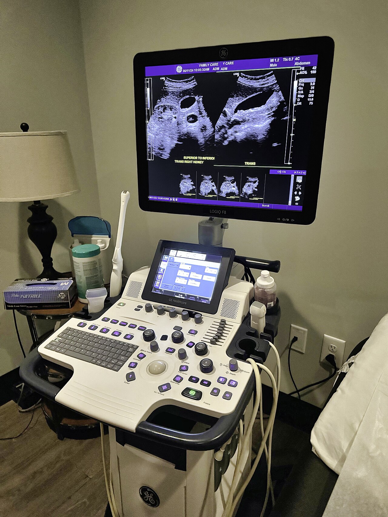
Image quality sells machines. The headline resolution figure means little on its own. What matters is whether the device shows the structure a clinician needs to see, at the depth they need to see it, in the room where they work. A demo in a quiet vendor suite flatters every machine. The honest test is a scan on a real patient, in the clinic’s own light, with the clinic’s own hands.
Ask for that test before buying.
Watch how the image holds up on a larger patient. Watch how fast it refreshes when the probe moves. Watch how it copes with a difficult window, a gassy abdomen, a deep vessel. A machine that looks crisp on a slim model in a demo can struggle on the patient who walks into the clinic on a busy afternoon. The picture that counts is the one the device makes under real load. The brochure shows a kinder one.
How it connects, and to what
A portable scanner shows its picture somewhere. Some carry their own screen. Many send the image to a phone or a tablet over a cable or a wireless link. The choice shapes daily use. A built-in screen needs nothing else. It adds weight and cost. A phone-paired probe is lighter, though it ties the clinic to the handsets the device supports.
Check the support list before assuming a fit.
A probe that pairs with one brand of phone may refuse another. A clinic that runs a mix of devices has to confirm each one. Confirm the operating system too. A model that supports a current tablet may drop an older one. The connection is easy to overlook in a demo. It becomes the first daily frustration if it was never checked.
Battery, weight, and the working day
Portability is the whole point. Weight and battery decide whether the device truly travels. A scanner that needs a wall socket every hour is a desk machine in disguise. Read the scan-time figure, then halve it in your head. Vendor numbers come from gentle modes. Color flow and continuous use drain a cell far faster.
Ask how long it scans under use. The idle figure flatters.
Weight tells a similar story. A handpiece that feels light in a showroom grows heavy across a long clinic list. Balance matters as much as mass. A device whose weight sits over the scanning face holds steady. One that drags at the wrist tires the hand. Hold the machine the clinic is considering. Hold it for longer than a minute. The wrist gives an honest answer the spec sheet cannot.
Durability and the things that break
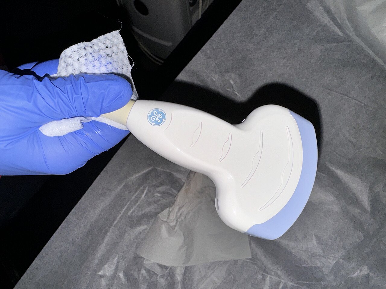
A clinic device lives a hard life. It gets dropped. It gets wiped with harsh disinfectant between patients. The probe cable, where one exists, flexes thousands of times. Ask what the housing is rated to survive. Ask whether the probe face tolerates the cleaning agents the clinic uses. A sealed, fluid-resistant body matters in a setting where spills happen.
The cheapest machine often costs the dearest in the end.
A device that fails in eight months, with a probe that costs half the system to replace, is no bargain. Read the warranty with care. Find out what a replacement probe costs, and how long a repair takes. A clinic with one scanner cannot afford weeks without it. Durability and service are part of the price, even when the sticker hides them.
Clearance, records, and the rules
A medical device has to clear the rules of the market it serves. A portable ultrasound is no exception. Confirm that the machine carries the clearance the clinic’s country requires. A device sold cheaply across a border may lack it. That gap becomes the clinic’s problem at an inspection.
The image also has to leave the device.
An exam belongs in the patient’s record. Check how the machine exports a study, and into what. A scanner that locks images inside an app, with no path to the clinic’s records, creates work and risk. A device that sends studies over the standard medical-imaging protocol folds into the systems a clinic already runs. Clean export, a quiet feature, counts for more than a flashy mode that goes unused.
Trial it in the clinic, not the showroom
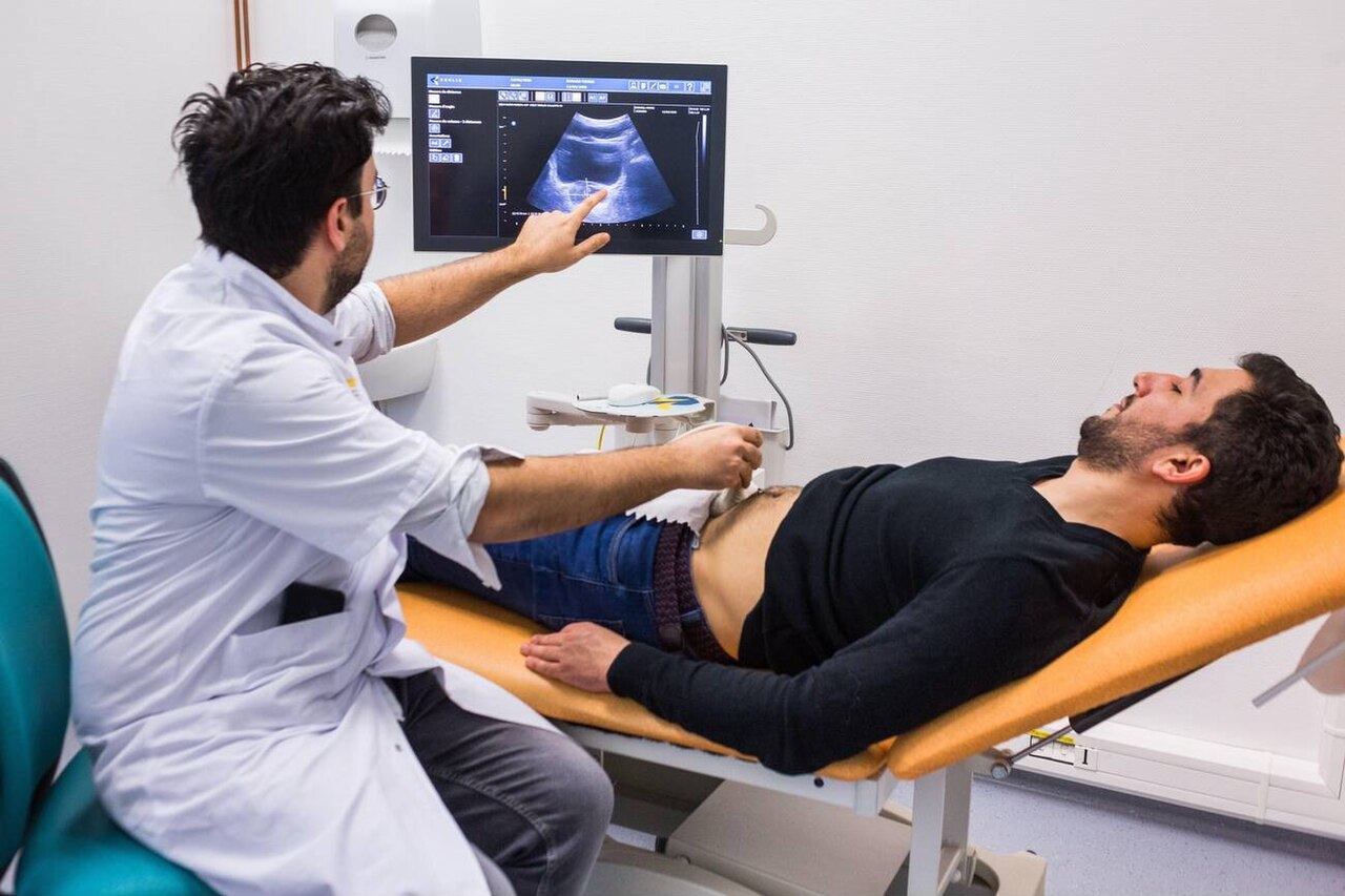
A vendor suite is built to flatter. The lighting is kind. The model is slim. The case is easy. None of that is the clinic’s Tuesday afternoon. The only honest trial happens on the clinic’s own patients, in the clinic’s own room, in the clinic’s own hands.
Ask for a loan unit. Serious vendors will usually provide one.
Run it for a week on the real caseload. Hand it to the people who will use it. The lead clinician is only one of them. A device the senior doctor loves can still gather dust if the nurses find it clumsy. Watch the small frictions. How long does it take to wake and pair? How many taps reach the mode a scan needs? Does the gel wipe clean from the seams? These tiny costs repeat hundreds of times a week. They decide whether a machine becomes part of the work or a chore avoided. A spec sheet never shows them. A week in the clinic shows nothing else.
Budget, without false economy
Price sets the shortlist. It should not end the thinking. The lowest number on a comparison page often hides the real cost. A bare probe with no warranty, no training, no local support, and a costly replacement part can outspend a dearer machine within a year. Build the true cost. Add the probes the caseload needs. Add the warranty. Add a spare, where one scanner is too few. Add the training that turns a box into a working tool.
Then compare the totals. The stickers mislead.
A clinic that buys on the headline price alone tends to buy twice. A clinic that prices the whole working system tends to buy once. The cheaper path is rarely the smaller number on the first page. It is the device that does the clinic’s real work, for years, without a drawer-full of regrets behind it.
Support, training, and the first month
A scanner arrives as a box. It becomes a clinical tool only when people can use it well. The gap between those two states is training. A clinic that skips it owns an expensive paperweight for far longer than it expects.
Ask what training comes with the purchase.
Some vendors send a specialist to the clinic for a day. Some offer video courses the staff work through at their own pace. Some hand over a manual and wish you luck. The difference shows up in the first month. A team taught to find the right preset, to optimise an image, to recognise a common artifact, starts producing useful scans within days. A team left to guess produces blurry pictures, blames the machine, then leaves it in the cupboard. The hardware was never the problem. The onboarding was.
Local support matters as much as the first lesson. A clinic in a large city may reach a service centre within hours. A clinic far from one waits days for every question. Ask where the nearest support sits. Ask how a fault gets handled, and how fast. A device with a local presence behind it keeps running. A grey-market unit with support an ocean away stops at the first problem and stays stopped. The picture quality fills the brochure. The support behind the device fills the years after the sale, and it decides whether the machine still scans on its third birthday.
A short way to decide
The choice comes down to a handful of honest questions. What does the clinic truly scan? Which probes does that work need? Does the picture hold up on a real patient, in a real room? Will the device connect to what the clinic owns? Can a hand hold it through a full list? Will it survive the cleaning, the drops, the years? Does it clear the rules and feed the records? Does the full price still make sense, the whole price and not the sticker alone?
Answer those, and the machine almost picks itself.
A portable ultrasound bought this way earns its place at the bedside. It scans what the clinic needs. It travels where the patient is. It lasts. The work it does, day after day, is the only review that matters. A clinic feels that verdict in quiet ways: a scan finished before the patient grows restless, a question answered without a referral, a tool reached for in place of one avoided.
About the Author
Julien Mercier
Senior R&D Engineer · Medical Ultrasound Transducer Development
Senior R&D Engineer with an M.S. in Applied Physics and over 15 years of experience in medical ultrasound transducer development, specializing in the design verification and performance testing of high-frequency imaging transducers. Currently leading the development and verification of the company’s next-generation high-frequency linear-array transducer, responsible for imaging performance evaluation and reliability analysis in preclinical testing. Brings extensive hands-on experience in piezoelectric element tuning, beamforming parameter optimization, and system-level performance testing.