





































Frequency selection
7.5 to 10 versus 10 to 13 MHz Linear Probe Frequency Selection
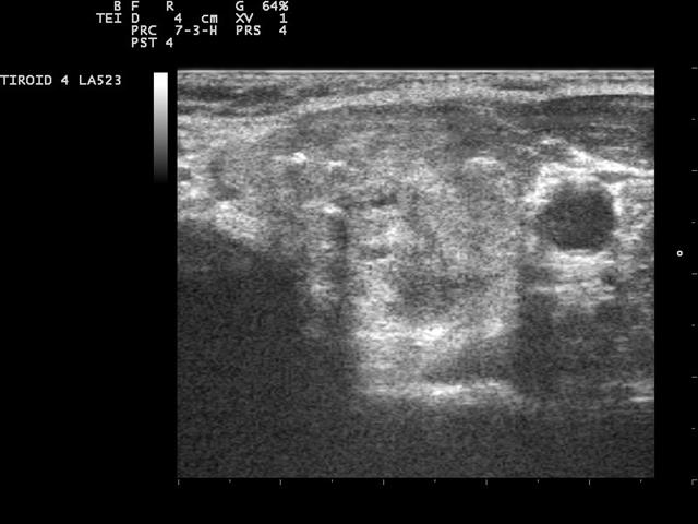
A linear probe is sold by its frequency band, and two bands cover the bulk of superficial work: a 7.5-to-10-megahertz range and a 10-to-13-megahertz range. The number decides two things at once. It sets how deep the beam reaches before it fades, and it sets how fine the detail is in the part of the field it does reach. The two bands sit a step apart on that scale, each suited to a depth and a structure, and a clinician picks between them by where the work sits under the skin. The choice is read off the body, not off the spec sheet.
What the frequency number controls
Frequency in ultrasound is a single dial that turns two outputs in opposite directions, and that one fact settles much of the choice. A higher number carries a shorter wavelength. Two structures a millimetre apart read as two when the wavelength is shorter than the gap between them and merge into one when it is longer, so the detail sharpens as the frequency climbs, down toward a tenth of a millimetre in the low teens of megahertz. The same higher number pays for that detail in depth. Tissue absorbs sound more strongly at a higher frequency, the beam losing energy faster as it travels, so it fades sooner and the field shrinks. A 13-megahertz beam runs out within three or four centimetres. A 7.5-megahertz beam, absorbed more gently, reaches five or six. A step up the scale sharpens the detail and shortens the depth. A step down lengthens the depth and coarsens the detail. No setting on any machine gives both at once. That is not a limit a better probe escapes; it is the physics of sound in tissue, the same for every ultrasound ever built. The band a probe carries is the band its crystal resonates across, since a piezoelectric element rings strongest at the frequency it was cut for and tapers off to each side of it. A 7.5-to-10-megahertz probe is built to ring through that span, a 10-to-13 probe through its own. Within the band the machine shifts the working frequency a step, dropping toward the floor of the band to reach a little further, lifting toward the ceiling to sharpen the near field. It cannot leave the band the crystal was built for, which is why the band printed on the probe, and not a software setting, fixes the depth and the detail the device can reach.
Where the 7.5 to 10 megahertz band reads
The 7.5-to-10-megahertz band reaches a little deeper, to around five or six centimetres, while holding a detail fine enough for the bulk of surface work. That reach suits the structures that sit a touch below the surface. A larger joint like a shoulder or a knee sits in this band, its tendons and its capsule a few centimetres down. The vessels of the limbs, a femoral artery in the groin or a larger arm vein, read at this depth. A thicker patient’s carotid, set deeper under a heavier neck, comes into range on this band.
The band also serves the structures that are large in their own right. A sizeable muscle belly, an abdominal wall in a focused look, a deep bursa, each sits far enough down that the extra reach matters more than the last increment of fineness. A clinician scanning across a range of patients, some lean and some heavy, leans on this band for the margin it keeps when the target sits deeper than the textbook.
The reach is what a heavier patient needs. A carotid that reads at two centimetres in a slim neck sits at four in a thick one, and the lower band carries the depth to reach it. A deep abscess, a haematoma in a bulky thigh, a hip set further down than a shoulder, each falls to this band for the reach. The grain comes a touch softer at that depth, the detail the body allows so far from the probe.
Where the 10 to 13 megahertz band reads
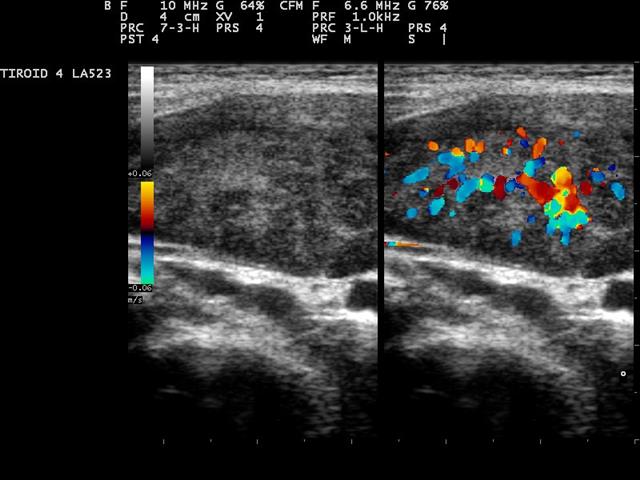
The 10-to-13-megahertz band draws a finer picture in a shallower field, reaching three or four centimetres with a detail toward a tenth of a millimetre. That fineness suits the structures that sit just under the skin and turn on small differences. A peripheral nerve shows its individual fascicles, the honeycomb a regional block is guided by. A tendon shows its fine parallel fibres, a small tear opening in the grain this band lays down clearly. The wall of a superficial vessel reads in clean layers, the intima-media thickness measured in fractions of a millimetre.
The thyroid sits squarely in this band on a normal neck, the gland’s even texture and a nodule’s fine calcium specks read at the resolution a structured score needs. A lymph node shows its internal architecture, the fatty centre a healthy node keeps. The salivary glands, the skin layers, a small foreign body lodged shallow, each reads at its sharpest here. For the structures that live in the first few centimetres and turn on fine detail, this band draws the picture that detail is read from.
The surface itself reads sharpest of all on this band. The eye, scanned through the closed lid, shows its chambers and its retina. The skin’s own layers separate cleanly enough to measure a lesion’s depth. Each sits in the first centimetre or two, where the higher band draws its finest grain.
The smallest-parts work turns on this band more than any other. A digital nerve in a finger, a tendon pulley in a hand, the fine septa inside a lymph node, each reads only when the resolution falls under a tenth of a millimetre. A foreign body a fraction of a millimetre across, a splinter of wood or a fleck of glass, throws its bright reflection on the higher band, a fleck the resolution here brings out clearly. For the finest structures at the surface, this is the band that resolves them.
What the frequency changes beyond detail and depth
Frequency shapes more than the depth and the grain. A high-frequency probe also drives harmonic imaging, where the machine listens not at the frequency it sent but at twice it, the harmonic the tissue itself generates as the sound passes through. The harmonic picture comes back cleaner, the haze and clutter of the near field cut away, so a superficial structure reads against a darker, quieter background. The higher the sent frequency, the higher the harmonic the probe must catch, one more way the band sets what the picture looks like.
The frequency also sets how the probe reads flow. The Doppler shift a moving target returns grows with the frequency sent, so a higher band lights up a slow flow and the fine vascularity of a small structure, a signal that stays faint on a lower band. The same higher frequency meets its sampling ceiling sooner, so a fast jet aliases at a lower velocity and the operator drops the scale to hold it. Frame rate enters here too, since a shallow high-frequency field is swept and refreshed quickly, the picture keeping up with a structure that moves under the probe.
Two bands, a step apart
The higher band draws the finer picture. The lower band reaches deeper. The two sit a step apart on one scale of frequency.
How a clinician reads the choice off the body
The pick follows the depth of the target. A reader asks how far under the skin the structure sits, then reaches for the band that draws its sharpest picture at that depth. A nerve in the wrist, a millimetre or two down, calls for the higher band. A nerve in the buttock, several centimetres down, calls for the lower one. The same structure at two depths takes two different bands.
The patient’s build shifts the same call. A lean patient brings every structure closer to the surface, so the higher band reaches it cleanly. A heavier patient pushes the same structure deeper, into the range the lower band is built to hold. A reader scanning a mixed list carries the depth of each patient in mind as much as the structure on the request.
The depth of the target is the whole of the question.
A reader who knows the two bands matches each to the work in front of it, the higher band for the finest detail in the first few centimetres, the lower for the reach to hold a deeper or a larger target in view. Many handheld probes now cover a broad span in one face and shift the working frequency by a preset, so a single device often spans both bands, the reader stepping the frequency to the depth instead of swapping a probe.
The choice is not made once and left. A reader who opens on the higher band and finds the target deeper than it looked steps the frequency down to bring it back, watching the picture clear as the band settles onto the depth. The frequency follows the structure through the study, set and reset as the depth read off the screen changes from one part of the scan to the next.
When one probe spans both bands
A broadband linear probe carries a span wide enough to work as both. It rings from the high single digits into the low teens of megahertz, and a preset sets where in that span it runs for a given job. A vascular preset holds the frequency high for a shallow vessel. A musculoskeletal preset drops it a step for a deeper joint. The reader picks the preset to the depth, and the one face covers the work two narrower probes once split between them.
That breadth changes how the choice is made without changing what it rests on. The depth of the target still decides the frequency. The difference is that the reader turns a dial, the other probe left in the drawer, the band following the structure under the skin in a single scan.
The span such a probe covers is wide by design. A crystal stack built from layered or composite material rings across a broader range than a plain element, so one face answers from the high single digits into the low teens of megahertz.
The reader sets it by the preset and confirms it on the image. A vascular preset opens the frequency high for a shallow vessel. When a vessel sits deeper than expected, a step down the band brings it back into a clean picture, the depth read off the screen and the frequency followed to it.
The breadth has its own character. A probe spanning a wide range tunes broadly across it. A crystal cut for a single point tunes sharply to that point and draws a touch finer there. A service that scans one depth all day may keep that dedicated probe. A service that ranges across depths takes the breadth and steps the frequency to each.
The width of that span has a name and a cost. A probe’s bandwidth, the spread of frequencies its crystal rings across, is what lets one face work as two, and a broadband element is engineered for it. The wider the bandwidth, the more depths a single probe covers, which is how a modern linear face stands in for the two or three single-band probes a cart once carried, the breadth bought with a more complex crystal and a higher price.
Common questions about linear probe frequency selection
What is the difference between a 7.5-to-10 and a 10-to-13 MHz linear probe?
The 7.5-to-10 band reaches a little deeper, to around five or six centimetres, with detail fine enough for the bulk of surface work. The 10-to-13 band draws a finer picture toward a tenth of a millimetre but fades at three or four centimetres. The higher band trades depth for detail.
Why does a higher frequency see less deep?
A higher frequency has a shorter wavelength, which resolves finer detail, and it is absorbed faster by tissue, so the beam fades sooner. Frequency turns detail and depth in opposite directions, so neither band gives both at once.
Which band suits the thyroid?
The 10-to-13 band on a normal neck, where the gland sits a centimetre or two down and its fine calcium specks and texture read at the resolution a structured score needs. A deeper or heavier neck may call for the lower band to keep the gland in view.
Which band suits nerves and tendons?
The higher band for a shallow nerve or tendon a millimetre or two down, where the fascicles and fibres read finest. A deeper nerve, several centimetres down, calls for the lower band to reach it.
How does patient build affect the choice?
A lean patient brings structures closer to the surface, so the higher band reaches them cleanly. A heavier patient pushes the same structure deeper, into the range where the lower band keeps its picture and the higher one fades.
Can one probe cover both bands?
Yes. A broadband linear probe rings across a wide span, and a preset sets where it runs for a given job, holding the frequency high for a shallow vessel and dropping it a step for a deeper joint. The depth of the target still decides the frequency.

About the Author
Julien Mercier
Senior R&D Engineer · Medical Ultrasound Transducer Development
Senior R&D Engineer with an M.S. in Applied Physics and over 15 years of experience in medical ultrasound transducer development, specializing in the design verification and performance testing of high-frequency imaging transducers. Currently leading the development and verification of the company’s next-generation high-frequency linear-array transducer, responsible for imaging performance evaluation and reliability analysis in preclinical testing. Brings extensive hands-on experience in piezoelectric element tuning, beamforming parameter optimization, and system-level performance testing.